Development of Axenic Culture and Astaxanthin Production in Microalgae
Min Chang Son1, Dong-Jun Lee1, Sejin Park1, Min Sung Kim2, Chul Won Lee2 and Won Gun An1,2*
1Division of Pharmacology, School of Korean Medicine, Pusan National University, Yangsan 626-870, Korea
2Institute of Marine BioTechnology Pusan National University, Busan 609-735, Korea Received April 29, 2015 /Revised June 30, 2015 /Accepted July 13, 2015
Microalgae are a renewable natural resource that requires only sunlight, carbon dioxide, phosphorus, and nitrogen for rapid growth. They produce a broad variety of basic chemical substances―such as vitamins, fatty acids and carotenoids-that have high added value potential for the pharmaceutical and food industries. The aim of this study was to develop axenic culture and to establish a cell growth assay for microalgae. A further experiment was carried out to determine the yield of astax- anthin derived from microalgae. The axenic culture was developed using a mixture of antibiotics [ampicillin (100 μg/ml), streptomycin (10 μg/ml), chloramphenicol (10 μg/ml), penicillin (10 μg/ml), neomycin (50 μg/ml), gentamycin (50 μg/ml), kanamycin (10 μg/ml), and nystatin (1.5 μg/ml)] and then used to extract a variety of useful components from the microalgae. The optimal concentration for the antibiotic mixture was 1-3 percent. A spectrophotometric cell growth assay was also established. Astaxanthin was extracted from Haematococus lacustris with a yield of 1.9×10⁻³ μg/l per 1 ml of culture medium. In conclusion, the axenic culture method developed here allows extraction of high-quality astaxanthin and other useful components from microalgae.
Key words : Antibiotics, astaxanthin, axenic culture, cell growth assay, microalgae
*Corresponding author
*Tel : +82-51-510-8455, Fax : +82-51-510-8420
*E-mail : [email protected]
This is an Open-Access article distributed under the terms of the Creative Commons Attribution Non-Commercial License (http://creativecommons.org/licenses/by-nc/3.0) which permits unrestricted non-commercial use, distribution, and reproduction in any medium, provided the original work is properly cited.
ISSN (Online) 2287-3406 Journal of Life Science 2015 Vol. 25. No. 7. 733~739 DOI : http://dx.doi.org/10.5352/JLS.2015.25.7.733
서 론
현대사회는 화석연료를 주된 에너지 공급원으로 사용하고 있으나 화석연료는 멀지 않은 장래에 고갈될 것으로 예측되고 있으며, 그 사용과정 중에 환경오염 문제를 발생시키고 있다.
특히 화석연료의 연소과정에서 발생하는 이산화탄소는 지구 온난화를 유발하는 것으로 알려져 있어 대기 중 이산화탄소의 농도를 감소시키기 위한 연구가 다양한 분야에서 전세계적으 로 폭넓게 연구되고 있다. 일례로, 화석연료가 가지는 문제점 을 해결하기 위한 방안의 하나로서 빛 에너지를 이용할 수 있는 미세조류에 관한 연구가 최근 활발하게 진행되고 있다 [10, 17, 23].
미세조류는 빛 에너지를 이용하여 이산화탄소를 고정화하 는 탄소동화능을 보유하고 있으며, 이를 통해 다양한 유용물 질을 생산할 수 있다[21]. 일부 미세조류는 에너지 함량이 높아 연료로의 사용 가능성이 타진되고 있어 이산화탄소의 저감과 유용물질 및 대체연료의 생산이라는 많은 장점을 가진다. 현 재 고부가가치의 의약품, 색소, 탄수화물 및 정밀화학약품 등
유용성분을 함유하고 있는 미세조류들을 유전자조작, 생육환 경조절 및 배양공정변수 최적화를 통해 인위적 대량배양이 가능토록 하는 미세조류 생명공학(microalgal biotechnology) 이 새로운 개척분야로 인식되고 있다. 또한 식품, 제약, 화장품 등 다양한 분야에서 천연물질의 수요가 급증하면서 세균, 곰 팡이 등 미생물 유래 유용물질 외에 천연물질 생산원으로서 미세조류에 대한 관심이 고조되고 있으며, 폐수처리와 농업으 로까지 그 이용범위가 확대되고 있다[7]. 이러한 미세조류가 다양한 유용물질을 생산할 수 있음에도 불구하고 그 생산량이 극히 적으며, 분리방법이 이미 정립된 상태여서 미세조류의 상업적 이용을 저해하는 요인으로 작용하고 있다. 따라서 미 세조류의 무균순수분리 및 유용물질의 검출방법을 개발할 필 요성이 있다.
미세조류는 군체를 형성하거나 점액성 물질에 둘러싸여 있 는데 세포 외부의 점액성 물질에는 많은 세균이 부착되어 있 다. 이러한 점액성 물질에 부착되어 있는 세균들은 쉽게 분리 되어 제거되지 않는다. 그러나 유용물질들을 함유하고 있는 미세조류를 이용한 유전학적 연구 및 생리학적 연구에서 세균 이 제거된 균주를 요구하고 있어 미세조류를 무균적으로 순수 분리시키는 것은 필수적이다[35]. 무균적 분리방법에는 원심 분리법[28], 초음파법[16], 희석법[29], 자외선법[27], 여과법 [11] 등의 물리적 방법과 페놀[22] 및 항생제를 이용하는 생물 화학적 방법[5, 33]들이 이용되고 있으나 항생제법이 보편적으 로 널리 사용되고 있다.
Isochrysis galbana H-002, H-007, H-008, H-009, H-010은 최 근 영양보충제로 각광받고 있는 docosahexaenoic acid (DHA),
Table 1. Microalgal species used for this study Microalga for
astaxanthine production
Microalgae for production of DHA and EPA Haematococuslacustris
- - - -
Isochrysisgalbana H-2 Isochrysisgalbana H-7 Isochrysisgalbana H-8 Isochrysisgalbana H-9 Isochrysisgalbana H-10
Table 2. The concentrations of antibiotics used for this study
Antibiotics Target Concentration
(μg/ml) Ampicillin
Streptomycin Chloramphenicol Penicillin Neomycin Gentamycin Kanamycin Nystatin
G(+), G(-) G(+), G(-)
G(-), virus, rickettsia G(+)
G(+), G(-)
G(+), G(-), mycoplasma G(+), G(-), mycoplasma Yeasts, molds
100 10 10 10 50 50 10 1.5 eicosapentaenoic acid (EPA) 등의 불포화 지방산을 합성하는
미세조류로 알려져 있다[1, 26]. 그리고 미세조류 Haematococcus lacustris는 생리활성물질인 astaxanthin (3,3'-dihydroxy-β, β' -carotene-4-4'-dione)을 생산하는 것으로 보고되고 있다[9, 15, 25]. Astaxanthin은 자연계에 널리 분포되어 있는 ketocar- otenoid로 polysoprenoid와 oxygen quenching 기능을 가진 benzenoid ring의 결합체이며[31], 물고기와 동물의 음식물 속 에 들어있는 색소의 원료일 뿐만 아니라, vitamin E와 β-car- otene 보다 더 강한 free radical 항산화 활성을 가진 잠재력이 있는 물질이다[8, 13]. 또한, astaxanthin은 세포내 환경이 과산 화 상태에 있을 때 막의 phospholipids와 다른 지질들을 보호 하는 작용을 가진 물질로 알려져 있으며[20, 24], astaxanthin 의 항산화 작용은 singlet oxygen 및 free radical을 소거함으로 써 항암 및 면역기능 활성에 효과가 있는 것으로 보고된 바 있다[3, 4, 12, 14, 34].
본 연구에서는 미세조류로부터 바이오매스 및 생리활성물 질의 대량생산을 최종목적으로 설계되었으며, 이에 따라 먼저 생리활성물질을 생산하는 미세조류의 무균순수분리 및 미세 조류 배양에 필요한 균체 생육도 측정법을 확립하고자 하였으 며, 또한 생리활성물질인 astaxanthin의 생성량을 파악하고자 하였다.
재료 및 방법
실험균주 및 배양
Astaxanthin과 DHA 및 EPA를 동시에 생산하는 미세조류 는 Table 1과 같다. Haematococus lacustris는 한국산업플랑크톤 소재은행으로부터, Isochrysis galbana 균주들은 한국 해양미세 조류은행으로부터 공급받았으며, 이들 미세조류들은 모두 순 수 분리되지 않은 단조주(uni-algal strain) 상태로 공급받았다.
또한 H. lacustris는 BBM 배지로, I. galbana 균주들은 f/2-Si 배지로 배양하여 실험을 진행하였다.
무균순수분리
단조주로부터 오염되어있는 세균을 제거하기 위하여 항생 제 처리법을 실시하여 무균 순수분리를 시도하였다. 항생물질 은 ampicillin (100 μg/ml), streptomycin (10 μg/ml), chlor- amphenicol (10 μg/ml), penicillin (10 μg/ml), neomycin (50
μg/ml), gentamycin (50 μg/ml), kanamycin (10 μg/ml), 그리 고 항진균제인 nystatin (1.5 μg/ml)을 사용하였으며, 각 항생 제를 지시 농도대로 각각 단독 처리하거나 항생제 혼합물을 조제하여 처리하였다. 미세조류 배양액을 항생제에 12시간 동 안 노출시킨 후, 항생제를 첨가하지 않은 배지에서 배양하면 서 미세조류의 생육도와 배양액의 무균성을 조사하였으며, 무 균성은 일정량의 배양액을 LB plate에 접종하여 30℃에서 배 양하면서 미생물 콜로니 생성 유무나 현미경으로 검경하여 확인하였다. 아래 Table 2에 표시한 항생제를 농도별로 처리한 후 생균수 측정법으로 오염유무를 평가하였다.
분광법을 이용한 측정
미세조류의 생성량, 성장속도 등을 정량적으로 파악하기 위 해 분광법을 이용하여 absorbance와 세포수의 상관관계를 조 사하였다.
Astaxanthin 생성을 위한 균주의 배양 조건
H. lacustris 순수균주를 이용하여 0.1 vvm, 5% CO2 조건에 서 250 ml elernmeyer flask에 130 ml working volume으로 전배양을 실시하였으며, 배양 15일 전후로 성장상태가 우수한 균주를 선발하여 실험에 사용하였다. 균주배양은 25˚C, 3000×
g, 10% inoculum, pH 7.0 조건에서 실시하였다. H. lacustris의 배양을 위해 60-70 μmol photon․m-2․s-의 조건에 맞는 형광 등 10개를 장착하고, 일정 광량을 조절할 수 있게 shaking in- cubator를 제작하여 사용하였다. 또한 이산화탄소 농도의 경 우 air와 고순도 이산화탄소를 일정비율로 혼합하여 조절하였 으며, 유속은 flowmeter를 이용하여 조절하였다.
Astaxanthin의 유도
배양된 세포를 원심분리하고 질소원이 배제된 배지(NIES- N 배지)에 재현탁 한 다음, 2,000× g에서 5분간 원심분리 하였 으며, 이 과정을 3회 반복하여 이전의 배지성분을 세척하였다.
세척이 완료 된 균주는 23℃, 150 rpm, 300 μmol photon․m-2
․s-1의 배지조건에서 astaxanthin induction을 실시하였다.
Table 3. The evaluation of bacterial contamination by treatment of antibiotics in the different microalgal species Microalgal
species
Mixture of antibioticsa
Cell survival (%)
Bacterial contamination
H. lacustris
Control 1%
2%
3%
4%
5%
100.0 50.0 16.7 13.3 13.3 6.7
+ - - - - -
I. galbanaH-8
Control 1%
2%
3%
4%
5%
100.0 50.0 62.5 62.5 37.5 25.0
+ - - - - -
I. galbanaH-7
Control 1%
2%
3%
4%
5%
100.0 100.0 60.0 20.0 4.0 4.0
+ - - - - - I. galbanaH-2 Control
1%
100.0 100.0
+ - I. galbana H-9 Control
1%
100.0 100.0
+ - I. galbana H-10 Control
1%
100.0 100.0
+ -
aMixture of antibiotics were composed of 100 μg/ml ampicillin, 10 μg/ml streptomycin, 10 μg/ml chloramphenicol, 10 μg/ml penicillin, 50 μg/ml neomycin, 50 μg/ml gentamycin, 10 μg/ml kanamycin, and 1.5 μg/ml nystatin.
Astaxanthin의 추출
Astaxanthin이 유도된 배양균체(H. lacustris)는 8,000× g, 4℃로 10분간 원심분리하여 상층액을 제거하고 균체는 증류 수로 반복 세척하였으며, 원심분리한 균체에 acetone을 첨가 하여 초음파 파쇄기(Branson 250, Dabbury, CT, USA)로 냉각 추출을 반복하였다. 그 후, 원심분리를 통하여 침전물의 균체 찌꺼기는 동결건조하여 고형분 회수율을 계산하였으며, 상층 액은 감압, 농축하여 용매를 제거하고, n-Hexane을 첨가하여 용해시켰다. 또한 분액깔때기에 물과 함께 첨가하여 흔들고, 수용액층을 제거(3회 반복)한 후, sodium sulfate anhydrous 를 이용하여 잔류수분을 제거시켜 astaxanthin을 추출하였다
Astaxanthin의 생성량 측정
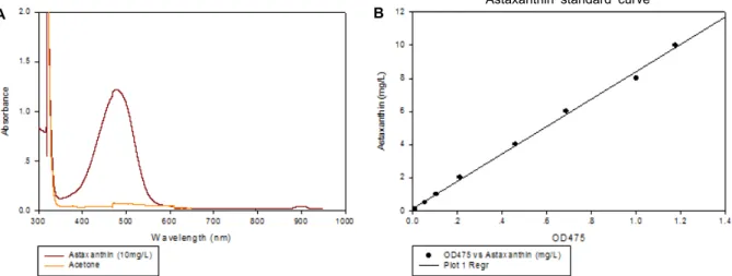
H. lacustris의 세포벽을 파쇄하여 astaxanthin을 추출하였 으며, 이를 이용하여 광범위한 파장에 걸쳐 UV spectrum을 조사하였고, 또한 확인된 파장에서 흡광도와 astaxanthin 농도 의 상관관계를 조사하여 생성량을 측정하였다. Astaxanthin standard curve를 그리기 위해 사용된 표준물질은 독일 슈트 트가르트 Subitec Gmbh 회사로부터 공급받았다.
결과 및 고찰
무균 순수분리
미세조류는 세포벽에 작은 구멍들이 존재하는데, 이곳에 여 러 세균들이 부착하여 공생한다[35]. 임과 이는 Gyrodinium을 순수분리하기 위해 ampicillin, cephalosporin, neomycin 등의 항생제 처리와 원심분리법을 이용하였고[33], 기 등은 Alexan- drium과 Peridinium의 무균배양을 위해 항생제와 미세필터를 사용하였다[11]. 또한, 조 등은 ampicillin, gentamycin, kana- mycin, neomycin, streptomycin 5종을 혼합한 항생제가 I. gal- bana의 무균화에 가장 효과적이라고 보고하였다[6]. 본 연구에 서 6종의 미세조류에 대해 혼합 항생제(ampicillin, streptomy- cin, chloramphenicol, penicillin, neomycin, gentamycin, ka- namycin, nystatin)을 처리한 후, 25℃ 광조건에서 120시간 정 치배양 한 결과는 Table 3과 같으며, 관찰 결과 모든 미세조류 가 순수분리 된 것을 확인할 수 있었다. 즉, 6종의 미세조류 순수분리를 위해 적합한 혼합 항생제의 농도는 H. lacustris가 1% 이었으며, I. galbanaH-8는 1-3% 이었고, I. galbanaH-7는 1-2% 이었으며, I. galbanaH-2, I. galbana H-9, I. galbana H-10는 각각 1% 이었다. 이는 1-3% 혼합 항생제의 농도가 미세조류의 치사효과는 줄이면서 또한 세균오염을 막을 수 있는 농도임을 의미한다.
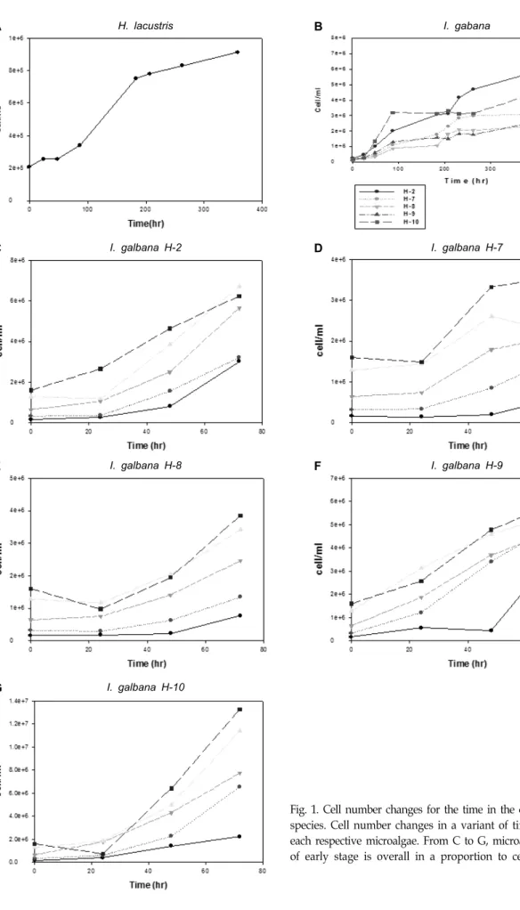
시간 및 초기 접종농도에 따른 미세조류의 증가량 비교 Fig. 1은 시간에 따른 미세조류의 증가량을 나타내었으며 또한 초기 접종농도에 따른 미세조류의 증가량을 비교하였다.
Fig. 1의 C-G에서 알 수 있듯이 미세조류량은 초기접종의 농도 에 대체적으로 비례하였다.
분광법을 이용한 측정
미세조류의 성장속도 생성량 등을 간편하게 정량적으로 측 정하기 위해 Melinda 등이 사용한 방법[18]에 따라, UV spec- trophotometer를 이용하여 세포수와 absorbance와의 관계를 알아보았다. Fig. 2에 제시한 바와 같이 680, 750 nm의 파장에 서 세포수와 absorbance 간에 강한 선형성이 나타남을 확인하 였다. 680, 750 nm에서의 흡광도는 미세조류의 chlorophyll a를 측정하는 파장이며, P. subcapitata의 경우 684 nm에서 최 대 흡광도를 나타낸다고 알려져 있는데 종에 따라 최대 흡광 도의 차이를 보인다는 보고와 일치하였다[19, 30]. 또한 세포수 와 건조중량과의 관계를 Fig. 3에 나타내었다. 강한 선형관계 가 관찰되었으며 그 비례상수는 2.0×10⁻⁸이었다. 따라서 UV spectrometer를 이용하여 세포의 생장속도를 확인할 수 있었 으며 이렇게 확립된 방법을 이용하여 항산화제 등의 유용한
A H. lacustris B I. gabana
C I. galbana H-2 D I. galbana H-7
E I. galbana H-8 F I. galbana H-9
G I. galbana H-10
Fig. 1. Cell number changes for the time in the different microalgal species. Cell number changes in a variant of time was shown for each respective microalgae. From C to G, microalgae concentration of early stage is overall in a proportion to cell number.
Fig. 2. The relationship between cell number and absorbance intensity at 680 and 750 nm, was shown, respectively.
Linear relationship was obtained.
Katsuda's method
Fig. 3. The relationship between dry weight and cell number was shown. Linear relationship was obtained.
A B Astaxanthin standard curve
Fig. 4. UV scanning for astaxanthin and its relationship with the intensity at 475 nm was shown. Lineal relationship was observed.
Fig. 5. Scanning for Haematococus lacustris and its medium (BBM) was shown.
용도로 사용이 가능한 astaxanthin을 배양, 추출 하는 데에 사 용하였다.
Astaxanthin의 생성량 측정
Astaxanthin은 vitamin E와 β-carotene 보다 강력한 free radical 항산화 활성을 가진 물질이며, 항암 및 면역기능 활성 에 효과가 있는 것으로 알려져 있다. 앞서 설명한 대로 H. la- custris로부터 생리활성 물질인 astaxanthin을 추출한 후, 이를 이용하여 넓은 파장에 걸쳐 UV spectrum을 얻었으며 472 nm 의 파장주위로 강한 흡수 피크를 확인하였다(Fig. 4A). 또한, 이렇게 확인된 472 nm의 파장에서 absorbance 양과 astax- anthin의 농도는 Fig. 4B에서 제시하는 바와 같이 선형성을 나타내었다. 이러한 방법은 Ana 등[2] 및 Strickland와 Parsons [32]이 사용한 분광학적인 방법에 따른 것이며, BBM 배지에서 배양된 H. lacustris를 이용하여 astaxanthin의 축적 정도를 확인하기 위하여 UV scanning을 실시한 결과, Fig. 5에 서 나타난 바와 같이 astaxanthin이 축적된 것을 관찰하였다.
이는 Ana 등, 이 등, 그리고 박 등이 H. lacustris로 부터 높은 항산화 효과를 갖는 천연색소인 astaxanthin의 축적을 확인한
후, 추출하였다는 보고와 일치하였다[2, 15]. 또한 배양액 1 ml 로부터 얻은 astaxanthin의 농도는 1.9×10⁻³±0.14×10⁻³ μg/l 이 었다.
결 론
미세조류로부터 유용성분 추출을 위한 무균 순수 분리법을 확립하기 위하여 항생제 혼합물을 이용하였으며, 그 결과 1-3% 혼합항생제의 농도가 미세조류의 치사 효과는 줄이면서 또한 세균오염을 막을 수 있는 최적농도임을 확인하였다. 또 한 미세조류의 양을 간편하게 측정하기 위하여 UV spectrum 상의 680, 750 nm의 파장에서 흡광도와 세포의 양을 비교한 결과, 선형성이 나타남을 확인하고 미세조류의 생장량을 평가 하였다. 더 나아가 H. lacustris 균주를 배양하여 천연 항산화제 등으로 이용할 수 있는 astaxanthin을 추출하였으며, H. la- custris 배양액 1 ml로부터 얻은 astaxanthin의 농도는 1.9×10⁻³ μg/l이었다. 따라서 개발된 무균 순수 분리법을 이용하여 미 세조류로부터 양질의 astaxanthin 및 유용성분들을 얻을 수 있을 것으로 사료된다.
감사의 글
본 연구는 한국연구재단(NRF-2013R1A2009593)과 부산광 역시 신성장산업과의 지원을 받아 수행하였음.
References
1. Aarab, L., Perez-Camacho, A., Viera-Toledo, M. dP., de Vicose, G. C., Fernandez-Palacios, H. and Molina, L. 2012.
Embryonic development and influence of egg density on early veliger larvae and effects of dietary microalgae on growth of brown mussel Pernaperna (L. 1758) larvae under laboratory conditions. Aquacult. Int. DOI 10.1007/s10499- 012-9612-7.
2. Ana, C., Mariela, G., Silvia, V., Maritza, H. and Nelson, G.
2003. Optimization of biomass, total carotenoids and astax- anthin production in Haematococcus pluvialis Flotow strain Steptoe (Nevada, USA) under laboratory conditions. Biol.
Res. 36, 343-357.
3. Bendich, A. 1991. Non vitamin a activity of carotenoids: im- muno enhancement. Food Sci. Technol. Res. 2, 127-130.
4. Bennedsen, M., Wang, X., Willen, R., Wadstrom, T. and Andersen, L. P. 1999. Treatment of H. pylori infected mice with antioxidant astaxanthin reduces gastric inflammation, bacterial load and modulates cytokine release by spleno- cytes. Immunol. Lett. 70, 185-189.
5. Campa-Córdova, A. I., Luna-González, A., Ascencio, F., Cortés-Jacinto, E. and Cáceres-Martínez, C. J. 2006. Effects of chloramphenicol, erythromycin, and furazolidone on growth of Isochrysisgalbana and Chaetocerosgracilis. Aquacul-
ture 260, 145-150.
6. Cho, J. Y., Choi, J. S., Kong, I. S., Park, S. I., Kerr, R. G.
and Hong, Y. K. 2002. A procedure for axenic isolation of the marine microalga Isochrysisgalbana from heavily con- taminated mass cultures. J. Appl. Phycol. 14, 385-390.
7. Choi, S. P. and Sim, S. J. 2012. Microalgal bioconversion to organic resources form CO2. KIC News 15, 11-24.
8. Guerin, M., Huntley, M. E. and Olaizola, M. 2003. Haemato- coccusastaxanthin: applications for human health and nutrition. Trends Biotechnol. 21, 210-216.
9. Hagen, C. H., Braune, W. and Greulich, F. 1993. Functional aspects of secondary carotenoids in Haematococcuslacust- ris [Girod] Rostafinski (Volvocales) IV.Protection from pho- todynamic damage.J. Photochem. Photobiol. 20, 153-160.
10. Jo, B. H. and Cha, H. J. 2010. Biodiesel production using microalgal marine biomass. KSBB J. 25, 109-115.
11. Ki, J. S., Cho, S. Y. and Han, M. S. 2006. Axenic Culture Method: A filtration technique to produce axenic cultures of the armoured Dinoflagellates. In: Hur S.B. (ed), Culture and application of useful microalgal. Life Science Publishing Co., 131-147.
12. Krinsky, N. I. 1989. Antioxidant functions of carotenoids.
Free Radical Bio. Med. 7, 617-635.
13. Kurashige, M., Okimasu, E., Inoue, M. and Utsumi, K. 1990.
Inhibition of oxidative injury of biological membranes by astaxanthin. Physiol. Chem. Phys. Med. NMR 22, 27-38.
14. Kurihara, H. 2002. Contribution of the antioxidative prop- erty of astaxanthin to its protective effect on the promotion of cancermetastasis in mice treated with restraint stress. Life Sci. 70, 2509-2520.
15. Lee, C. G. and Park, J. K. 2008. Immobilization of astax- anthin extracted from photosynthetic micro algae Haematococcus lacustris. J. Chitin Chitosan 13, 210-214.
16. Lim, M., Ong, B. L. and Wee, Y. C. 1992. A method of ob- taining axenic cultures of Trentepohlia spp. (Chlorophyta).
J. Phycol. 28, 567-569.
17. Mclaren, J. S. 2005. Crop biotechnology provides an opportu- nity to develop a sustainable future. Trends Biotechnol. 23, 339-342.
18. Melinda, G., Clive, G., Robert, H. and Susan, H. 2011.
Interference by pigment in the estimation of microalgal bio- mass concentration by optical density. J. Microbiol. Methods.
85, 119-123
19. Millie, D., Schofield, O., Kirkpatrick, G., Johnsen, G. and Evens, T. 2002. Using absorbance and fluorescence spectra to discriminate microalgae. Eur. J. Phycol. 37, 313-322.
20. Naguib, Y. M. A. 2000. Antioxidant activities of astaxanthin and related carotenoids. J. Agr. Food Chem. 48, 1150-1154.
21. Oh, H. M. and Ahn, C. Y. 2009. CO2 Fixation and biodiesel production using microalgae. KIC News 12, 12-20.
22. Olivier, S., Scragg, A. H. and Morrison, J. 2003. The effect of chlorophenols on the growth of Chlorella VT-1. Enzyme Micro. Techno. 32, 837-842.
23. Park, J. I., Woo, H. C. and Lee, J. H. 2008. Production of bio-energy from marine algae: Status and perspectives. Kor.
Chem. Eng. Res. 46, 833-844.
초록:미세조류를 이용한 무균분리법 개발 및 astaxanthin 생산
손민창1․이동준1․박세진1․김민성2․이철원2․안원근1,2*
(1부산대학교 한의학전문대학원 약물의학부, 2부산대학교 해양생물기술연구소)
미세조류는 천연재생자원으로서 단순히 빛, 이산화탄소, 인 및 질소만이 존재하는 환경에서도 빠른 속도로 자 라며 동시에 다양한 기본 화학물질(예를 들면 제약 및 식품산업에서 높은 부가가치를 창출 할 수 있는 비타민, 지방산, 카로티노이드 등)들을 생산한다. 본 연구에서는 생리활성물질을 생산하는 미세조류의 무균순수분리방법 의 개발 및 미세조류 배양에 필요한 균체 생육도 측정법을 확립하고자 하였다. 미세조류에서 유용성분 추출을 위하여 항생제 혼합물 [ampicillin (100 μg/ml), streptomycin (10 μg/ml), chloramphenicol (10 μg/ml), penicillin (10 μg/ml), neomycin (50 μg/ml), gentamycin (50 μg/ml), kanamycin (10 μg/ml), nystatin (1.5 μg/ml)]을 이용 한 결과 1-3%의 혼합 항생제의 농도범위에서 최적의 결과를 얻었으며 분광광도법을 이용한 균체생육도 측정법 또한 확립하였다. Haematococus lacustris 미세조류를 배양하여 astaxanthin을 추출하였으며, H. lacustris 배양액 1 ml로부터 얻은 astaxanthin의 농도는 1.9x10⁻³ μg/l이었다. 따라서 개발된 무균 순수분리법을 이용하여 미세조류로 부터 양질의 astaxanthin 및 유용성분들을 얻을 수 있을 것으로 사료된다.
24. Palozza, P. and Krinsky, N. I. 1992. Astaxanthin and can- thaxanthin are potent antioxidants in a membrane model.
Arch. Biochem. Biophys. 297, 291-295.
25. Park, J. K., Tran, P. N., Kim, J. D., Hong, S. J. and Lee, C. G. 2009. Carotenogenesis in Haematococcus lacustris: role of protein tyrosine phosphatases. J. Microbiol. Biotechnol. 19, 918-921.
26. Patil, V., Kallqvist, T., Olsen, E., Vogt, G. and Gislerød, H.
R. 2007. Fatty acid composition of 12 microalgae for possible use in aquaculture feed. Aquacult. Int. 15, 1-9.
27. Rappé, M. S., Connon, S. A., Vergin, K. L. and Giovannoni, S. J. 2002. Cultivation of the ubiquitous SAR 11 marine bac- terioplankton clade. Nature 418, 630-633.
28. Reardon, E. M., Price, C. A. and Guillard, R. R. L. 1979.
Harvest of marine microalgae by centrifugation in density gradients of "Percoll," a modified silica sol. In: Reed E. (ed), Methodological Surveys in Biochemistry, Vol. 8. Ellis Norwood Publishing, Chichester, U.K., 171-175.
29. Rippka, R., Coursin, T., Hess, W., Lichtlé, C., Scanlan, D.
J., Palinska, K.A., Iteman, I., Partensky, F., Houmard, J. and Herdman, M. 2000. Prochlorococcusmarinus Chisholm, et al.
1992 subsp. pastoris subsp. nov.strain PCC 9511, the first ax-
enic chlorophyll a2/b2-containing cyanobacterium (Oxypho- tobacteria). Int. J. Syst. Evol. Microbiol .50, 1833-1847.
30. Rodrigues, L. H. R., Arenzon, A., Raya-Rodriguez, M. T.
and Fontoura, N. F. 2011. Algal density assessed by spec- trophotometry: A calibration curve for the unicellular algae Pseudokirchneriella subcapitata. J. Environ. Chem. Ecotoxicol. 3, 225-228.
31. Shimidzu, N., Goto, M. and Miki, W. 1996. Carotenoids as singlet oxygen quenchers in marine organisms. Fish. Sci. 62, 134-137.
32. Strickland, J. and Parsons, T. 1972. A manual of seawater analysis. Bull. Fish Res. Bd. Can. 125, 1-310.
33. Yim, J. H. and Lee, H. K. 2004. Axenic culture of Gyrodiniu- mimpudicum strain KG03, a marine red-tide microalga that produces exopolysaccharide. J. Microb. 42, 305-314.
34. Yonouchi, J. 1993. Astaxanthin enhances in vitro antibody production to T-dependent antigens without facilitating pol- yclonal B-cell activation. Nutr. Cancer 19, 269-280.
35. Youn, J. Y. and Hur, S. B. 2007. Antibiotics and their opti- mum concentration for axenic culture of marine microalgae.
Algae 22, 229-234.