Natural Origin Polymers: Applications as Wound Care Materials
Fatih Karadeniz, Hye Kyeong Sung and Han Seong Kim*
Department of Organic Material Science and Engineering, Pusan National University, Busan 46241, Korea Received February 26, 2019 /Revised March 25, 2019 /Accepted March 26, 2019
Wound care is a health industry concern affecting millions worldwide. Recent increase in metabolic disorders such as diabetes comes with elevated risk of wound-based complications. Treatment and management of wounds are difficult practices due to complexity of the wound healing process. Con- ventional wound dressings and treatment applications only provide limited benefits which are mainly aimed to keep wound protected from external factors. To improve wound care, recent developments make biopolymers to be of high interest and importance to researchers and medical practitioners.
Biopolymers are polymers or natural origin produced by living organisms. They are credited to be highly biocompatible and biodegradable. Currently, studies reported biopolymers to exhibit various health beneficial properties such as antimicrobial, anti-inflammatory, hemostatic, cell proliferative and angiogenic activities which are crucial for effective wound management. Several biopolymers, namely chitosan, cellulose, collagen, hyaluronic acid and alginic acid have been already investigated and ap- plied as wound dressing agents. Different derivatives of biopolymers have also been developed by cross-linking with other molecules, grafting with other polymers, and loading with bioactive agents or drugs which showed promising results towards wound healing without any undesired outcome such as scarring and physiological abnormalities. In this review, current applications of common bio- polymers in wound treatment industry are highlighted to be a guide for further applications and studies.
Key words : Chitosan, collagen, extracellular matrix, tissue repair, wound care
*Corresponding author
*Tel : +82-51-510-2409, Fax : +82-51-512-8175
*E-mail : [email protected]
This is an Open-Access article distributed under the terms of the Creative Commons Attribution Non-Commercial License (http://creativecommons.org/licenses/by-nc/3.0) which permits unrestricted non-commercial use, distribution, and reproduction in any medium, provided the original work is properly cited.
Journal of Life Science 2019 Vol. 29. No. 3. 382~393 DOI : https://doi.org/10.5352/JLS.2019.29.3.382
Background
Part of the skin which is damaged or disrupted in struc- ture and/or function by various factors including but not limited to disease symptoms, infections, external stress, and thermal factors is defined as wound [33]. These complica- tions are often ignored, however, according to recent data published by World Health Organization (WHO), above 5 million people out of 50 million fatally wounded die each year which also leave millions of people in need of urgent treatment for their wounds [99]. As the term wound com- prises very different injuries or damages, it is usually classi- fied according to different variables such nature of the wound, occurrence of tissue loss and damaged skin layers.
Current market consists of high amount of wound treatment products, some of designed for specific types of wounds,
and new ways to treat or care wounds are being developed consistently. Despite the efforts and available options, wound treatment is still a field that needs cost efficient, envi- ronmentally friendly, effective and safe healing enhancers to relieve the burden for healthcare services and individuals suffering from different types of wounds [32, 58, 78].
Wound healing
Healing of the skin wounds requires a specific set of proc- esses initiated with the injury via an intricate cascade of bio- logical interactions leading to regeneration of the damaged tissue. Whole process of wound healing is a unique network and interactions among different type of cells (e.g. fibro- blasts, keratinocytes, monocytes, macrophages, etc.), ex- tracellular matrix (ECM) components, and cytokines [29].
Normal skin is protected from environmental factors by the protective barrier of epidermis and dermis layers [87].
Wound healing process is concurrently started with damage which breaks the equilibrium state of the protective barrier.
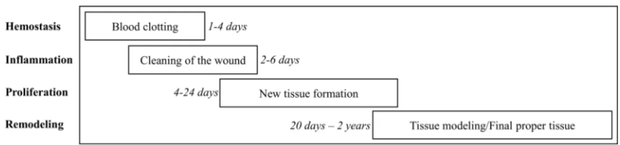
Typically, wound healing occurs naturally and follows four or five step process according to different wound healing characterization models (Fig. 1) [12, 86]. These steps overlap each other and consists of hemostasis, inflammation, migra-
- Review -
Fig. 1. Stages and approximate time span of wound healing process.
tion/proliferation, and remodeling (maturation) [12, 54].
Rate of these steps, hence the wound healing period, depend on several contributing factors, i.e. type of the wound (acute, chronic, superficial, etc.) and the status of individual during healing process (immunological and hormonal well-being) [31].
First step of the wound healing process is called hemo- stasis and targets to form blood clots with cross-linked fibrin protein as a first barrier against external factors, blood loss and to retain moisture [12]. Repairing the damage starts promptly after the injury and follows a strictly regulated closely linked biochemical signaling cascade at each step.
First step, hemostasis, starts with the accumulation of the thrombocyte platelets at the site of the injury to form the clot using fibrin protein which cross-links with itself and act as a mesh barrier [50]. This barrier is mainly composed of fibrin along with some blood cells and accumulated thrombocytes. Conventional wound treatment with tradi- tional products such as sponges, bandages, and gauze dress- ings find their use in this first step of wound healing and act as extra barrier that prevent bleeding and keep the wound moist for further steps to take place seamlessly [21].
Inflammation is the next step overlapping the fibrin mesh clot formation by release of pro-inflammatory cytokines from cells in injured tissue. Inflammatory response following the release of cytokines is the movement of leukocytes and monocytes to the injury site and clean the area of bacteria and remove the debris [11, 64]. In the inflammation step, growth factors are also secreted by activated monocytes in order to initiate the next overlapping wound healing step, migration/proliferation [49]. In this stage angiogenesis oc- curs simultaneously with granulating tissue formation, ECM component synthesis and deposition, and epithelization and contraction of the injured area. During granulating tissue formation new fibroblasts mature and secrete collagen and fibronectin as the first steps of forming new ECM. New ves-
sels are formed by angiogenesis and reepithelization of the epidermis layer is observed as the epithelial cells migrate and place themselves on top of each other, creating a cover for the newly organized tissue [28]. This stage is also respon- sible for scars and esthetical abnormalities due to excess for- mation of the granulating tissue and in return more than necessary collagen synthesis [23, 61]. Also, during this stage and the overlapping last stage of remodeling wounded tis- sue regains its functional state along anatomical and physio- logical properties via several activated pathways such Wnt/
β-catenin, MAPK, and PI3/Akt [88, 105, 108]. Last stage of the wound healing process is the remodeling the newly formed tissue. This step overlaps the synthesis of the colla- gen as the newly produced collagen is amassed randomly.
Remodeling of the new tissue includes the degradation of the irregularly deposited collagen and reorganization of the collagen fibrils regularly in bundles along the required lines [86].
Complications and unwanted esthetical outcomes are common during wound healing if any of the overlapping steps is altered in some way. As the wound healing process is complex, closely linked and tightly regulated, a compre- hensive care is of utmost importance during the healing without small to none alterations in patients’ daily life.
Wound care agents
Several applications are available to provide the care and
treatment effects that wound requires, however with their
limitations to overcome the challenges of complex wound
healing process. Failure to deliver adequate care may result
in serious undesirable aftereffects such as loss of function,
scars, chronic wounds, and fibrosis [27, 31, 102]. These appli-
cations, called wound dressing, target the injured area as
they cover the wound to serve as a scaffold for new cells
and ECM to form upon, and produced from suitable materi-
als which are biodegradable, allowing oxygen and vapor
Fig. 2. Chemical structures of biopolymers used in wound dressings.
permeation while blocking the external impacts and internal leaks [19]. One other important factor to be taken into con- sideration is the prevention of the infection of the wounded area to hasten and smooth the healing process. Several natu- ral and synthetic materials have been developed by collabo- rative researches of different fields such as biochemistry, tex- tile, and organic material engineering [85, 91]. These pro- moted wound dressing materials have been shown to pos- sess promising beneficial effects towards the uninterrupted and complication-free wound healing. Owing to recent im- provements in skin tissue engineering and wound healing studies, bioactive substances have gained much attention and acclaim. They have been positively influential in in- trinsic and dynamic healing process through direct inter- action with responsible pathways or building blocks of the new ECM [22, 47]. Natural products have been studied in detail for decades and suggested to exhibit numerous health beneficial properties that can be utilized as wound treatment agents. Due to their bioactivities, natural product-based wound care products can interact with inflammation, pro- liferation and remodeling steps of healing [10, 85]. As pre- vious studies showed, natural bioactive substances can en- hance or intervene cellular signaling pathways to increase the rate and efficacy of differentiation, specialization, and function of significant cell types of wound healing, fibro- blasts, macrophages, and keratinocytes [37, 68, 75, 94]. Apart from intracellular interaction, bioactive substances can also regulate inflammatory response, eliminate infectious threats, and aid the ECM building. Among the beneficial materials that have been used in wound care, polymers from different sources are widely employed with a broad scope as poly- mers have been exploited as durable and highly biocompat-
ible materials with ideal characteristics beneficial to crucial wound healing steps including cell proliferation, ECM or- ganization and antimicrobial action [10, 45]. In this context, current review targets and underlines the current trends in using natural origin biopolymers as wound dressing and treatment materials.
Natural origin biopolymers in wound treatment
Polymers in wound dressing
Polymers are big organic molecules formed from smaller repeating units called monomers. Polymers have been used in different fields of health care system for a long time.
Hemorrhage prevention is one of those fields where bio- polymers are mainly employed as most of natural polymers present favorable combination of beneficial properties, i.e.
biodegradable, non-inflammatory and non-irritant [60, 65].
Some synthetic polymers, that are polymers created through chemical reactions in laboratory conditions, have been broadly utilized. Among them polylactic acid and poly- glycolic acid are part of implant development studies and skin tissue engineering while in bone tissue engineering hy- droxyapatite is widely exploited as hemostatic agent against bone sternums [6, 30, 67, 97]. Liquid and glycol wound seal- ants also largely contain synthetic polymers. Aside from syn- thetic polymers, proteins and polysaccharides are also poly- mers of natural origin that are heavily used to prevent bleed- ing when applied as wound dressing.
Natural biopolymers are polymers that are built in living organisms using organic molecules as their building blocks/
monomers such as amino acids and monosaccharides (Fig. 2).
Broad range of sources varying from plants to bacteria have been utilized to obtain different biopolymers with different advantages to be put use in wound care products [2, 65].
Fibrinogen, collagen, albumin, and gelatin are proteins known for their hemostatic properties along commonly ap- plied polysaccharides such as chitosan, cellulose, and poly-N-acetyl glucosamine [79, 80, 104]. Due to their natural origin, natural biopolymer wound care agents are exten- sively biodegradable and biocompatible which makes them favorable over synthetic polymers. They have been applied on wounds in different forms varying from sponges to liquids. Natural polymers also have been observed to be re- absorbed readily by the body without causing inflammatory response when applied on injured areas. Coupling these fea- tures with hydrophilicity and their supporting presence dur- ing new tissue formation has generated much attention to- wards the use of natural polymers in skin tissue engineering, especially in wound care and treatment [10, 15].
Chitosan
Chitin is naturally abundant and simple β-(1→4) glycan composed of 2-acetoamido-2-deoxy-d-glucopyranose units.
It is the major constituent of shells of arthropods such as crabs, shrimps, lobsters, insects, and it also is produced ex- tracellularly by fungi and some brown alga. Chitin is a by- product or a waste from crab, shrimp, and crawfish process- ing industries and a highly water-insoluble compound.
Chitosan is a functional and basic linear polysaccharide pre- pared by N-deacetylation of chitin in the presence of alkaline.
Chitin and chitosan are known to exhibit antitumor, hypo- cholesterolemic, and antihypertensive activity [71, 107]. The main motive for the development of new applications for chitosan lies in the fact that it is a very abundant poly- saccharide, as well as nontoxic and biodegradable. Among all reported biologically active properties of chitosan, hemo- stasis stimulation and tissue generation acceleration made it a widely chosen polymer agent to be introduced in wound treatment [3, 14]. To add its already advantageous character- istics, its ability to prevent bacterial and fungal growth is crucial for efficient wound healing [18]. In addition to anti- microbial activities, chitosan is also reported to promote cell proliferation along fibroblast activation at a degree which is directly correlated with chitosan deacetylation levels [63, 95].
Initially chitosan has been used similar to that of other polymers in wound treatment as a dressing material to serve
as a scaffold for new tissue generation and as hydrogels for stimulate the wound healing [3]. Chitosan-based dressings provided the necessary conditions for efficient wound care;
a moist environment, protected from secondary infections and hastened tissue regeneration. Using sheets of N-carbox- ybutyl chitosan on surgery wounds of plastic surgery pa- tients expressed faster and more organized healing with minimal anomaly compared to that of traditional dressings [9]. Other studies by Ishihara et al. [42, 43] revealed that application of chitosan hydrogels notably enhanced the wound healing and contraction as well as accelerated the closure of incisions in vivo. Stone et al. [89] demonstrated that skin grafting splits returned to normal color earlier with chitosan dressings compared to conventional ones. Chitosan also advanced the reepithelization, angiogenesis and nerve regeneration. Further, mixing chitosan with other polymers produced promising results towards better wound manage- ment. Particularly, blending chitosan with polyethylene gly- col to form a wound treatment film expressed encouraging results [35]. Wounds applied with chitosan-polyethylene glycol film showed stimulated protein adsorption, increased cell proliferation and ECM formation along with sterile con- ditions free of any secondary infection. Photo-crosslinkable chitosan hydrogels bound with fibroblast growth factor-2 (bFGF) and epidermal growth factor (EGF) were shown to significantly stimulate wound healing through wound con- traction and reepithelization in healing-impaired mice and rats with burn wounds, respectively [4, 72]. In another study reported by Paul and Sharma [74], a wound dressing con- taining a combination of antibiotics and analgesics with chi- tosan, alginate and polyethylene glycol base helped to main- tain chronic non-healing ulcers on human subjects and showed beneficial effects towards faster healing and control of infection.
Owing to its cationic characteristics, chitosan is very suit-
able to be used as a drug carrier polymer. This nature of
chitosan was also employed in studies for better wound
treatment. Collagen synthesis was increased in both in vitro
and in vivo evaluations when the wounds were treated with
polyethylene glycol-grafted chitosan composite film loaded
with curcumin [56]. Ong et al. [73] showed that chitosan
wound dressings impregnated with polyphosphate and sil-
ver exhibited significant hemostatic and anti-infective effects
on wounds, particularly against Staphylococcus and Pseudo-
monas sp. Other studies loaded chitosan-based wound dress-ing materials with cerium oxide [41], taurine [20], and neuro-
peptide neurotensin [69] to obtain downregulated inflamma- tory signaling, increased tissue regeneration, and stimulated fibroblast migration and collagen deposition, respectively.
Overall, studies showed that chitosan acts as suitable ma- trix for new tissue growth while activating macrophages, fi- broblast migration and proper organization of ECM. Also, chitosan was reported to act as a notable hemostatic agent with pain reducing properties. N-Acetylglucosamine, mono- mer of chitosan, is able to stimulate fibroblast proliferation and aids to orderly clustered collagen deposition when re- leased by depolymerization of chitosan wound dressing films, foams or hydrogels [9].
Alginate
Alginate, a predominantly brown alga sourced poly- saccharide, is comprised of (1-4)-linked β-D-mannuronate and α-L-guluronate monomers. Alginate is quite effective in absorbing the wound excretion and preventing undesired odor and pain [44]. Alginate dressings turn into gels through absorbing wound excretions via ionic exchange of alginate calcium and wound, or blood, sodium [93]. As expected, al- ginate also provides necessities for a proper wound treat- ment such as moist environment, limiting infection and ex- ternal interference, and stimulating tissue regeneration. Also, monocytes were shown to produce elevated levels of IL-6 and TNF-α, which are important cytokines for wound heal- ing, following alginate introduction [1, 103]. Cell adhesive- ness to the alginate is the main drawback of alginate use in wound treatment. This lack of scaffolding support for new ECM formation was overcome through the addition of pep- tide sequences in order to obtain cell-interactive alginates [5, 51]. Cell-interactive alginates were observed to mimic ECM features to accelerate the wound healing process by stimulated cellular response to alginate. In addition, mod- ifications on alginate was regarded to be more attainable compared to some other polymers which leaded to develop- ment of alginate-based combined wound treatment agents [52]. Improved wound healing results were achieved by blending alginate with curcumin or silver, silk fibroin, and chitosan [55, 83, 98]. Hydrogen films formed by sodium algi- nate blended with Aloe vera prior to UV-crosslinking showed favorable wound protection along protection from UV and light-induced damages [76]. Promising results were ob- served on wounds of healing-impaired mice when a hydro- gel blend of alginate, chitosan, and fucoidan was applied [70]. In another study by Xie et al. [101], chitosan-colla-
gen-alginate composite dressing expressed increased fibro- blast migration, and upregulated expression of bFGF, EGF.
Rats treated with composite dressing showed accelerated and smooth wound healing compared to rats treated with gauze or chitosan-only dressings. Alginate-chitosan compo- site dressings were also tested for their ability to be carriers for beneficial molecules. Hu et al. [39] reported that amor- phous hydrogels based on chitosan/alginate composite im- pregnated by EGF showed significantly better healing out- comes on rat wounds. Likewise, alginate was also shown to be feasible for mineral crosslink, particularly zinc. Cros- slinked zinc-sodium alginate polyacrylamide hydrogels showed superior antibacterial and wound care properties [109].
Collagen
Collagen is the most abundant protein in mammals as
it is the dominant component of the connective tissue. It is
formed by repeating amino acids bound by peptide bonds
and constitute the substantial part of ECM. Although there
are more than 20 types of collagen, human body consists
of mainly type I collagen followed by type II and III which
comprise less than 10% of total collagen [7]. Collagen is de-
graded into small fragments, particularly gelatin, during the
inflammatory response to an injury. Collagen degradation
is followed by macrophage migration to injured area and
fibroblast proliferation in order to form the new tissue. These
processes are initiated by the specific cleaved parts (Arg-Gly-
Asp) of the collagen, hence, collagen degradation is one of
the most important parts of the wound healing [8]. In a sim-
ilar fashion, presence of gelatin induces keratinocytes to lose
cell adhesion and gain their mobility, therefore stimulating
migration for tissue regeneration [77]. However, deteriorated
regulation of collagen production versus the collagen cleav-
age results in unsuccessful healing, undesired outcomes, or
chronic wounds. When the collagen degradation is not cou-
pled with sufficient collagen synthesis due to dysregulated
enzymatic activity (especially MMPs), newly formed colla-
gens are steadily cleaved keeping wound healing process
in the inflammatory step [82]. This problem was shown to
be eliminated via external collagen supplement. Collagen-
based wound dressings play important role in this context,
providing the excess collagen by means of inert collagen and
gelatin which in turn hinder the MMP activity and further
the wound healing process from inflammatory step to pro-
liferation [13]. Structural modification (cross-linking, blend-
ing, loading, etc.) of collagen supplied by wound dressings can help to maintain the degradation rates, hence, accelerat- ing the wound healing.
Studies exhibited that wound healing action of collagen can be significantly improved by modifications such as de- polymerization, binding with anti-inflammatory and anti- bacterial molecules, etc. Use of exogenous collagen for skin repair by remodeling the collagen fibrils with electrospinning and crosslinking with bioactive ingredients showed im- proved wound healing benefits. Rho et al. [81] suggested that mimicking the ECM collagen could be achieved by elec- trospinning collagen nanofibers coated with laminin. Using electrospun collagen-laminin composite expressed improve- ments in cell adhesion and proliferation during tissue repair.
Diabetic wounds improved when collagen linked with quer- cetin was introduced, due to remarkable reactive oxygen species scavenging [17]. A similar study also suggested that curcumin loaded collagen matrix showed healing properties on diabetic wounds with improved reepithelization [46].
Electrospinning collagen fibrils in a similar way to that of natural skin also showed increased wound healing efficiency for collagen-based wound treatment products. Sun et al. [90]
reported that producing nanofibrous collagen scaffolds elec- trospun in a basket weave pattern which resembles the colla- gen in native skin, enhanced wound healing in diabetic wounds of rats.
Hyaluronan
Hyaluronan is a biopolymer found in human body com- monly throughout connective, epithelial, and neural tissues.
It is made of disaccharide repeats; D-glucuronic acid and N-acetyl-D-glucosamine linked by alternating glycosidic bonds of β-1,4 and β-1,3. Present in epithelial tissue, the role of hyaluronan in efficient wound healing is critical. Migra- tion/proliferation and remodeling stages of wound healing are stimulated by the presence of hyaluronan [16]. When degraded, the cleaved parts of hyaluronan was showed to exhibit angiogenesis enhancing properties [25]. Also, pro- liferation of keratinocytes was enhanced following degrada- tion of hyaluronan as the degradation products bound to CD44 receptors [26].
In the same way to collagen supplement, providing exog- enous hyaluronan during the healing process of an injury expressed beneficial effects, although hyaluronan is natively present in skin tissue. Following post-injury hyaluronan in- troduction, reduced scarring was observed with healthy tis-
sue repair. Hu et al. [38] developed a hyaluronan scaffold which downregulated the TGF-β1 expression and provided an environment for promoted wound healing. Huang et al.
[40] suggested that treating non-healing wounds with hya- luronan-chitosan hydrogels loaded with vancomycin carry- ing poly(lactic-co-glycolic acid) microspheres significantly reduced microbial load of the injured area and stimulated the proliferation of endothelial cells. Likewise, hyaluronan- pullulan composite wound films improved wound healing by means of stimulated hemostasis and improved non-enzy- matic debridement [53]. Similar results were reported with wound healing studies using hyaluronan linked with active molecules or other polymers. Hyaluronan conjugated with chitosan and edaravone exhibited anti-inflammatory effects during wound healing both in vitro and in vivo [92] while hyaluronic acid, bisphosphonate and silver conjugation re- sulted in promoted healing with minimal microbial load in vivo [84]. Also, a randomized clinical trial conducted by Yildirim et al. [106] reported that topically applied hyalur- onan presented improved palatal epithelial wound healing with reduced pain.
Cellulose
Cellulose is a biopolymer containing repeating β-d-glu-
cose monomers linked with β-1, 4-glycosidic bonds and
present in cell walls of plant and bacteria. The porous struc-
ture of cellulose resembles the ECM of human skin and is
suggested to be beneficial as a scaffold for tissue generation
[36]. Due to its chemical structure, cellulose is mainly used
to keep the wounds moist and remove the wound excretions
via absorbing the dead tissue molecules and fibers. Keeping
wound moist is of high importance to wound healing, as
the moist environment is needed for supplying growth fac-
tors, migrating macrophages and proliferating fibroblasts
[48]. Other than wound protection activities cellulose does
not exhibit any beneficial effects in healing process. However,
modification of cellulose by linking bioactive agents and
therapeutic molecules or conjugating with other polymers
generated promising results for cellulose to be used as base
for wound treatment products. Blending cellulose with anti-
microbials such as silver nanoparticles and myostatin re-
sulted in wound dressing materials that show notable anti-
bacterial effects against E. coli and S. aureus when applied
on open wounds [62, 100]. Reepithelization of burn wounds
was achieved by wound dressing hydrogels produced from
cellulose UV cross-linked with acylic acid [66]. Similar re-
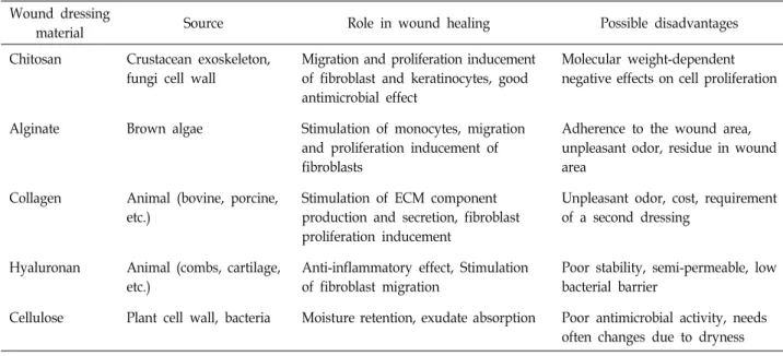
Table 1. Sources, biological roles and possible drawbacks of natural polymer wound dressings Wound dressing
material Source Role in wound healing Possible disadvantages
Chitosan Crustacean exoskeleton, fungi cell wall
Migration and proliferation inducement of fibroblast and keratinocytes, good antimicrobial effect
Molecular weight-dependent negative effects on cell proliferation
Alginate Brown algae Stimulation of monocytes, migration
and proliferation inducement of fibroblasts
Adherence to the wound area, unpleasant odor, residue in wound area
Collagen Animal (bovine, porcine, etc.)
Stimulation of ECM component production and secretion, fibroblast proliferation inducement
Unpleasant odor, cost, requirement of a second dressing
Hyaluronan Animal (combs, cartilage, etc.)
Anti-inflammatory effect, Stimulation of fibroblast migration
Poor stability, semi-permeable, low bacterial barrier
Cellulose Plant cell wall, bacteria Moisture retention, exudate absorption Poor antimicrobial activity, needs often changes due to dryness
sults were observed with bacteria-derived cellulose mem-
brane conjugated with chitosan [57]. Wound dressings with antibacterial and anti-inflammatory activities that can accel- erate tissue regeneration were obtained by conjugating cellu- lose with tungsten oxide and polydopamine [24, 59].
Some cellulose modifications such as creating nanoscale fibrillary, oxidized, and methylated cellulose produced promising results. Nano-fibrillar cellulose showed improved healing on skin graft donor wounds [34] while methylated and oxidized cellulose stimulated critical cellular responses such as cell migration and proliferation in injured area with improved hemostasis [96].
Cellulose provide perfect environment for keeping wound moist and protected and with efficiently modifiable structure cellulose-based wound treatment agents and tissue repair applications can be developed with improved benefits.
Summary and future trends
The biophysical process of wound healing is quite com- plex and regulated with diverse signaling and physi- ochemical pathways. The desirable outcome of wound heal- ing with minimal scar development, healthy tissue re- generation and organized ECM deposition is closely linked to the interactions between transcription factors, extracellular stimuli and cellular signaling. Natural polymers provide un- matched biodegradability, promising bioactivities and cost- efficient sourcing with minimal to none side effects to devel-
opment of wound treatment applications. Chitosan, alginate,
hyaluronan, collagen and cellulose are main biopolymers
that are currently main actors of wound treatment and skin
engineering studies and industry. Although current market
comprises of various wound treatment options based on nat-
ural biopolymers each providing specific advantages to-
wards different types of wounds, improvements are needed
as the attaining perfect healing is a condition yet to be
achieved. Biological roles and drawbacks of the reviewed
polymers summarized in Table 1. Future wound dressings
and tissue repair agents are needed to come with improved
abilities to provide antimicrobial, hemostatic, anti-inflamma-
tory, ECM mimicking properties, all in a suitable environ-
ment where cellular responses are also stimulated and
regulated. Future studies to understand the chemical, me-
chanical and biophysical aspects of biopolymers will ad-
vance the modifications of biopolymers to better suit the
wound care needs. Also, recent developments in nano-
technology and bioengineering will enable loading bio-
polymers with growth factors, drugs, or bioresponsive mole-
cules for enhancing tissue repair in nano-level with max-
imum cellular interaction. Developments in electrospinning
and 3D bio-printing will also enable to produce scaffolds
that mimic ECM in an elevated level to provide the perfect
microenvironment for tissue formation, cell adhesion, skin
repair, etc. Furthermore, recent technologies pave the way
for discoveries that will yield knowledge and know-how to
produce efficient cell-based therapies and tissue engineering
to obtain ECM substitutes. With technological improvements
in bioengineering and advantageous structures of bio- polymers, wound treatment applications are also improving significantly to obtain accelerated wound healing for both acute and chronic wounds with none undesirable outcome.
Acknowledgement
This work was supported by the BB21+ Project in 2019.
References
1. Aderibigbe, B. A. and Buyana, B. 2018. Alginate in wound dressings. Pharmaceutics 10, 42.
2. Agrawal, P., Soni, S., Mittal, G. and Bhatnagar, A. 2014. Role of polymeric biomaterials as wound healing agents. Int. J.
Low. Extrem. Wounds 13, 180-190.
3. Ahmed, S. and Ikram, S. 2016. Chitosan based scaffolds and their applications in wound healing. Achiev. Life Sci. 10, 27- 37.
4. Alemdaroğlu, C., Değim, Z., Çelebi, N., Zor, F., Öztürk, S.
and Erdoğan, D. 2006. An investigation on burn wound healing in rats with chitosan gel formulation containing epi- dermal growth factor. Burns 32, 319-327.
5. Alsberg, E., Anderson, K. W., Albeiruti, A., Franceschi, R.
T. and Mooney, D. J. 2001. Cell-interactive alginate hydro- gels for bone tissue engineering. J. Dent. Res. 80, 2025-2029.
6. Athanasiou, K. A., Niederauer, G. G. and Agrawal, C. M.
1996. Sterilization, toxicity, biocompatibility and clinical ap- plications of polylactic acid/polyglycolic acid copolymers.
Biomaterials 17, 93-102.
7. Avila Rodríguez, M. I., Rodríguez Barroso, L. G. and Sánchez, M. L. 2018. Collagen: a review on its sources and potential cosmetic applications. J. Cosmet. Dermatol. 17, 20-26.
8. Bellis, S. L. 2011. Advantages of RGD peptides for directing cell association with biomaterials. Biomaterials 32, 4205-4210.
9. Biagini, G., Bertani, A., Muzzarelli, R., Damadei, A., DiBene- detto, G., Belligolli, A., Riccotti, G., Zucchini, C. and Rizzoli, C. 1991. Wound management with N-carboxybutyl chitosan.
Biomaterials 12, 281-286.
10. Boateng, J. and Catanzano, O. 2015. Advanced therapeutic dressings for effective wound healing-a review. J. Pharm.
Sci. 104, 3653-3680.
11. Bodnar, R. J. 2015. Chemokine regulation of angiogenesis during wound healing. Adv. Wound Care 4, 641-650.
12. Braiman-Wiksman, L., Solomonik, I., Spira, R. and Tennen- baum, T. 2007. Novel insights into wound healing sequence of events. Toxicol. Pathol. 35, 767-779.
13. Brett, D. 2008. A review of collagen and collagen-based wound dressings. Wounds 20, 347-356.
14. Chan, L. W., Kim, C. H., Wang, X., Pun, S. H., White, N.
J. and Kim, T. H. 2016. PolySTAT-modified chitosan gauzes for improved hemostasis in external hemorrhage. Acta Biomater. 31, 178-185.
15. Chaudhari, A., Vig, K., Baganizi, D., Sahu, R., Dixit, S.,
Dennis, V., Singh, S., Pillai, S., Chaudhari, A. A., Vig, K., Baganizi, D. R., Sahu, R., Dixit, S., Dennis, V., Singh, S. R.
and Pillai, S. R. 2016. Future prospects for scaffolding meth- ods and biomaterials in skin tissue engineering: a review.
Int. J. Mol. Sci. 17, 1974.
16. Chen, W. Y. and Abatangelo, G. 1999. Functions of hyalur- onan in wound repair. Wound Repair Regen. 7, 79-89.
17. Chu, J., Shi, P., Yan, W., Fu, J., Yang, Z., He, C., Deng, X.
and Liu, H. 2018. PEGylated graphene oxide-mediated quer- cetin-modified collagen hybrid scaffold for enhancement of mscs differentiation potential and diabetic wound healing.
Nanoscale 10, 9547-9560.
18. Cremar, L., Gutierrez, J., Martinez, J., Materon, L., Gilkerson, R., Xu, F. and Lozano, K. 2018. Development of antimicro- bial chitosan based nanofiber dressings for wound healing applications. Mashhad Univ. Med. Sci. 5, 6-14.
19. Dabiri, G., Damstetter, E. and Phillips, T. 2016. Choosing a wound dressing based on common wound characteristics.
Adv. Wound Care 5, 32-41.
20. Değim, Z., Çelebi, N., Sayan, H., Babül, A., Erdoğan, D. and Take, G. 2002. An investigation on skin wound healing in mice with a taurine-chitosan gel formulation. Amino Acids 22, 187-198.
21. Dhivya, S., Padma, V. V. and Santhini, E. 2015. Wound dressings - a review. BioMedicine 5, 22.
22. Dias, A. M. A., Braga, M. E. M., Seabra, I. J., Ferreira, P., Gil, M. H. and de Sousa, H. C. 2011. Development of natu- ral-based wound dressings impregnated with bioactive compounds and using supercritical carbon dioxide. Int. J.
Pharm. 408, 9-19.
23. Ehrlich, H. 2000. Collagen considerations in scarring and re- generative repair, pp. 99-113. In: Garg, H. G. and Longaker, M. T. (eds.), Scarless Wound Healing. CRC Press: Boca Raton, FL, USA.
24. El Fawal, G. F., Abu-Serie, M. M., Hassan, M. A. and Elnouby, M. S. 2018. Hydroxyethyl cellulose hydrogel for wound dressing: fabrication, characterization and in vitro evaluation. Int. J. Biol. Macromol. 111, 649-659.
25. Gao, F., Liu, Y., He, Y., Yang, C., Wang, Y., Shi, X. and Wei, G. 2010. Hyaluronan oligosaccharides promote excisional wound healing through enhanced angiogenesis. Matrix Biol.
29, 107-116.
26. Gao, F., Yang, C. X., Mo, W., Liu, Y. W. and He, Y. Q. 2008.
Hyaluronan oligosaccharides are potential stimulators to an- giogenesis via RHAMM mediated signal pathway in wound healing. Clin. Investig. Med. 31, 106.
27. Ghatak, S., Hascall, V. C., Rodriguez, R. M., Markwald, R.
R. and Misra, S. 2017. Inflammation, wound healing, and fibrosis, pp. 195-209. In: Turksen, K. (ed.), Wound healing:
Stem Cells Repair and Restorations, Basic and Clinical Aspects. Wiley-Blackwell: Hoboken, NJ, USA.
28. Golebiewska, E. M. and Poole, A. W. 2015. Platelet secretion:
from haemostasis to wound healing and beyond. Blood Rev.
29, 153-162.
29. Gonzalez, A. C. de O., Costa, T. F., Andrade, Z. de A., Medrado, A. R. A. P., Gonzalez, A. C. de O., Costa, T. F.,
Andrade, Z. de A. and Medrado, A. R. A. P. 2016. Wound healing - a literature review. An. Bras. Dermatol. 91, 614-620.
30. Gunatillake, P. A., Adhikari, R. and Gadegaard, N. 2003.
Biodegradable synthetic polymers for tissue engineering.
Eur. Cells Mater. 5, 1-16.
31. Guo, S. and DiPietro, L. A. 2010. Factors affecting wound healing. J. Dent. Res. 89, 219-229.
32. Gupta, S., Andersen, C., Black, J., de Leon, J., Fife, C., Lantis Ii, J. C., Niezgoda, J., Snyder, R., Sumpio, B., Tettelbach, W., Treadwell, T., Weir, D. and Silverman, R. P. 2017. Manage- ment of chronic wounds: diagnosis, preparation, treatment, and follow-up. Wounds a Compend. Clin. Res. Pract. 29, S19- S36.
33. Gurtner, G. C., Werner, S., Barrandon, Y. and Longaker, M.
T. 2008. Wound repair and regeneration. Nature 453, 314- 321.
34. Hakkarainen, T., Koivuniemi, R., Kosonen, M., Escobedo- Lucea, C., Sanz-Garcia, A., Vuola, J., Valtonen, J., Tammela, P., Mäkitie, A., Luukko, K., Yliperttula, M. and Kavola, H.
2016. Nanofibrillar cellulose wound dressing in skin graft donor site treatment. J. Control. Release 244, 292-301.
35. Hashemi Doulabi, A., Mirzadeh, H., Imani, M. and Samadi, N. 2013. Chitosan/polyethylene glycol fumarate blend film:
physical and antibacterial properties. Carbohydr. Polym. 92, 48-56.
36. Hoenich, N. A. 2007. Cellulose for medical applications:
past, present, and future. BioResources 1, 270-280.
37. Houghton, P. J., Hylands, P. J., Mensah, A. Y., Hensel, A.
and Deters, A. M. 2005. In vitro tests and ethnopharmaco- logical investigations: wound healing as an example. J.
Ethnopharmacol. 100, 100-107.
38. Hu, M., Sabelman, E. E., Cao, Y., Chang, J. and Hentz, V.
R. 2003. Three-dimensional hyaluronic acid grafts promote healing and reduce scar formation in skin incision wounds.
J. Biomed. Mater. Res. 67B, 586-592.
39. Hu, Y., Zhang, Z., Li, Y., Ding, X., Li, D., Shen, C. and Xu, F. J. 2018. Dual-crosslinked amorphous polysaccharide hy- drogels based on chitosan/alginate for wound healing applications. Macromol. Rapid Commun. 39, 1800069.
40. Huang, J., Ren, J., Chen, G., Li, Z., Liu, Y., Wang, G. and Wu, X. 2018. Tunable sequential drug delivery system based on chitosan/hyaluronic acid hydrogels and plga micro- spheres for management of non-healing infected wounds.
Mater. Sci. Eng. C 89, 213-222.
41. Huang, X., Li, L. D., Lyu, G. M., Shen, B. Y., Han, Y. F., Shi, J. L., Teng, J. L., Feng, L., Si, S. Y., Wu, J. H., Liu, Y.
J., Sun, L. D. and Yan, C. H. 2018. Chitosan-coated cerium oxide nanocubes accelerate cutaneous wound healing by curtailing persistent inflammation. Inorg. Chem. Front. 5, 386- 393.
42. Ishihara, M., Nakanishi, K., Ono, K., Sato, M., Kikuchi, M., Saito, Y., Yura, H., Matsui, T., Hattori, H., Uenoyama, M.
and Kurita, A. 2002. Photocrosslinkable chitosan as a dress- ing for wound occlusion and accelerator in healing process.
Biomaterials 23, 833-840.
43. Ishihara, M., Ono, K., Sato, M., Nakanishi, K., Saito, Y.,
Yura, H., Matsui, T., Hattori, H., Fujita, M., Kikuchi, M. and Kurita, A. 2001. Acceleration of wound contraction and healing with a photocrosslinkable chitosan hydrogel. Wound Repair Regen. 9, 513-521.
44. Jones, V., Grey, J. E. and Harding, K. G. 2006. Wound dressings. BMJ 332, 777-780.
45. Kamoun, E. A., Kenawy, E. R. S. and Chen, X. 2017. A re- view on polymeric hydrogel membranes for wound dress- ing applications: PVA-based hydrogel dressings. J. Adv. Res.
8, 217-233.
46. Karri, V. V. S. R., Kuppusamy, G., Talluri, S. V., Mannemala, S. S., Kollipara, R., Wadhwani, A. D., Mulukutla, S., Raju, K. R. S. and Malayandi, R. 2016. Curcumin loaded chitosan nanoparticles impregnated into collagen-alginate scaffolds for diabetic wound healing. Int. J. Biol. Macromol. 93, 1519- 1529.
47. Koehler, J., Brandl, F. P. and Goepferich, A. M. 2018.
Hydrogel wound dressings for bioactive treatment of acute and chronic wounds. Eur. Polym. J. 100, 1-11.
48. Korting, H., Schöllmann, C. and White, R. 2011. Management of minor acute cutaneous wounds: importance of wound healing in a moist environment. J. Eur. Acad. Dermatol.
Venereol. 25, 130-137.
49. Landén, N. X., Li, D. and Ståhle, M. 2016. Transition from inflammation to proliferation: a critical step during wound healing. Cell. Mol. Life Sci. 73, 3861-3885.
50. Laurens, N., Koolwijk, P. and De Maat, M. P. M. 2006. Fibrin structure and wound healing. J. Thromb. Haemost. 4, 932-939.
51. LeBaron, R. G. and Athanasiou, K. A. 2000. Extracellular matrix cell adhesion peptides: functional applications in or- thopedic materials. Tissue Eng. 6, 85-103.
52. Lee, K. Y. and Mooney, D. J. 2012. Alginate: properties and biomedical applications. Prog. Polym. Sci. 37, 106-126.
53. Li, H., Xue, Y., Jia, B., Bai, Y., Zuo, Y., Wang, S., Zhao, Y., Yang, W. and Tang, H. 2018. The preparation of hyaluronic acid grafted pullulan polymers and their use in the for- mation of novel biocompatible wound healing film. Carbo- hydr. Polym. 188, 92-100.
54. Li, J., Chen, J. and Kirsner, R. 2007. Pathophysiology of acute wound healing. Clin. Dermatol. 25, 9-18.
55. Li, X., Chen, S., Zhang, B., Li, M., Diao, K., Zhang, Z., Li, J., Xu, Y., Wang, X. and Chen, H. 2012. In situ injectable nano-composite hydrogel composed of curcumin, n,o-car- boxymethyl chitosan and oxidized alginate for wound heal- ing application. Int. J. Pharm. 437, 110-119.
56. Li, X., Nan, K., Li, L., Zhang, Z. and Chen, H. 2012. In vivo evaluation of curcumin nanoformulation loaded methoxy poly(ethylene glycol)-graft-chitosan composite film for wound healing application. Carbohydr. Polym. 88, 84-90.
57. Lin, W. C., Lien, C. C., Yeh, H. J., Yu, C. M. and Hsu, S.
2013. Bacterial cellulose and bacterial cellulose–chitosan membranes for wound dressing applications. Carbohydr.
Polym. 94, 603-611.
58. Lindholm, C. and Searle, R. 2016. Wound management for the 21st century: combining effectiveness and efficiency. Int.
Wound J. 13, 5-15.
59. Liu, Y., Sui, Y., Liu, C., Liu, C., Wu, M., Li, B. and Li, Y.
2018. A physically crosslinked polydopamine/nanocellulose hydrogel as potential versatile vehicles for drug delivery and wound healing. Carbohydr. Polym. 188, 27-36.
60. Lloyd, L. L., Kennedy, J. F., Methacanon, P., Paterson, M.
and Knill, C. J. 1998. Carbohydrate polymers as wound management aids. Carbohydr. Polym. 37, 315-322.
61. Martin, P. and Nunan, R. 2015. Cellular and molecular mechanisms of repair in acute and chronic wound healing.
Br. J. Dermatol. 173, 370-378.
62. Miao, J., Pangule, R. C., Paskaleva, E. E., Hwang, E. E., Kane, R. S., Linhardt, R. J. and Dordick, J. S. 2011. Lysostaphin- functionalized cellulose fibers with antistaphylococcal activ- ity for wound healing applications. Biomaterials 32, 9557- 9567.
63. Minagawa, T., Okamura, Y., Shigemasa, Y., Minami, S. and Okamoto, Y. 2007. Effects of molecular weight and deacety- lation degree of chitin/chitosan on wound healing. Carbohydr.
Polym. 67, 640-644.
64. Minutti, C. M., Knipper, J. A., Allen, J. E. and Zaiss, D. M.
W. 2017. Tissue-specific contribution of macrophages to wound healing. Semin. Cell Dev. Biol. 61, 3-11.
65. Mogoşanu, G. D. and Grumezescu, A. M. 2014. Natural and synthetic polymers for wounds and burns dressing. Int. J.
Pharm. 463, 127-136.
66. Mohamad, N., Mohd Amin, M. C. I., Pandey, M., Ahmad, N. and Rajab, N. F. 2014. Bacterial cellulose/acrylic acid hy- drogel synthesized via electron beam irradiation: accel- erated burn wound healing in an animal model. Carbohydr.
Polym. 114, 312-320.
67. Moran, J. M., Pazzano, D. and Bonassar, L. J. 2003. Characte- rization of polylactic acid–polyglycolic acid composites for cartilage tissue engineering. Tissue Eng. 9, 63-70.
68. Morgan, C. and Nigam, Y. 2013. Naturally derived factors and their role in the promotion of angiogenesis for the heal- ing of chronic wounds. Angiogenesis 16, 493-502.
69. Moura, L. I. F., Dias, A. M. A., Leal, E. C., Carvalho, L., de Sousa, H. C. and Carvalho, E. 2014. Chitosan-based dress- ings loaded with neurotensin―an efficient strategy to im- prove early diabetic wound healing. Acta Biomater. 10, 843- 857.
70. Murakami, K., Aoki, H., Nakamura, S., Nakamura, S., Taki- kawa, M., Hanzawa, M., Kishimoto, S., Hattori, H., Tanaka, Y., Kiyosawa, T., Sato, Y. and Ishihara, M. 2010. Hydrogel blends of chitin/chitosan, fucoidan and alginate as heal- ing-impaired wound dressings. Biomaterials 31, 83-90.
71. Muxika, A., Etxabide, A., Uranga, J., Guerrero, P. and de la Caba, K. 2017. Chitosan as a bioactive polymer: processing, properties and applications. Int. J. Biol. Macromol. 105, 1358- 1368.
72. Obara, K., Ishihara, M., Ishizuka, T., Fujita, M., Ozeki, Y., Maehara, T., Saito, Y., Yura, H., Matsui, T., Hattori, H., Kikuchi, M. and Kurita, A. 2003. Photocrosslinkable chito- san hydrogel containing fibroblast growth factor-2 stim- ulates wound healing in healing-impaired db/db mice.
Biomaterials 24, 3437-3444.
73. Ong, S. Y., Wu, J., Moochhala, S. M., Tan, M. H. and Lu, J. 2008. Development of a chitosan-based wound dressing with improved hemostatic and antimicrobial properties.
Biomaterials 29, 4323-4332.
74. Paul, W. and Sharma, C. P. 2004. Chitosan and alginate wound dressings: a short review. Trends Biomater. Artif.
Organs 18, 18-23.
75. Pereira, R. F. and Bártolo, P. J. 2016. Traditional therapies for skin wound healing. Adv. Wound Care 5, 208-229.
76. Pereira, R., Mendes, A. and Bártolo, P. 2013. Alginate/aloe vera hydrogel films for biomedical applications. Procedia CIRP 5, 210-215.
77. Pilcher, B. K., Dumin, J. A., Sudbeck, B. D., Krane, S. M., Welgus, H. G. and Parks, W. C. 1997. The activity of collage- nase-1 is required for keratinocyte migration on a type i collagen matrix. J. Cell Biol. 137, 1445-1457.
78. Powers, J. G., Higham, C., Broussard, K. and Phillips, T. J.
2016. Wound healing and treating wounds: chronic wound care and management. J. Am. Acad. Dermatol. 74, 607-625.
79. Rathi, S., Saka, R., Domb, A. J. and Khan, W. 2019. Protein- based bioadhesives and bioglues. Polym. Adv. Technol. 30, 217-234.
80. Revelli, L., Tempera, S. E., Bellantone, C., Raffaelli, M. and Lombardi, C. P. 2016. Topical hemostatic agents, pp. 249- 259. In: Lombardi, C. P. and Bellantone R. (eds.), Minimally Invasive Therapies for Endocrine Neck Diseases. Springer Publishing: Cham, Switzerland.
81. Rho, K. S., Jeong, L., Lee, G., Seo, B. M., Park, Y. J., Hong, S. D., Roh, S., Cho, J. J., Park, W. H. and Min, B. M. 2006.
Electrospinning of collagen nanofibers: effects on the behav- ior of normal human keratinocytes and early-stage wound healing. Biomaterials 27, 1452-1461.
82. Rodríguez, D., Morrison, C. J. and Overall, C. M. 2010. Matrix metalloproteinases: what do they not do? new substrates and biological roles identified by murine models and proteomics. Biochim. Biophys. Acta 1803, 39-54.
83. Roh, D. H., Kang, S. Y., Kim, J. Y., Kwon, Y. B., Young Kweon, H., Lee, K. G., Park, Y. H., Baek, R. M., Heo, C.
Y., Choe, J. and Lee, J. H. 2006. Wound healing effect of silk fibroin/alginate-blended sponge in full thickness skin defect of rat. J. Mater. Sci. Mater. Med. 17, 547-552.
84. Shi, L., Zhao, Y., Xie, Q., Fan, C., Hilborn, J., Dai, J. and Ossipov, D. A. 2018. Moldable hyaluronan hydrogel enabled by dynamic metal-bisphosphonate coordination chemistry for wound healing. Adv. Healthc. Mater. 7, 1700973.
85. Simões, D., Miguel, S. P., Ribeiro, M. P., Coutinho, P., Mendonça, A. G. and Correia, I. J. 2018. Recent advances on antimicrobial wound dressing: a review. Eur. J. Pharm.
Biopharm. 127, 130-141.
86. Singer, A. J. and Clark, R. A. F. 1999. Cutaneous wound healing. N. Engl. J. Med. 341, 738-746.
87. Slominski, A. T., Zmijewski, M. A., Semak, I., Kim, T. K., Janjetovic, Z., Slominski, R. M. and Zmijewski, J. W. 2017.
Melatonin, mitochondria, and the skin. Cell. Mol. Life Sci.
74, 3913-3925.
88. Squarize, C. H., Castilho, R. M., Bugge, T. H. and Gutkind,
J. S. 2010. Accelerated wound healing by mtor activation in genetically defined mouse models. PLoS One 5, e10643.
89. Stone, C. A., Wright, H., Devaraj, V. S., Clarke, T. and Powell, R. 2000. Healing at skin graft donor sites dressed with chitosan. Br. J. Plast. Surg. 53, 601-606.
90. Sun, L., Gao, W., Fu, X., Shi, M., Xie, W., Zhang, W., Zhao, F. and Chen, X. 2018. Enhanced wound healing in diabetic rats by nanofibrous scaffolds mimicking the basketweave pattern of collagen fibrils in native skin. Biomater. Sci. 6, 340- 349.
91. Tamayol, A., Mohammadi, M. H., Bagherifard, S., Khadem- hosseini, A., Akbari, M., Serex, L., Faramarzi, N. and Mostafalu, P. 2016. Textile technologies and tissue engineer- ing: a path toward organ weaving. Adv. Healthc. Mater. 5, 751-766.
92. Tamer, T. M., Valachová, K., Hassan, M. A., Omer, A. M., El-Shafeey, M., Mohy Eldin, M. S. and Šoltés, L. 2018.
Chitosan/hyaluronan/edaravone membranes for anti-in- flammatory wound dressing: in vitro and in vivo evaluation studies. Mater. Sci. Eng. C 90, 227-235.
93. Thomas, S. 2000. Alginate dressings in surgery and wound management-part 1. J. Wound Care 9, 56-60.
94. Tsala, D. E., Amadou, D. and Habtemariam, S. 2013. Natural wound healing and bioactive natural products. Phytopharma- cology 4, 532-560.
95. Ueno, H., Mori, T. and Fujinaga, T. 2001. Topical for- mulations and wound healing applications of chitosan. Adv.
Drug Deliv. Rev. 52, 105-15.
96. Wagenhäuser, M. U., Mulorz, J., Ibing, W., Simon, F., Spin, J. M., Schelzig, H. and Oberhuber, A. 2016. Oxidized (non)- regenerated cellulose affects fundamental cellular processes of wound healing. Sci. Rep. 6, 32238.
97. Wahl, D. A., Sachlos, E., Liu, C. and Czernuszka, J. T. 2007.
Controlling the processing of collagen-hydroxyapatite scaf- folds for bone tissue engineering. J. Mater. Sci. Mater. Med.
18, 201-209.
98. Wang, L., Khor, E., Wee, A. and Lim, L. Y. 2002. Chitosan-al- ginate pec membrane as a wound dressing: assessment of incisional wound healing. J. Biomed. Mater. Res. 63, 610-618.
99. WHO 2010. Injuries and violence: The facts. Available at:
http://www.who.int/violence_injury_prevention/key_facts/
en/ (Accessed: 12 February 2019).
100. Wu, J., Zheng, Y., Song, W., Luan, J., Wen, X., Wu, Z., Chen, X., Wang, Q. and Guo, S. 2014. In situ synthesis of silver-nanoparticles/bacterial cellulose composites for slow- released antimicrobial wound dressing. Carbohydr. Polym.
102, 762-771.
101. Xie, H., Chen, X., Shen, X., He, Y., Chen, W., Luo, Q., Ge, W., Yuan, W., Tang, X., Hou, D., Jiang, D., Wang, Q., Liu, Y., Liu, Q. and Li, K. 2018. Preparation of chitosan-colla- gen-alginate composite dressing and its promoting effects on wound healing. Int. J. Biol. Macromol. 107, 93-104.
102. Xue, M. and Jackson, C. J. 2015. Extracellular matrix re- organization during wound healing and its impact on ab- normal scarring. Adv. Wound Care 4, 119-136.
103. Yang, D. and Jones, K. S. 2009. Effect of alginate on innate immune activation of macrophages. J. Biomed. Mater. Res.
Part A 90A, 411-418.
104. Yang, X., Liu, W., Li, N., Wang, M., Liang, B., Ullah, I., Luis Neve, A., Feng, Y., Chen, H. and Shi, C. 2017. Design and development of polysaccharide hemostatic materials and their hemostatic mechanism. Biomater. Sci. 5, 2357-2368.
105. Yew, T. L., Hung, Y. T., Li, H. Y., Chen, H. W., Chen, L.
L., Tsai, K. S., Chiou, S. H., Chao, K. C., Huang, T. F., Chen, H. L. and Hung, S. C. 2011. Enhancement of wound healing by human multipotent stromal cell conditioned medium: the paracrine factors and p38 mapk activation.
Cell Transplant. 20, 693-706.
106. Yıldırım, S., Özener, H. Ö., Doğan, B. and Kuru, B. 2017.
Effect of topically-applied hyaluronic-acid on pain and pal- atal epithelial wound healing: an examiner-blind, random- ized, controlled clinical trial. J. Periodontol. 89, 1-14.
107. Younes, I., Rinaudo, M., Younes, I. and Rinaudo, M. 2015.
Chitin and chitosan preparation from marine sources. struc- ture, properties and applications. Mar. Drugs 13, 1133-1174.
108. Zhang, D. L., Gu, L. J., Liu, L., Wang, C. Y., Sun, B. S., Li, Z. and Sung, C. K. 2009. Effect of wnt signaling path- way on wound healing. Biochem. Biophys. Res. Commun.
378, 149-151.
109. Zhou, Q., Kang, H., Bielec, M., Wu, X., Cheng, Q., Wei, W.
and Dai, H. 2018. Influence of different divalent ions cross- linking sodium alginate-polyacrylamide hydrogels on anti- bacterial properties and wound healing. Carbohydr. Polym.
197, 292-304.
초록:자연 고분자 : 상처 치료 재료로 활용
파티 카라데니즈․성혜경․김한성*
(부산대학교 유기소재시스템공학과)