Value of temporary stents for the management of
perivaterian perforation during endoscopic retrograde cholangiopancreatography
Sang Min Lee, Kwang Bum Cho
Sang Min Lee, Kwang Bum Cho, Department of Internal Medi- cine and Institute for Medical Science, Keimyung University School of Medicine, Daegu 700-712, South Korea
Author contributions: Lee SM and Cho KB contributed to this paper.
Supported by The research promoting grant from the Keimyung University Dongsan Medical Center in 2004
Correspondence to: Kwang Bum Cho, MD, PhD, Department of Internal Medicine and Institute for Medical Science, Keimy- ung University School of Medicine, 194 Dongsan-dong, Jung-gu, Daegu 700-712, South Korea. [email protected]
Telephone: +82-53-2507088 Fax: +82-53-2507442 Received: July 28, 2014 Revised: August 25, 2014 Accepted: September 16, 2014
Published online: November 16, 2014
Abstract
Endoscopic retrograde cholangiopancreatography (ERCP) has become the mainstay of treatment in hepato-pancre- ato-biliary disease. However, ERCP requires a high level of technical skills and experience in therapeutic endos- copy, there is always a risk of complications. Especially, the perforation per se affects the patient adversely, and the clinical course may lead to a poor prognosis, even with appropriate management. The treatments for ERCP- related perforation are diverse, depending on the location and mechanism of the bowel perforation and the time of diagnosis. Thus, we reviewed the appropriate surgical and non-surgical management options for therapeutic ERCP-related perforations, especially, evaluating metallic stenting as a treatment modality in perivaterian perfora- tion.
© 2014 Baishideng Publishing Group Inc. All rights reserved.
Key words: Endoscopic retrograde cholangiopancrea- tography; Perforation; Self-expandable metallic stent;
Duodenum; Perivaterian
MINIREVIEWS
Submit a Manuscript: http://www.wjgnet.com/esps/
Help Desk: http://www.wjgnet.com/esps/helpdesk.aspx DOI: 10.12998/wjcc.v2.i11.689
World J Clin Cases 2014 November 16; 2(11): 689-697 ISSN 2307-8960 (online)
© 2014 Baishideng Publishing Group Inc. All rights reserved.
World Journal of Clinical Cases
W J C C
Core tip: Although the evidence supporting the use of fully covered self-expandable metallic stent in periva- terian perforations is still insufficient, the clinical out- comes were encouraging.
Lee SM, Cho KB. Value of temporary stents for the management of perivaterian perforation during endoscopic retrograde cholan- giopancreatography. World J Clin Cases 2014; 2(11): 689-697 Available from: URL: http://www.wjgnet.com/2307-8960/full/
v2/i11/689.htm DOI: http://dx.doi.org/10.12998/wjcc.v2.i11.689
INTRODUCTION
Endoscopic retrograde cholangiopancreatography (ERCP) has become the mainstay of treatment in hepato-pancre- ato-biliary disease since its introduction in 1968
[1]. In the past, ERCP had been used as a diagnostic tool in choledo- cholithiasis presenting with jaundice, dilated common bile duct, acute pancreatitis, and cholangitis, but recently ERCP combined with sphincterotomy and stone removal has become a valuable therapeutic procedure
[2].
Because ERCP requires a high level of technical skills and experience in therapeutic endoscopy, there is always a risk of complications, such as bleeding, perforation, pan- creatitis, and cholangitis. Indeed, complication rates range from 5.4% to 11.2%
[3-11], among which the rate of perfo- ration, a potentially fatal complication, is 0.3%-1.0%
[3,12,13], and the rate of mortality in perforated patients is high (8%-23%)
[3,12-14]. Moreover, perforation
per se affects thepatient adversely, and the clinical course may lead to a poor prognosis, even with appropriate management. De- layed diagnosis and management can further affect clini- cal outcomes adversely
[15,16].
The treatments for ERCP-related perforation are di-
verse, depending on the location and mechanism of the
bowel perforation and the time of diagnosis
[17,18]. Previ- ously, most ERCP-related perforations, regardless of the above factors, were managed using surgery, and the mor- tality rate with such surgery was generally high. However, after the introduction of treatment strategies according to the type of perforation, nonsurgical management, such as radiologic interventions using percutaneous transhepatic biliary drainage (PTBD) and endoscopic management us- ing endoscopic nasobiliary drainage (ENBD), endoscopic retrograde biliary drainage (ERBD), endoclips, and fibrin glue, have been developed. Consequently, treatment out- comes have improved greatly over time
[12,15-24].
Now, nonsurgical techniques are being used in suit- able select patients more than often than surgery. Among the various nonsurgical options, several recent studies have reported that fully covered self-expandable metallic stents (SEMSs) could be used in ERCP-related perfora- tion, especially in periampullary perforations
[25-28]. Thus, we reviewed the appropriate surgical and non-surgical management options for therapeutic ERCP-related per- forations, especially, evaluating metallic stenting as a treat- ment modality in perivaterian perforation.
CLASSIFICATION OF ERCP-RELATED PERFORATION
The treatment modality in ERCP-related perforations is associated with the type of the perforation (Table 1). Stapfer
et al[18]classified perforations into four types according to anatomical location and severity. Type
Ⅰduodenal injuries are perforations of the lateral or medial wall, caused by the endoscope itself. Type
Ⅱduodenal in- juries are perforations of the medial wall. These are peri- vaterian or periampullary perforations, and most occur during endoscopic sphincterotomies. Type
Ⅲduodenal injuries are perforations of the distal bile duct, typically due to wire or basket instrumentation. Type
Ⅳduodenal injuries are diminutive retroperitoneal perforations due to excessive use of compressed air to retain a patent bowel lumen.
Similar to Stapfer’s classification, Howard et al
[17]report- ed three types of ERCP-related perforations in accordance with the mechanism of injury. Group
Ⅰperforations are guidewire perforations of the duct, group
Ⅱperforations
are periampullary perforations, and group
Ⅲperforations are duodenal perforations remote from the ampulla.
Regarding incidence by type of perforation, gener- ally, periampullary perforations caused by endoscopic sphincterotomies are most common, 15%-55%
[12,17-19]. Polydorou
et al[23]reported incidences using a modified classification of ERCP-related perforation. Type
Ⅰ, and type
Ⅱinjuries are identical with Stapfer’s type
Ⅰand
Ⅱinjuries, but type
Ⅲinjuries are ductal or duodenal perfo- rations caused by endoscopic instruments, but not guide- wires, and type
Ⅳinjuries are guidewire perforations with the presence of retroperitoneal air on X-ray examination.
They showed incidences of 68% for type
Ⅱ, 16% for type
Ⅰ, 11% for type
Ⅲ, and 4% for type
Ⅳperfora- tions. Another study showed that guidewire-related per- forations were most common (32%)
[19]. Moreover, 15%
were sphincterotomy-related perforations, 11% occurred during passage of the endoscope, and 9% occurred due to stent migration. Morgan et al
[22]reported that 12 of 24 cases of ERCP-related perforations were related to sphincterotomy, and the other 12 cases were perfora- tions remote from the papilla. Although the incidence of ERCP-related perforations varied slightly among the previous studies, sphincterotomy-related perforations tend to be most common, followed by guidewire-related perforations and free wall perforations.
RISK FACTORS FOR ERCP-RELATED PERFORATIONS
Several studies have reported risk factors for ERCP- related perforations. Overall risk factors, regardless of the type of ERCP-related perforation, include old age and a longer ERCP procedure time. Enns et al
[12]demon- strated that patients older than 65 years had a greater risk of ERCP-related perforation. Longer procedure times are often accompanied by repeated cannulation or more invasive methods to achieve “good” results. Thus, there tends to also be a greater risk of perforation. Addition- ally, ERCP-related perforation may be increased when performed by a trainee endoscopist. However, experts in the therapeutic ERCP field operate frequently and espe- cially with severe and difficult cases; thus, there is always a risk of perforation during the procedure regardless of the surgeon’s experience.
Risk factors for Stapfer’s type
Ⅰperforation are ab- normal anatomical structures, such as gastrojejunostomy, pancreaticoduodenectomy, duodenal diverticulum or stricture, and situs inversus
[22,29-31]. With anatomical fea- tures that differ from those of normal situations, it may be difficult to penetrate the bowel lumen using a side- viewing endoscope, increasing the risk of perforation by the endoscope itself.
Risk factors for Stapfer’s type
Ⅱ,
Ⅲ, and
Ⅳperfora- tions are similar and overlapping. They include sphincter of Oddi dysfunction, precut sphincterotomy, and a di- lated common bile duct on abdominal imaging
[12]. Precut sphincterotomy has been reported as a known risk factor
Table 1 Classification of endoscopic retrograde cholangiop- ancreatography-related perforations
Ref. Type
Stapfer et al[18] Type Ⅰ, duodenal perforation of medial or lateral wall
Type Ⅱ, perivaterian perforation Type Ⅲ, perforation of distal bile duct Type Ⅳ, retroperitoneal air alone Howard et al[17] Group Ⅰ, guidewire perforation
Group Ⅱ, periampullary retroperitoneal perforation Group Ⅲ, duodenal perforation remote from the ampulla
for pancreatitis
[6,7,32]. However, several studies have dem- onstrated that precut sphincterotomy also increases the risk of perforation compared with a conventional sphinc- terotomy. In fact, the risk of perforation increases if the incision for the sphincterotomy is outside of the usually recommended sector (11 to 1 o’clock position)
[6,33-35]. A previous report demonstrated that 7 of 13 sphincterot- omy-related perforations were associated with precut- ting
[12]. Since a dilated common bile duct is associated with distal common bile duct stricture, the risk of perfo- ration may be related to the deep manipulation needed to achieve a deep cannulation. Additionally, an ampullec- tomy can increase the risk of perforations. Alfieri et al
[15]reported that ampullectomy had been performed in 7 of 30 (23%) cases of ERCP-related perforations.
CLINICAL MANIFESTATIONS AND DIAGNOSIS
Patients with ERCP-related perforations may complain mainly of epigastric pain and tenderness, but obviously these complaints are very nonspecific. Other symptoms and signs include fever, tachycardia, leukocytosis, and mildly elevated serum amylase levels. Several studies have reported rare complications after ERCP, such as pneumo- mediastinum, pneumothorax, and gas in the portal sys-
tem
[36-42], whereas patients with retroperitoneal air present
on abdominal imaging, after an endoscopic sphincteroto- my, can be asymptomatic clinically. Generally, the patients did not require intervention but only conservative man- agement. Genzlinger
et al[43]showed that asymptomatic patients with retroperitoneal air evident on a computed tomography (CT) scan did not require surgical inter- vention. As the range is diverse, from asymptomatic to severe signs of peritonitis, to suspect and recognize the possibility of perforation early, during and after ERCP is most important. For early detection of perforation, it is necessary to check the patient’s condition immediately after ERCP. If the patient complains severe abdominal pain, abdominal X-ray and CT are good methods to identify ERCP-related perforation. If retroperitoneal air is visible during the procedure, abdominal X-ray and CT are also useful.
ERCP-related perforation can sometimes be diag- nosed readily by imaging if suspected. Typically, an ab- dominal X-ray may show retroperitoneal air around the right kidney. Suspected perforation may not be confirmed by an abdominal X-ray, but a contrast CT scan or upper gastrointestinal oral contrast evaluation should be help- ful. However, this could be delayed unless the physician suspects a perforation. Furthermore, if the patient has elevated serum amylase levels and complaints of epigas- tric pain, it is difficult to distinguish between perforation and pancreatitis. Gottlieb et al
[44]reported that post-ERCP pancreatitis can be excluded if their values of amylase and lipase 2 h after ERCP are below 276 U/L and 1000 U/L, respectively. Another study demonstrated that post- ERCP pancreatitis cases showed serum amylase levels
greater than five-fold the normal level
[45]. Thus, labora- tory findings, especially serum amylase and lipase levels, may be important clues for differentiating perforation from pancreatitis.
Although a physical examination is frequently useful in suspected patients, not all perforated patients show signs of acute peritonitis
[43]. Bell
et al[46]demonstrated positive physical findings in 75% of the included patients, but no specific finding of perforations. Thus, it is im- portant to consider not only a physical examination and laboratory findings but also abdominal imaging, such as abdominal X-ray and abdominal CT scans, for an accu- rate diagnosis.
TREATMENT OF ERCP-RELATED PERFORATION
Traditionally, ERCP-related perforation has been man- aged surgically. The objectives of such surgical manage- ment include control of infection and inflammation (drainage of the retroperitoneal/intraperitoneal fluid and air and drainage of the biliary system) and closure of the perforation, with or without bypass
[2]. However, the recent trend has been towards a selective approach according to the type of perforation and, more recently, according to the overall status of the patient, considering issues such as age, vital signs, peritoneal signs, comorbidi- ties, and CT images.
Duodenal free wall perforations (Stapfer type
Ⅰor Howard Group
Ⅲ) tend to be larger and located remote- ly from the ampulla and to cause substantial collections in the peritoneal or retroperitoneal space. Thus, these perfo- rations should be subjected to prompt surgical interven- tion. One study reported three of four cases of type I perforation that underwent surgery immediately
[12]. One case had abnormal anatomy, a gastrojejunostomy, and the others cases had duodenal diverticulum and stricture.
All three patients were suspected and diagnosed imme- diately; there was no mortality. The one patient without surgical management had severe comorbidities, thus re- ceiving only conservative management but died 2 d later.
Polydorou
et al[23]reported that 83% (6/7) of Stapfer’
s type
Ⅰperforation cases underwent surgery. Three pa- tients with type
Ⅰperforation showed abnormal anatomy due to Billroth
Ⅱgastrectomies. Among the patients who underwent surgery, two died as a result of respira- tory insufficiency and aspiration pneumonia. One patient with a type
Ⅰperforation, caused by rupture of the diver- ticulum, was managed conservatively. The patient had no fever or signs of peritonitis but only complained of mild abdominal pain.
In the studies mentioned above, in general most
of the type
Ⅰperforations were treated using surgery
due to the large size of the perforation. Recently, some
studies have introduced endoscopic management using
simple metallic endoclips or an endoloop with multiple
endoclips and fibrin glue for free wall duodenal perfora-
tions
[28,34,47-50]. In addition, an over-the-scope-clip, used
primarily in gastrointestinal bleeding or perforation, could be considered for use in post-ERCP perforations
[51,52].
Distal bile duct injuries (Stapfer type
Ⅲor How- ard Group
Ⅰ), caused by penetration of the guidewire through the bile duct during cannulation in a narrow or obstructed duct, tend to be smaller than duodenal free wall perforations. Commonly, these perforations tend to become obstructed spontaneously, and these pa- tients can be ‘cured’ by conservative management with intravenous antibiotics, hydration, pain control, or nil- by-mouth
[12,17,18,53]. However, since some patients have ongoing bile leakage, endoscopic management to prevent bile leakage to the retroperitoneum may be necessary. To prevent such leakage, ENBD or ERBD together with insertion of a plastic or metallic stent can be used. If endoscopic management is not possible, PTBD can be performed.
Diminutive duodenal perforation (Stapfer type
Ⅳ) is not a true perforation; generally, it can be treated suf- ficiently with conservative management alone. In fact, Genzlinger et al
[43]reported that retroperitoneal air alone, with no abnormal clinical signs or symptoms, does not require surgical intervention. However, regardless of the type of perforation, patients with retained stones or unal- leviated biliary obstruction should undergo surgery
[18].
Perivaterian perforations (Stapfer type
Ⅱor Howard group
Ⅱ) occurring after endoscopic sphincterotomy are controversial issues in the treatment field presently because of variation in clinical outcomes
[14,18,34,41,53-57]. Surgical intervention is not an issue but is an abstruse problem, because it seems that conservative management alone, including biliary drainage, may aggravate or fail to cure the perforation. Several studies have demonstrated that conservative management with or without biliary drainage was successful in peri-ampullary perforation pa-
tients
[18,53-55].
Wu et al
[16]reported that 55% (6/11) of patients with type II perforations were treated with conservative man- agement with or without biliary drainage. In all patients, clinical signs and symptoms improved rapidly. However, 80% (4/5) of the patients who underwent surgery died, due to delayed diagnosis and operation as well as sepsis.
The surgical indications for those patients were large retroperitoneal fluid collections, liver abscess on abdomi- nal CT scan, and severe abdominal pain. Enns
et al[12]demonstrated that 46% (6/13) of patients with type II perforations were treated using conservative management with or without nasogastric suction. In 38% (5/13) of the patients, biliary drainage (stent insertion with three, PTBD with two) was performed. The mortality rate was zero in patients managed conservatively and with biliary drainage. Alfieri
et al[15]showed that 40% (6/15) of pa- tients with type
Ⅱperforations were treated successfully by conservative management with biliary drainage (PTBD or nasobiliary drainage).
The rates of nonsurgical management vs surgical in- tervention in peri-ampullary perforation vary widely (Ta- ble 2)
[12,15-18,22,23,58,59]. Thus, the appropriate choice of treat- ment modality for peri-ampullary perforation remains an important issue. Most surgical indications in peri-ampul- lary perforations include hemodynamic instability, signs of peritonitis, continuing leakage, septic conditions, and a perforation of large size. One author also suggested that patients with a large amount of fluid collection in the peritoneum or retroperitoneum on abdominal CT should be treated aggressively
[12], because the possibility of con- tinuing leakage is high. If there is no surgical indication, the essential aspects of nonsurgical management consist of diversion of duodenal, biliary, and pancreatic drain- age
[21]. A nasogastric or nasoduodenal tube for duodenal
Table 2 Treatment of periampullary perforations (Stapfer’s type II perforations) Ref. Patients (n) Patients according to
treatment method, n (%) Treatment method Mortality n (%)
Alfieri et al[15] 15 6 (40.0) Conservative management + ENBD ± PTBD 0 (0.0)
9 (60.0) Surgery 1 (11.1)
Wu et al[16] 11 6 (54.5) Conservative management 0 (0.0)
5 (45.5) Surgery 4 (80.0)
Kim et al[58] 5 2 (40.0) Conservative management 0 (0.0)
3 (60.0) Surgery 0 (0.0)
Enns et al[12] 13 11 (84.6) Conservative management ± biliary drainage (PTBD, ERBD) 0 (0.0)
2 (13.4) Surgery 0 (0.0)
Polydorou et al[23] 30 24 (80.0) Conservative management ± biliary drainage (PTBD, ERBD) 0 (0.0)
6 (20.0) Surgery 0 (0.0)
Stapfer et al[18] 6 3 (50.0) Conservative management 0 (0.0)
3 (50.0) Surgery 0 (0.0)
Howard et al[17] 22 18 (81.8) Conservative management ± biliary drainage (ENBD, ERBD) 0 (0.0)
4 (18.2) Surgery 1 (25)
Morgan et al[22] 12 12 (100.0) Conservative management 0 (0.0)
0 (0.0) Surgery 0 (0.0)
Kim et al[59] 9 8 (88.8) Conservative management ± ENBD 0 (0.0)
1 (11.2) Surgery 0 (0.0)
Conservative management: intravenous antibiotics, fluids, pain control, nil-by-mouth, close monitoring, and nasogastric drainage. ENBD: Endoscopic naso- biliary drainage; PTBD: Percutaneous transhepatic biliary drainage; ERBD: Endoscopic retrograde biliary drainage.
decompression can be used. ENBD or ERBD can be used in internal biliary drainage to prevent leakage of bile juice into the perforation site. For external biliary drain- age, PTBD is used as well.
However, some conditions, such as severe common bile duct dilatation or a large perforation hole, may re- duce the diversion of biliary drainage using ENBD or ERBD
[27]. Thus, several studies have reported that fully covered self-expandable metallic stents (SEMS) can be useful in biliary stenting for perivaterian perforations
[25-28]. Initially, most periampullary perforation patients receive conservative management with or without bili- ary drainage, according to most previous studies. If conservative management failed and a delayed operation was then performed, the subsequent clinical course was found to be poor in some studies
[15,16]. Thus, there is a need to treat using active conservative management. Tak- ing advantage of biliary drainage by ENBD, ERBD, use of plastic or metallic stents, PTBD, duodenal drainage via a nasogastric or nasoduodenal tube, pancreatic drainage, and inflammation control are essential. “Conservative management” indicating only intravenous antibiotics, hydration, pain control, and nil-by-mouth is inadequate.
Indeed, it is important to combine methods to prevent bile leakage into the perforation site.
ARE FULLY COVERED SEMS IN ERCP- RELATED PERFORATIONS VALUABLE?
As mentioned above, it is important to divert biliary drainage to prevent leakage of bile juice into the perito- neum in a peri-ampullary perforation. For such diversion, fully covered SEMS occlude the perforation site by radial force, and the perforation site can heal quickly. That is, re- covery of the epithelium in the injury site, stent-associat- ed reepithelialization, is achieved. A similar procedure has been performed previously in esophageal perforations.
Siersema et al
[60,61]reported that a fully covered SEMS was useful in nonmalignant and traumatic esophageal perfora- tions; however, in general fully covered SEMS have been used for malignant perforations or fistulas for palliative management. A fully covered SEMS enabled the sealing of an esophageal perforation and prevented mediastinal
infection.
Some case series have reported the use of SEMS in ERCP-related perforations (Table 3)
[25-28]. Vezakis
et al[28]reported a case of a persistent high-volume duodenal fistula, caused during an endoscopic sphincterotomy, that was treated successfully using a partially covered SEMS.
Jeon
et al[26]also reported the use of a fully covered SEMS in a sphincterotomy-related duodenal perforation.
Although this patient had retroperitoneal fluid collections and peritonitis, she was not considered a candidate for surgery. She was treated with multiple plastic stents for internal biliary drainage and with PTBD for external bili- ary drainage due to her poor medical condition and old age (82 years). However, because of persistent percutane- ous catheter drainage (> 150 mL/d) and contrast leak- age from a distal common bile duct on tubogram, a fully covered SEMS was inserted after removing the previous plastic stents. She then recovered completely, and the fully covered SEMS was removed 1 mo later.
Park et al
[27]also considered duodenal perforation af- ter endoscopic sphincterotomy, similar to the above two studies. Their case was a 61-year-old female complaining of right upper quadrant pain. Biliary duct dilatation had been detected on an abdominal CT scan, and therefore ERCP with sphincterotomy was performed. The day after ERCP, she developed severe abdominal pain, fever, and leukocytosis according to laboratory findings. An ab- dominal CT showed retroperitoneal air and fluid collec- tion, and the diagnosis was peri-ampullary perforation. A fully covered SEMS (5-cm-long, 10 mm in diameter) was inserted immediately after identifying the perforation, and the patient recovered completely. The retroperitoneal fluid collection seen on the abdominal CT scan resolved.
The stent was then removed 10 d after insertion.
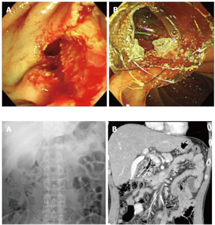
In another previously unpublished case, a 46-year- old male was referred to the hospital for right quadrant abdominal pain. He had previously undergone a Billroth I operation for gastric ulcer perforation. Because the patient developed abnormal liver functioning and gall- bladder stones on abdominal CT scan, ERCP was per- formed to identify the biliary duct stone. However, after endoscopic sphincterotomy, a peri-ampullary perforation was detected, and a fully covered SEMS was placed im- mediately during the ERCP (Figures 1 and 2). After stent-
Table 3 Temporary self-expandable metallic stent used for peri-ampullary perforations
Ref. Age/sex ERCP indication Abdominal CT scan Stent indication Type of stent/duration (d) Vezakis et al[28] 61/F Stones or sphincter of
Oddi dysfunction
Retroperitoneal air Duodenal fistula, Continuing leakage
Partially covered SEMS/14 Jeon et al[26] 82/F Stones Retroperitoneal air and fluid Continuing leakage Fully covered SEMS/28 Canena et al[25] 55/F Stones Retroperitoneal air and fluid Perforation Fully covered SEMS/21
29/F Stones Retroperitoneal air and fluid Perforation Fully covered SEMS/30
31/M Stones Retroperitoneal air and fluid Perforation Fully covered SEMS/30
76/F Stones Retroperitoneal air and fluid Perforation Fully covered SEMS/29
Park et al[27] 61/F Biliary tree dilatation Retroperitoneal air and fluid Perforation Fully covered SEMS/10 Unpublished 46/M Stones Retroperitoneal air Perforation Fully covered SEMS/ spontaneously fell out CT: Computed tomography; SEMS: Self-expandable metallic stent; ERCP: Endoscopic retrograde cholangiopancreatography.
ing, the patient was stable and was discharged without complications. Although he underwent endoscopy for removal of the stent on day 28 after insertion, the stent had already fallen out spontaneously.
Because fully covered SEMS are available in large diameters, they can be used in a dilated common bile duct without stent migration. They are capable of main- taining long-term patency of the lumen, in contrast to plastic stents. Also, in comparison with uncovered SEMS, fully covered SEMS have several benefits. Uncovered SEMS tend to embed readily in the duct, making it dif- ficult to remove the stent
[62-64]. Thus, it is inappropriate to use them for benign conditions, such as strictures, obstructions, and traumatic perforations. The fully cov- ered SEMS overcomes these disadvantage, and can be removed readily
[64-66].
However, the optimal duration of stenting has not been established. Bakken et al
[67]reported that the mean duration of stent placement was 67 (range, 0-279) d for benign strictures and 59 (range, 1-601) d for leaks, fistulas, and perforations. Another study showed that the mean duration was 37 (range, 4-84) d for benign conditions
[68]. While discrepancies exist among studies, the time until stent removal is approximately 2 mo for benign esopha- geal conditions. Several studies have reported stenting durations in peri-ampullary perforations ranging from 10 to 30 d
[25-28]. Moreover, because the treatment outcome did not seem to depend on the duration of stenting, and the stent was removed according to the status of the pa- tient, a stent should be removed when the patient shows improved perforation-related symptoms, signs, and imag- ing results, such as simple abdominal X-rays and abdomi-
nal CT scans, even after 1 wk of stenting.
Although several studies have demonstrated good outcomes using temporary fully covered SEMS in ERCP- related perforations, clinically, the situation has not been clarified entirely. Because treatment failure after non-sur- gical treatment, including the insertion of plastic stents and fully covered SEMS, can cause high mortality and morbidity, close attention must be paid to the decision on treatment modality. A decision taking into consideration the surgery time, while performed non-surgical treat- ment, is also important. Unrelieved abdominal pain, con- tinued leakage on abdominal CT scans, or hemodynamic instability despite non-surgical management are consid- erations relevant to surgical intervention. Thus, frequent physical examinations and serial follow-up using abdomi- nal CT scans are helpful in checking for adverse events or treatment failure. However, a patient’s condition, such as cardiopulmonary comorbidity, hemodynamic instabil- ity, and old age, is also highly relevant to postoperative mortality. If a patient with peri-ampullary perforation has an inoperable condition due to high postoperative risks, a fully covered SEMS can be attempted for palliative treat- ment
[26]. First, it is better to use a fully covered SEMS, especially for a major leakage and large perforation, be- cause ENBD and ERBD may not prevent bile flow into the perforation site completely. Although it is essential to select cases according to their condition, optimal conser- vative management using a fully covered SEMS may be a good treatment option.
In conclusion, early diagnosis of ERCP-related duo- denal perforation is important, and according to the type of perforation, its treatment varies from conservative
Figure 1 Insertion of fully covered self-expandable metal- lic stent for the management of periampullary perforation immediately after endoscopic sphincterotomy. A: A peri- ampullary perforation was seen after endoscopic sphincter- otomy; B: A fully covered self-expandable metallic stent (5-cm- long, 10 mm in diameter) inserted into the common bile duct to prevent bile entering the perforation site can be seen at the ampulla of vater.
A B
A B Figure 2 Deployed fully covered self-expandable metallic
stent on (A) abdominal X-ray and (B) abdominal computed tomography scan.
management to surgical intervention. Although conserva- tive management is the mainstay for all types of perfora- tions, except type
Ⅰperforations, the most appropriate treatment modality should be established by performing a comprehensive evaluation of the patient. In particular, a fully covered SEMS for perivaterian perforations was used in selected cases, and the clinical outcomes were encouraging. However, the evidence supporting the use of fully covered SEMS in perivaterian perforations is still insufficient, and further studies are required.
REFERENCES
1 McCune WS, Shorb PE, Moscovitz H. Endoscopic cannula- tion of the ampulla of vater: a preliminary report. Ann Surg 1968; 167: 752-756 [PMID: 5646296]
2 Lai CH, Lau WY. Management of endoscopic retrograde cholangiopancreatography-related perforation. Surgeon 2008; 6: 45-48 [PMID: 18318088]
3 Christensen M, Matzen P, Schulze S, Rosenberg J. Compli- cations of ERCP: a prospective study. Gastrointest Endosc 2004; 60: 721-731 [PMID: 15557948]
4 Coppola R, Riccioni ME, Ciletti S, Cosentino L, Coco C, Magistrelli P, Picciocchi A. Analysis of complications of en- doscopic sphincterotomy for biliary stones in a consecutive series of 546 patients. Surg Endosc 1997; 11: 129-132 [PMID:
9069143]
5 Halme L, Doepel M, von Numers H, Edgren J, Ahonen J.
Complications of diagnostic and therapeutic ERCP. Ann Chir Gynaecol 1999; 88: 127-131 [PMID: 10392249]
6 Loperfido S, Angelini G, Benedetti G, Chilovi F, Costan F, De Berardinis F, De Bernardin M, Ederle A, Fina P, Fratton A.
Major early complications from diagnostic and therapeutic ERCP: a prospective multicenter study. Gastrointest Endosc 1998; 48: 1-10 [PMID: 9684657]
7 Masci E, Toti G, Mariani A, Curioni S, Lomazzi A, Dinelli M, Minoli G, Crosta C, Comin U, Fertitta A, Prada A, Passoni GR, Testoni PA. Complications of diagnostic and thera- peutic ERCP: a prospective multicenter study. Am J Gas- troenterol 2001; 96: 417-423 [PMID: 11232684 DOI: 10.1111/
j.1572-0241.2001.03594.x]
8 Vaira D, D’Anna L, Ainley C, Dowsett J, Williams S, Bail- lie J, Cairns S, Croker J, Salmon P, Cotton P. Endoscopic sphincterotomy in 1000 consecutive patients. Lancet 1989; 2:
431-434 [PMID: 2569609]
9 Vandervoort J, Soetikno RM, Tham TC, Wong RC, Fer- rari AP, Montes H, Roston AD, Slivka A, Lichtenstein DR, Ruymann FW, Van Dam J, Hughes M, Carr-Locke DL.
Risk factors for complications after performance of ERCP.
Gastrointest Endosc 2002; 56: 652-656 [PMID: 12397271 DOI:
10.1067/mge.2002.129086]
10 Zinsser E, Hoffmann A, Will U, Koppe P, Bosseckert H.
Success and complication rates of diagnostic and therapeu- tic endoscopic retrograde cholangiopancreatography--a prospective study. Z Gastroenterol 1999; 37: 707-713 [PMID:
10494605]
11 Zissin R, Shapiro-Feinberg M, Oscadchy A, Pomeranz I, Leichtmann G, Novis B. Retroperitoneal perforation during endoscopic sphincterotomy: imaging findings. Abdom Imag- ing 2000; 25: 279-282 [PMID: 10823451]
12 Enns R, Eloubeidi MA, Mergener K, Jowell PS, Branch MS, Pappas TM, Baillie J. ERCP-related perforations: risk fac- tors and management. Endoscopy 2002; 34: 293-298 [PMID:
11932784 DOI: 10.1055/s-2002-23650]
13 Pungpapong S, Kongkam P, Rerknimitr R, Kullavanijaya P. Experience on endoscopic retrograde cholangiopancrea- tography at tertiary referral center in Thailand: risks and
complications. J Med Assoc Thai 2005; 88: 238-246 [PMID:
15962677]
14 Booth FV, Doerr RJ, Khalafi RS, Luchette FA, Flint LM. Sur- gical management of complications of endoscopic sphinc- terotomy with precut papillotomy. Am J Surg 1990; 159:
132-135; discussion 132-135 [PMID: 2294790]
15 Alfieri S, Rosa F, Cina C, Tortorelli AP, Tringali A, Perri V, Bellantone C, Costamagna G, Doglietto GB. Management of duodeno-pancreato-biliary perforations after ERCP: outcomes from an Italian tertiary referral center. Surg Endosc 2013; 27:
2005-2012 [PMID: 23299135 DOI: 10.1007/s00464-012-2702-9]
16 Wu HM, Dixon E, May GR, Sutherland FR. Management of perforation after endoscopic retrograde cholangiopancrea- tography (ERCP): a population-based review. HPB (Oxford) 2006; 8: 393-399 [PMID: 18333093 DOI: 10.1080/13651820600 700617]
17 Howard TJ, Tan T, Lehman GA, Sherman S, Madura JA, Fogel E, Swack ML, Kopecky KK. Classification and man- agement of perforations complicating endoscopic sphincter- otomy. Surgery 1999; 126: 658-663; discussion 664-665 [PMID:
10520912]
18 Stapfer M, Selby RR, Stain SC, Katkhouda N, Parekh D, Jabbour N, Garry D. Management of duodenal perforation after endoscopic retrograde cholangiopancreatography and sphincterotomy. Ann Surg 2000; 232: 191-198 [PMID:
10903596]
19 Fatima J, Baron TH, Topazian MD, Houghton SG, Iqbal CW, Ott BJ, Farley DR, Farnell MB, Sarr MG. Pancreaticobiliary and duodenal perforations after periampullary endoscopic procedures: diagnosis and management. Arch Surg 2007;
142: 448-454; discussion 454-455 [PMID: 17515486 DOI:
10.1001/archsurg.142.5.448]
20 Lee TH, Bang BW, Jeong JI, Kim HG, Jeong S, Park SM, Lee DH, Park SH, Kim SJ. Primary endoscopic approxima- tion suture under cap-assisted endoscopy of an ERCP- induced duodenal perforation. World J Gastroenterol 2010;
16: 2305-2310 [PMID: 20458771]
21 Lee TH, Han JH, Park SH. Endoscopic treatments of en- doscopic retrograde cholangiopancreatography-related duodenal perforations. Clin Endosc 2013; 46: 522-528 [PMID:
24143315 DOI: 10.5946/ce.2013.46.5.522]
22 Morgan KA, Fontenot BB, Ruddy JM, Mickey S, Adams DB.
Endoscopic retrograde cholangiopancreatography gut per- forations: when to wait! When to operate! Am Surg 2009; 75:
477-483; discussion 483-484 [PMID: 19545095]
23 Polydorou A, Vezakis A, Fragulidis G, Katsarelias D, Va- gianos C, Polymeneas G. A tailored approach to the man- agement of perforations following endoscopic retrograde cholangiopancreatography and sphincterotomy. J Gastroin- test Surg 2011; 15: 2211-2217 [PMID: 22005896 DOI: 10.1007/
s11605-011-1723-3]
24 Sebastian S, Byrne AT, Torreggiani WC, Buckley M. En- doscopic closure of iatrogenic duodenal perforation dur- ing endoscopic ultrasound. Endoscopy 2004; 36: 245 [PMID:
14986227 DOI: 10.1055/s-2004-814257]
25 Canena J, Liberato M, Horta D, Romão C, Coutinho A. Short- term stenting using fully covered self-expandable metal stents for treatment of refractory biliary leaks, postsphinc- terotomy bleeding, and perforations. Surg Endosc 2013; 27:
313-324 [PMID: 22806507 DOI: 10.1007/s00464-012-2368-3]
26 Jeon HJ, Han JH, Park S, Youn S, Chae H, Yoon S. Endo- scopic sphincterotomy-related perforation in the common bile duct successfully treated by placement of a covered metal stent. Endoscopy 2011; 43 Suppl 2 UCTN: E295-E296 [PMID: 21915834 DOI: 10.1055/s-0030-1256464]
27 Park WY, Cho KB, Kim ES, Park KS. A case of ampul- lary perforation treated with a temporally covered metal stent. Clin Endosc 2012; 45: 177-180 [PMID: 22866262 DOI:
10.5946/ce.2012.45.2.177]
28 Vezakis A, Fragulidis G, Nastos C, Yiallourou A, Poly-
dorou A, Voros D. Closure of a persistent sphincterotomy- related duodenal perforation by placement of a covered self-expandable metallic biliary stent. World J Gastroenterol 2011; 17: 4539-4541 [PMID: 22110286 DOI: 10.3748/wjg.v17.
i40.4539]
29 Faylona JM, Qadir A, Chan AC, Lau JY, Chung SC. Small- bowel perforations related to endoscopic retrograde chol- angiopancreatography (ERCP) in patients with Billroth II gastrectomy. Endoscopy 1999; 31: 546-549 [PMID: 10533739 DOI: 10.1055/s-1999-61]
30 Lin LF, Siauw CP, Ho KS, Tung JC. ERCP in post-Billroth II gastrectomy patients: emphasis on technique. Am J Gas- troenterol 1999; 94: 144-148 [PMID: 9934745 DOI: 10.1111/
j.1572-0241.1999.00785.x]
31 Wilkinson ML, Engelman JL, Hanson PJ. Intestinal perfora- tion after ERCP in Billroth II partial gastrectomy. Gastroin- test Endosc 1994; 40: 389-390 [PMID: 8056259]
32 Freeman ML. Complications of endoscopic biliary sphinc- terotomy: a review. Endoscopy 1997; 29: 288-297 [PMID:
9255535 DOI: 10.1055/s-2007-1004193]
33 Boender J, Nix GA, de Ridder MA, van Blankenstein M, Schütte HE, Dees J, Wilson JH. Endoscopic papillotomy for common bile duct stones: factors influencing the complica- tion rate. Endoscopy 1994; 26: 209-216 [PMID: 8026367 DOI:
10.1055/s-2007-1008945]
34 Cotton PB, Lehman G, Vennes J, Geenen JE, Russell RC, Meyers WC, Liguory C, Nickl N. Endoscopic sphincter- otomy complications and their management: an attempt at consensus. Gastrointest Endosc 1991; 37: 383-393 [PMID:
2070995]
35 Trap R, Adamsen S, Hart-Hansen O, Henriksen M. Severe and fatal complications after diagnostic and therapeutic ERCP: a prospective series of claims to insurance cover- ing public hospitals. Endoscopy 1999; 31: 125-130 [PMID:
10223360 DOI: 10.1055/s-1999-13659]
36 Alexiou K, Sakellaridis T, Sikalias N, Karanikas I, Econo- mou N, Antsaklis G. Subcutaneous emphysema, pneumo- mediastinum and pneumoperitoneum after unsuccessful ERCP: a case report. Cases J 2009; 2: 120 [PMID: 19192290 DOI: 10.1186/1757-1626-2-120]
37 Ciaccia D, Branch MS, Baillie J. Pneumomediastinum after endoscopic sphincterotomy. Am J Gastroenterol 1995; 90:
475-477 [PMID: 7872289]
38 Colemont LJ, Pelckmans PA, Moorkens GH, Van Maercke YM. Unilateral periorbital emphysema: an unusual compli- cation of endoscopic papillotomy. Gastrointest Endosc 1988;
34: 473-475 [PMID: 3234686]
39 Merine D, Fishman EK. Uncomplicated portal venous gas associated with duodenal perforation following ERCP: CT features. J Comput Assist Tomogr 1989; 13: 138-139 [PMID:
2910933]
40 Savides T, Sherman S, Kadell B, Cryer H, Derezin M. Bilat- eral pneumothoraces and subcutaneous emphysema after endoscopic sphincterotomy. Gastrointest Endosc 1993; 39:
814-817 [PMID: 8293908]
41 Scarlett PY, Falk GL. The management of perforation of the duodenum following endoscopic sphincterotomy: a pro- posal for selective therapy. Aust N Z J Surg 1994; 64: 843-846 [PMID: 7980259]
42 Tam F, Prindiville T, Wolfe B. Subcutaneous emphysema as a complication of endoscopic sphincterotomy of the am- pulla of Vater. Gastrointest Endosc 1989; 35: 447-449 [PMID:
2551769]
43 Genzlinger JL, McPhee MS, Fisher JK, Jacob KM, Helzberg JH. Significance of retroperitoneal air after endoscopic ret- rograde cholangiopancreatography with sphincterotomy.
Am J Gastroenterol 1999; 94: 1267-1270 [PMID: 10235205 DOI:
10.1111/j.1572-0241.1999.00996.x]
44 Gottlieb K, Sherman S, Pezzi J, Esber E, Lehman GA. Early recognition of post-ERCP pancreatitis by clinical assessment
and serum pancreatic enzymes. Am J Gastroenterol 1996; 91:
1553-1557 [PMID: 8759660]
45 Testoni PA, Bagnolo F. Pain at 24 hours associated with amylase levels greater than 5 times the upper normal limit as the most reliable indicator of post-ERCP pancreatitis.
Gastrointest Endosc 2001; 53: 33-39 [PMID: 11154486 DOI:
10.1067/mge.2001.111390]
46 Bell RC, Van Stiegmann G, Goff J, Reveille M, Norton L, Pearlman NW. Decision for surgical management of perfo- ration following endoscopic sphincterotomy. Am Surg 1991;
57: 237-240 [PMID: 2053743]
47 Marano BJ, Bonanno CA. Metallic biliary endoprosthesis causing duodenal perforation and acute upper gastrointes- tinal bleeding. Gastrointest Endosc 1994; 40: 257-258 [PMID:
8013842]
48 Matsuda T, Fujii T, Emura F, Kozu T, Saito Y, Ikematsu H, Saito D. Complete closure of a large defect after EMR of a lateral spreading colorectal tumor when using a two-chan- nel colonoscope. Gastrointest Endosc 2004; 60: 836-838 [PMID:
15557972]
49 Mutignani M, Iacopini F, Dokas S, Larghi A, Familiari P, Tringali A, Costamagna G. Successful endoscopic closure of a lateral duodenal perforation at ERCP with fibrin glue.
Gastrointest Endosc 2006; 63: 725-727 [PMID: 16564890 DOI:
10.1016/j.gie.2005.11.028]
50 Nakagawa Y, Nagai T, Soma W, Okawara H, Nakashima H, Tasaki T, Hisamatu A, Hashinaga M, Murakami K, Fujioka T.
Endoscopic closure of a large ERCP-related lateral duodenal perforation by using endoloops and endoclips. Gastrointest Endosc 2010; 72: 216-217 [PMID: 20304402 DOI: 10.1016/
j.gie.2009.10.040]
51 Buffoli F, Grassia R, Iiritano E, Bianchi G, Dizioli P, Staiano T. Endoscopic “retroperitoneal fatpexy” of a large ERCP- related jejunal perforation by using a new over-the-scope clip device in Billroth II anatomy (with video). Gastrointest Endosc 2012; 75: 1115-1117 [PMID: 21820111 DOI: 10.1016/
j.gie.2011.05.029]
52 Doğan ÜB, Keskın MB, Söker G, Akın MS, Yalaki S. Endo- scopic closure of an endoscope-related duodenal perfora- tion using the over-the-scope clip. Turk J Gastroenterol 2013;
24: 436-440 [PMID: 24557968]
53 Martin DF, Tweedle DE. Retroperitoneal perforation during ERCP and endoscopic sphincterotomy: causes, clinical fea- tures and management. Endoscopy 1990; 22: 174-175 [PMID:
2209500 DOI: 10.1055/s-2007-1012833]
54 Chung RS, Sivak MV, Ferguson DR. Surgical decisions in the management of duodenal perforation complicating endoscopic sphincterotomy. Am J Surg 1993; 165: 700-703 [PMID: 8506969]
55 Ciostek P, Bielska H, Myrcha P, Jarosz O, Milewski J, No- szczyk W. [Surgical tactics in treatment of duodenal injuries after endoscopic sphincterotomy]. Wiad Lek 1997; 50 Suppl 1 Pt 2: 421-424 [PMID: 9424916]
56 Neri V, Ambrosi A, Fersini A, Valentino TP. Duodenal per- foration in course of endoscopic retrograde cholangiopan- creatography-endoscopic sphincterotomy. Therapeutic con- siderations. Ann Ital Chir 2006; 77: 161-164 [PMID: 17147091]
57 Sarr MG, Fishman EK, Milligan FD, Siegelman SS, Cameron JL. Pancreatitis or duodenal perforation after peri-Vaterian therapeutic endoscopic procedures: diagnosis, differentia- tion, and management. Surgery 1986; 100: 461-466 [PMID:
3738765]
58 Kim BS, Kim IG, Ryu BY, Kim JH, Yoo KS, Baik GH, Kim JB, Jeon JY. Management of endoscopic retrograde cholangiopancreatography-related perforations. J Korean Surg Soc 2011; 81: 195-204 [PMID: 22066121 DOI: 10.4174/
jkss.2011.81.3.195]
59 Kim JH, Yoo BM, Kim JH, Kim MW, Kim WH. Manage- ment of ERCP-related perforations: outcomes of single insti- tution in Korea. J Gastrointest Surg 2009; 13: 728-734 [PMID:
19130154 DOI: 10.1007/s11605-008-0786-2]
60 Siersema PD, Schrauwen SL, van Blankenstein M, Steyerberg EW, van der Gaast A, Tilanus HW, Dees J. Self-expanding metal stents for complicated and recurrent esophagogastric cancer. Gastrointest Endosc 2001; 54: 579-586 [PMID: 11677473]
61 Siersema PD, Homs MY, Haringsma J, Tilanus HW, Kuipe- rs EJ. Use of large-diameter metallic stents to seal traumatic nonmalignant perforations of the esophagus. Gastrointest Endosc 2003; 58: 356-361 [PMID: 14528208]
62 Baron TH. Covered self-expandable metal stents for benign biliary tract diseases. Curr Opin Gastroenterol 2011; 27: 262-267 [PMID: 21248636 DOI: 10.1097/MOG.0b013e3283438a26]
63 Dumonceau JM, Devière J, Delhaye M, Baize M, Cremer M.
Plastic and metal stents for postoperative benign bile duct strictures: the best and the worst. Gastrointest Endosc 1998;
47: 8-17 [PMID: 9468417]
64 García-Cano J, Taberna-Arana L, Jimeno-Ayllón C, Mar- tínez-Fernández R, Serrano-Sánchez L, Reyes-Guevara AK, Viñuelas-Chicano M, Gómez-Ruiz CJ, Morillas-Ariño MJ, Pérez-García JI, Pérez-Vigara G, Pérez-Sola A. Use of fully covered self-expanding metal stents for the management of benign biliary conditions. Rev Esp Enferm Dig 2010; 102:
526-532 [PMID: 20883068]
65 Mahajan A, Ho H, Sauer B, Phillips MS, Shami VM, Ellen K, Rehan M, Schmitt TM, Kahaleh M. Temporary placement of fully covered self-expandable metal stents in benign bili- ary strictures: midterm evaluation (with video). Gastrointest Endosc 2009; 70: 303-309 [PMID: 19523620 DOI: 10.1016/
j.gie.2008.11.029]
66 Wang AY, Ellen K, Berg CL, Schmitt TM, Kahaleh M. Fully covered self-expandable metallic stents in the management of complex biliary leaks: preliminary data - a case series.
Endoscopy 2009; 41: 781-786 [PMID: 19693751 DOI: 10.1055/
s-0029-1215050]
67 Bakken JC, Wong Kee Song LM, de Groen PC, Baron TH. Use of a fully covered self-expandable metal stent for the treatment of benign esophageal diseases. Gastrointest Endosc 2010; 72: 712-720 [PMID: 20883848 DOI: 10.1016/
j.gie.2010.06.028]
68 Senousy BE, Gupte AR, Draganov PV, Forsmark CE, Wagh MS. Fully covered Alimaxx esophageal metal stents in the endoscopic treatment of benign esophageal diseases. Dig Dis Sci 2010; 55: 3399-3403 [PMID: 20859687 DOI: 10.1007/
s10620-010-1415-y]
P- Reviewer: ElGeidie AAR, Murata A S- Editor: Ji FF L- Editor: A E- Editor: Lu YJ