∙Received: March 28, 2014. Accepted: April 21, 2014.
∙Corresponding author : Jae Il Kim
Department of Nuclear Medicine, Seoul National University Hospital, 101 Daehak-ro, Jongno-gu, Seoul 110-744, Korea
Tel: +82-2-2072-2535, Fax: +82-2-747-0208 E-mail: [email protected]
Original Article
PET/MRI에 있어 MRI 조영제가 PET에 미치는 영향
서울대학교병원 핵의학과1, 서울대학교병원 영상의학과2 김재일1⋅김인수2⋅이홍재1⋅김진의1
Effect of MRI Media Contrast on PET/MRI
Jae Il Kim1, In Soo Kim2, Hong Jae Lee1 and Jin Eui Kim1
1Department of Nuclear Medicine, Seoul National University Hospital, Seoul, Korea
2Department of Diagnostic Radiology, Seoul National University Hospital, Seoul, Korea
Purpose: Integrated PET/MRI has been developed recently has become a lot of help to the point oncologic, neological, cardiological nuclear medicine. By using this PET/MRI, a μ-map is created some special MRI sequence which may be divided parts of the body for attenuation correction. However, because an MRI contrast agent is necessary in order to obtain an more MRI information , we will evaluate to see an effect of SUV on PET image that corrected attenuation by MRI with contrast agent. Materials and Methods: As PET/MRI machine, Biograph mMR (Siemens, Germany) was used. For phantom test, 1mCi 18F-FDG was injected in cylinderical uniformity phantom, and then acquire PET data about 10 minutes with VIBE-DIXON, UTE MRI sequence image for attenuation correction. T1 weighted contrast media, 4 cc DOTAREM (GUERBET, FRANCE) was injected in a same phatnom, and then PET data, MRI data were acquired by same methodes. Using this PET, non-contrast MRI and contrast MRI, it was reconstructed attenuation correction PET image, in which we evanuated the difference of SUVs. Additionally, for let a high desity of contrast media, 500 cc 2 plastic bottles were used. We injected 18F-FDG with 5 cc DOTAREM in first bottle. At second bottle, only 18F-FDG was injected. and then we evaluated a SUVs reconstructed by same methods. For clinical patient study, rectal caner-pancreas cancer patients were selected. we evaluated SUVs of PET image corrected attenuastion by contrast weighted MRI and non-contrast MRI. Results: For a phantom study, although VIBE DIXON MRI signal with contrast media is 433% higher than non-contrast media MRI, the signals intensity of μ-map, attenuation corrected PET are same together. In case of high contrast media density, image distortion is appeared on μ-map and PET images. For clinical a patient study, VIBE DIXON MRI signal on lesion portion is increased in 495% by using DOTAREM. But there are no significant differences at μ-map, non AC PET, AC-PET image whether using contrast media or not. In case of whole body PET/MRI study, %diff between contras and non contrast MRAC at lung, liver, renal cortex, femoral head, myocardium, bladder, muscle are -4.32%, -2.48%, -8.05%, -3.14%, 2.30%, 1.53%, 6.49% at each other. Conclusion: In integrated PET/MRI, a segmentation μ-map method is used for correcting attenuation of PET signal. although MRI signal for attenuation correciton change by using contrast media, μ-map will not change, and then MRAC PET signal will not change too. Therefore, MRI contrast media dose not affect for attenuation correction PET. As well, not only When we make a flow of PET/MRI protocol, order of PET and MRI sequence dose not matter, but It's possible to compare PET images before and after contrast agent injection. (Korean J Nucl Med Technol 2014;18(1):19-25)
Key Words : PET/MR, μ-map, Attenuation correction, Contrast media, PET/CT, SUV, Attenuation correction
서 론
2012년에 설치가 되고, 임상적인 부분과 연구적인 부분 에 많이 활성화가 되고 있는 PET/MRI는 현대 핵의학에 많은 도움을 주고 있다. 기존의 PET/CT에 비해 PET/MRI
Fig. 1. This patient has osteosarcoma at right femur shaft. T1 weighted image (A), T1 weighted image with contrast media (B), positron emission tomograph (C), PET/MRI fusion image (D).
Fig. 2. A procedure of attenuation correction of PET/MRI. The μ-map that has four segmentation is made by MRI for attenuation cor- rection of PET raw data.
종축 이완시간인 T1의 차이를 커지게 하는 T1 weighted media contrast를 사용을 한다. 이 조영제를 사용하게 되면 해부학적 정보가 많이 담겨 있는 T1 weighted 영상에 병리 학적 정보를 담을 수 있는데, 조영 증강의 효과를 보여, 보 다 많은 진단적 정보를 얻을 수 있게 된다(Fig. 1).
하지만, PET/MRI의 경우, PET 영상의 보정을 위해 MRI 영상을 이용하게 되는데, 이러한 보정용 MRI 영상은 T1 weighted 영상이여서, 조영 증강을 하게 되면 PET의 보 정에 영향 줄 것이라 예상되었다. 기존의 PET/CT의 경우 CT 조영제를 사용하게 되면 보정용 CT의 HousField unit 값이 틀려져 PET의 신호가 올라가 SUV에서도 영향을 주 는 게 확인되었다. 특히 Ba enema와 같이 밀도가 높은 조 영제를 쓰는 경우 PET/CT 후에 시행토록 권고를 할 정도로 조영제에 의한 영향이 아주 크다. 이러한 생각에 PET/MRI 에서도 조영증강은 보정용 MRI 영상을 모두 획득 후 시행 토록 권고를 하고 있다(Fig. 2).
특히 PET 보정용 MRI의 경우 Siemens 사는 ‘segmenta- tion MRI 보정법’을 사용하고 있어 각 장기에서 나오는 MRI 신호의 강도에 μ-map이 직접적으로 영향을 미치고 있다. 아래 그림에서처럼 MRI의 신호 강도에 따라 보정 계 수가 정해지게 된다. 즉, 공기와 폐, 지방과 연부 조직의 경 우 MRI 신호의 변화에 따라 다른 장기로 인식할 수 있는 경우가 커지게 된다(Fig. 3).
사용하였고, Uniformity phantom에는 18F-FDG를 31±0.54 MBq 20 mL를 채워 10분간 교반 후, 사용하였다.
추가적으로 조영제의 밀도를 높여서 팬텀 테스트를 하기 위해 500 cc 생수병을 사용하여, 18F-FDG를 18±0.3 MBq 20 mL를 채워 3분간 교반 후, 사용하였다.
환자는 직장암, 췌장암을 가진 환자였고, 평균 연령은 58±12 y이고, 몸무게는 61±15 kg, 주입된 18F-FDG 량은 314±52 MBq이였다(Fig. 5).
2. PET/MRI, 조영제
사용된 PET/MRI 장비로는 Siemens 사의 mMR을 사용
A B C D
Fig. 3. To make a μ-map (B) need a VIBE-DIXON sequence which include in-phase, opposite-phase, water image, fat image (A). According to MRI signal intensity, μ-value is assigned 4 segmentation - air, lungs, fat, soft tissue (C).
Fig. 4. This image is for pancreatic head Ca. Pancreas head cancer is not distincted at T1 image (A). But we can recognize alegion por- tion at contrast enhanced T1 image (B).
Fig. 5. Uniformity phantom was used for normal density contrast media (A). Plastic bottle was used for high density contrast me- dia (B).
하였다. 3Tesla의 고자장 MRI 장비는 기존의 MRI 장비에 비해 전신을 검사할 수 있는 전용 코일이 이용하고 있으며
고성능 경사자장과 보다 발전된 BOLD, Diffusion, Spectro- scopy 가능하였다. LSO crystal 28672개를 사용한 PET 장 비는 고자장에서 전기적 신호를 인식하기 위해 기존의 PMT 는 사용하지 못하고, 반도체 검출기인 APD (Avalanched Photo Detector)를 사용하였다. 그래서 TOF나 PSF (point spread function) 기술은 사용되지 못하고 있어, 기존의 PET/CT의 PET 영상에 비해 해상도가 다소 떨어지고 있다. 하지만 이 mMR 장비의 경우, 기존의 PET/CT와 달리 gantry 속에 main magnet-shimming coil-gradient coil-PET detector-RF coil이 동축상에 내장이 되어 있어, MRI 신호를 받는 동시 에 PET 신호를 받을 수 있게 설계가 되어 있어, 완전한 동 시적인 PET과 MRI의 퓨전이 가능케 되었다(Fig. 6).
MRI 조영제로는 프랑스의 Guerbet사의 dotarem (0.5 mmol/mL)을 사용하였다. Uniformity phantom test에는 8 mL
A B
A B
A B
Fig. 6. PET detector and MRI coil component are integrated in same gantry by locate at same axis.
Fig. 7. Dotarem made by Guerbet Co. Ltd. France, is T1 weight- ed contrast media. So we will diagnose a pathological change in anatomical T1 image.
Fig. 8. Cylinderical uniformity phantom is on the spinal coil, sur- face coil with 2 MRI phantom at both sides.
를 넣어 물과의 비율을 1 : 0.00082로 맞추었어며, 고밀도 테 스트를 위해 생수병에는 4 mL를 넣어 물과의 비율은 1 : 0.02 로 uniformity phantom에 비해 조영제 밀도를 23.4배 증가 시켰다.
환자에게는 실제 조영제를 사용하는 검사에 대해 10 mL/
60 kg를 주사하여, 체중에 대한 조영제의 비율을 1 : 0.0002 정도로 맞추었다(Fig. 7).
3. 방법
1) Uniformity phantom: 실린더형 uniformity phantom에
18F-FDG 31MBq를 넣고 교반 후, PET/MRI 테이블에 올 려 놓은 후, spine coil과 surface coil로 덮고, 1bed 스캔을 하였다. PET 데이터는 10분간 받고, iterative로 reconstruc- tion하였다. 감쇠 보정을 위해 VIBE-DIXON sequence와
UTE sequence 영상을 얻었고, 정확한 위치 잡이를 위해 axial T1 영상을 얻어 fusion을 하였다. Non contrast PET/
MRI 스캔이 끝나면 바로, dotarem 8 cc를 넣고 교반 후, 이 전 스캔이랑 똑같은 조건에서 PET, VIBE DIXON, UTE, T1 영상을 얻어 contrast enhanced PET/MRI 영상을 획득 한다. Reconstruction이 완료되면 조영제 사용 전후의 VIBE DIXON, UTE, T1, 감쇠 보정 된 PET 영상에서 ROI를 5 군데 설정을 하여 SUV를 비교, 평가하였다. 동일한 방법 으로 생수병에 물을 채우고, 18F-FDG 18MBq를 넣고 교반 후, non-contrast PET/MRI 데이터를 얻는다. 그리고 나서 dotarem 4 mL를 넣고 contrast PET/MRI 영상을 얻고 조 영제 사용 전후의 SUV의 차이를 비교, 평가하였다(Fig. 8).
2) 환자: 직장암과 췌장암을 가진 환자 중 조영제 사용하 는 PET/MRI 검사를 하는 환자를 선택하였다. 금식된 상태 에서 환자가 도착하면, MRI 조영제를 위한 동의서를 작성 하고, 팔에 카테타와 프레셔 라인을 잡는다. 그리고 18F-FDG 를 몸무게(kg) × 0.14 [mCi] 주입을 하고, 1시간 동안 안정실
Fig. 9. Left image is coronal μ-map for applying attenuation correction of PET signal (A). Right image is an attenuation corrected PET im- age by using MRI μ-map (B).
Fig. 10. MRI signal with contrast media is 433% higher than non contrast image (A). But signal of both u-map (B) and attenuation correc- tion PET image(C) are same together.
에서 기다린다. 우선 PET/MRI 검사실에 들어와서 mMR 전용 코일인 ‘head and neck coil’, ‘spine coil’, ‘surface coil’
을 환자에게 덮고, bed당 3분씩 전신 PET/MRI 데이터를 얻는다. 그리고 바로 부분 검사를 하는데, PET 데이터는 병소가 있는 구역에서 10분간 데이터를 받고, 조영제 사용 하기 전에 VIBE-DIXON, UTE를 얻고 감쇠보정을 한다.
그리고 조영제를 사용한 VIBE-DIXON, UTE를 얻어 동일 한 PET 데이터를 감쇠 보정-reconstruction을 하였으며, MRI, μ-map, non-MRAC, MRAC PET 영상에서 폐, 간, 신장 피질, 대퇴골두, 심근, 방광, 근육의 SUV 차이를 비교, 평가하 였다(Fig. 9).
결 과
1. 팬텀-저밀도
Uniformity phantom에 조영제를 1 : 0.00082 비율로 맞추 었을 경우, VIBE-DIXON 영상의 MRI 신호 강도는 조영 전 59.69, 조영 후 315.45로 433% 증가하였다. 하지만 PET 의 감쇠를 보정하기 위해 μ-map으로 변환하였더니 팬텀의 내부를 모두 연부조직으로 인식을 하여 조영 전-후의 차이 는 없었다. 그래서 이 μ-map으로 감쇠 보정을 한 MRAC PET 영상에서도 차이가 없었다(Fig. 10).
A B
C
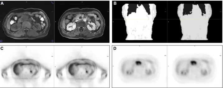
A B
Fig. 12. This images are regional PET/MRI. Even if MRI signal increase (A), signal of μ-map (B), non-AC PET (C), AC PET (D) is same together like phantom test.
Fig. 11. Some distortion of bottle shape appeared. At high den- sity contrast media MRI μ-map.
VIBE-DIXON 영상에서 MRI 신호는 조영 증강 전후로 495% 증가하였다. 하지만 μ-map에서의 차이는 보이지 않 았으며, 감쇠 보정 전-후 PET 데이터도 7.14%로 그 차이가 미미하였다(Fig. 12).
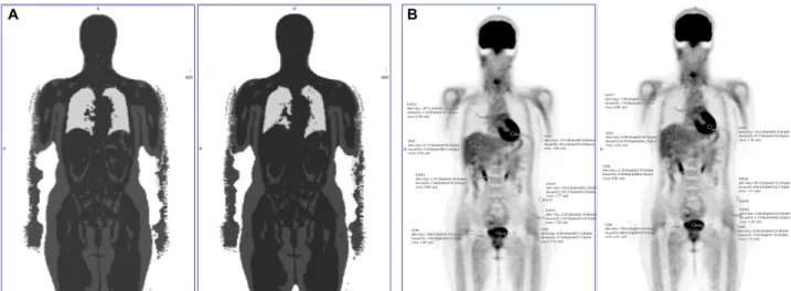
4. 환자-전신 검사
는 감쇠 보정용 μ-map과 감쇠 보정된 PET 데이터에는 큰 영향을 주지 않음을 알 수 있었다. 왜냐하면, MRI 신호로 인체를 4구역으로 구획 구분하여 μ-value를 산출을 하다 보 니, 실제로 MRI 신호가 바뀌더라도 구획 구분을 같은 영역 으로 해석을 하기 때문이다. 특히 MRI 신호가 특정한 역치 값 이상을 넘게 되면, 그 영역에서의 μ-value는 연부 조직 으로 인식하게끔 되어 있어, 아무리 MRI 신호가 조영제로 강하게 증강이 되더라도, 감쇠 보정엔 큰 영향을 미치니 않 게 된다.
이러한 이유로, 다양한 종류의 sequence를 검사해야 하 는 MRI의 경우, PET 데이터 sequence 전에 조영제를 사용 해도 되고, 그 이후에 사용해도 감쇠 보정되는 PET에는 영 향이 없음을 알 수 있었다. 더불어 조영제 사용하긴 전후를 직접으로 비교가 가능하고, 조영제 사용 후 시간이 지나고 나서 추가 PET 검사를 하더라도 감쇠 보정된 PET 영상을 이용할 수 있을 것이다.
A B
C D
Table 1. SUV of contrast PET/MRI is average 1.10% higher than non-contrast one. This score is meaning no difference significantly kBq/cm2 Lung Liver Renal cortex Femoral head Myocardium Bladder Muscle Total average
Pre 1.39 8.08 4.1 1.91 60.9 13.1 3.39
Post 1.33 7.88 3.77 1.36 62.3 13.3 3.61
%diff -4.32% -2.48% -8.05% -3.14% 2.30% 1.53% 6.49% -1.10%
요 약
PET/MRI에서는 MRI의 진단적 가치를 높이기 위해 T1 조영제를 사용하고 있다. PET의 감쇠 보정을 위해 T1 시 컨스 계열인 VIBE DIXON은 조영제에 직접적으로 영향 을 미치지만, 실제 μ-map과 감쇠 보정된 PET 영상에는 큰 변화가 없었다. 그러므로 PET/MRI 검사시 조영제 사용은 PET 데이터 얻기 전 후 언제든 사용할 수 있을 것이다.
REFERENCES
1. 정준기, 이명철. 고창순 핵의학. 제3판. 고려의학; 2008. ISBN=
978-89-7043-659-3.
2. Drzezga A, Souvatzoglou M, Eiber M, Beer AJ, Fürst S, Martinez-Möller A, et al. First clinical experience with in- tegrated whole-body PET/MR: comparison to PET/CT in pa- tients with oncologic diagnoses. J Nucl Med 2012;53:845-855.
3. Delso G, Fürst S, Jakoby B, Ladebeck R, Ganter C, Nekolla SG, et al. Performance measurements of the Siemens mMR in- tegrated whole-body PET/MR scanner. J Nucl Med 2011;52:
1914-1922.
4. Hofmann M, Bezrukov I, Mantlik F, Aschoff P, Steinke F, Beyer T, et al. MRI-based attenuation correction for whole-body PET/MRI: quantitative evaluation of segmenta- tion- and atlas-based methods. J Nucl Med 2011;52:1392-1399.
5. Lois C, Bezrukov I, Schmidt H, Schwenzer N, Werner MK, Kupferschläger J, et al. Effect of MR contrast agents on quanti- tative accuracy of PET in combined whole-body PET/MR imaging. Eur J Nucl Med Mol Imaging 2012;39:1756-1766.