大韓獸醫學會誌 (2013) 第 53 卷 第 1 號 Korean J Vet Res(2013) 53(1) : 65~68
65
<단례보보>
Seroprevalence of hepatitis E virus in zoo animal species in Korea
Young-Jo Song
1, Bo-Sook Kim
2, Woo-Jung Park
1, Byung-Joo Park
1, Seul-Kee Lee
1, Jong-Il Shin
1, Nak-Hyung Lee
1, Joong-Bok Lee
1, Seung-Yong Park
1, Chang-Seon Song
1, Kun-Ho Seo
3, In-Soo Choi
1,4*
Departments of
1Infectious Diseases, and
3Public Health, and
4Veterinary Science Research Institute, College of Veterinary Medicine, Konkuk University, Seoul 143-701, Korea
2
Seoul Zoo, Gwacheon 427-702, Korea
(Received: October 15, 2012; Revised: December 8, 2012; Accepted: December 11, 2012)
Abstract : Hepatitis E virus (HEV) can infect not only human but also several animals. This study has been conducted to evaluate the comprehensive anti-HEV seroprevalence in zoo animals in Korea. Anti-HEV antibodies were identified in 14 of 64 zoo animal species. HEV antibodies were detected for the first time in Eurasian Lynx, Setland Pony, Fallow Deer, Ezo Sika, Formosa Deer, East Wapitis, Barasingha, Corriedale, American Bison, Guanacos, Reticulated Giraffe, and Saanen. These results indicate that the several zoo animal species were exposed to HEV.
Keywords : hepatitis E virus, seroprevalence, zoo animal species
Hepatitis E virus (HEV) is an emerging zoonotic agent.
HEV is the only member of the genus Hepevirus in the fam- ily Hepeviridae. HEV is a non-enveloped virus that has an approximately 7.2 kb single-stranded genome comprised of positive-sense RNA [7]. HEV strains have been classified into four major genotypes based on phylogenetic analysis of full genome sequences or highly conserved partial sequences [7]. HEV infections by genotypes 1 and 2 are restricted to humans and cause mainly waterborne outbreak of hepatitis in developing countries. However, HEV genotypes 3 and 4 are detected in both humans and animals, and induce sporadic cases of acute hepatitis in developed countries. Human infec- tions by HEV genotypes 3 and 4 are possibly transmitted from animal species including pigs and wild boar [8]. Previ- ous serological studies in human, pig, and cat populations in Korea reported seroprevalence of anti-HEV antibody as 18%, 15%, and 8.1%, respectively [3, 12]. However, no reports have addressed the seroprevalence study in zoo animal spe- cies. This study was conducted to evaluate the comprehen- sive anti-HEV seroprevalence status in zoo animal species in Korea.
This study was conducted to evaluate comprehensively the anti-HEV seroprevalence in zoo animal species. A total of 201 serum samples were obtained from zoo animals at a Seoul Zoo, Korea from 2005 to 2010. The samples were stored at –80
oC until analysis. The samples were all tested in duplicate for anti-HEV antibody with a direct sandwich enzyme immunoassay kit (Wantai Biopharmaceutical, China).
The assay was carried out by following the manufacturer’s
instructions. Briefly, 50 µL of the serum sample was added to each well containing 50 µL of the diluent, and the micro- plate was incubated for 30 min at 37
oC. The microplate was washed five times with 350 µL of a wash solution. 100 µL of horseradish peroxidase-conjugated recombinant HEV anti- gen was added to each well, and the microplate was incu- bated for 30 min at 37
oC. The microplate was washed five times with 350 µL of a wash solution. 50 µL of chromogen A and chromogen B solution were added into each well, and the microplate was incubated at 37
oC for 15 min by avoiding light. The color-developing reaction was stopped by adding 50 µL of the stop solution to each well. The absorbance for each well was determined at 450 nm. The cutoff value was calculated to be 0.12 plus the mean absorbance of negative control according to the manufacturer’s instructions. Serum samples showing an absorbance value greater than the cutoff value were determined to be positive.
Among the zoo animal species tested in this study, HEV antibodies were found in 1 of 1 Eurasian Lynx, 1 of 1 Setland Pony, 6 of 14 Fallow Deer, 2 of 8 Ezo Sika, 5 of 7 Formosa Deer, 7 of 11 Red Deer, 9 of 13 East Wapitis, 1 of 8 Barasingha, 1 of 5 Corriedale, 2 of 3 Domestic Goats, 2 of 2 American Bison, 1 of 5 Guanacos, 1 of 3 Reticulated Giraffe, and 1 of 7 Saanen (Table 1). Previous studies reported that anti-HEV antibodies were detected in different animal spe- cies including pigs, cattle, dogs, cats, horse, rabbits, and rodents [1, 12]. These results indicate that these animal species might be exposed to HEV or a HEV-like agent. These studies also suggest that several animals such as pigs may play an impor-
*Corresponding author
Tel: +82-2-2049-6055, Fax: +82-2-3436-5880 E-mail: [email protected]
66 Young-Jo Song, Bo-Sook Kim, Woo-Jung Park, Byung-Joo Park, Seul-Kee Lee, Jong-Il Shin, Nak-Hyung Lee, Joong-Bok Lee, Seung-Yong Park, Chang-Seon Song, Kun-Ho Seo, In-Soo Choi
Table 1. Prevalence of anti-HEV antibodies in zoo animal species
English name Scientific name No. of tested samples No. of positive samples (%) Order Diprotodontia
Wallaroo Macropus robustus 1 0 (0)
Parma Wallaby Macropus parma 1 0 (0)
Red Kangaroo Macropus rufus 1 0 (0)
American Black Bear Ursus americanus 1 0 (0)
Ezo Brown Bear Ursus arctos yesoensis 1 0 (0)
Asian black Bear Ursus thibetanus 1 0 (0)
European Brown Bear Ursus arctos arctos 1 0 (0)
Polar Bear Ursus maritimus 1 0 (0)
Red Fox Vulpes vulpes 1 0 (0)
Fennec Fox Vulpes zerda 1 0 (0)
Leopard Panthera pardus 2 0 (0)
Siberian Tiger Panthera tigris altaica 1 0 (0)
Eurasian Lynx Lynx lynx 1 1 (100)
Caracal Caracal caracal 1 0 (0)
Eurasian Badger Meles meles 1 0 (0)
Harbor Seal Phoca vitulina 1 0 (0)
Order Perissodactyla
Africa Pony Equus caballus 1 0 (0)
Setland Pony Equus przewalskii caballus 1 1 (100)
Grant’s Zebra Equus burchellii 1 0 (0)
Grevy’s Zebra Equus grevyi 1 0 (0)
Order Artiodactyla
Fallow Deer Dama dama 14 6 (42.9)
Hog Deer Axis porcinus 3 0 (0)
Japanese Sika Cervus nippon nippon 6 0 (0)
Ezo Sika Cervus nippon yezoensis 8 2 (25)
Yak Sika Cervus nippon yakusimae 11 0 (0)
Formosa Deer Cervus nippon taiouanus 7 5 (71.4)
Red Deer Cervus elaphus 11 7 (63.6)
East Wapiti Cervus canadensis 13 9 (69.2)
Pere David’s Deer Elaphurus davidianus 6 0 (0)
Korean Water Deer Hydropotes inermis argyropus 2 0 (0)
Barasingha Cervus duvaucelii 8 1 (12.5)
Rocky Mountain Big-horn Sheep Ovis canadensis 1 0 (0)
Corriedale Ovis aries 5 1 (20)
Thinhorn Sheep Ovis dalli 5 0 (0)
Nyala Tragelaphus angasii 2 0 (0)
Sitatunga Tragelaphus spekeii 4 0 (0)
Markhor Capra falconeri 6 0 (0)
Himalayan Tahr Hemitragus jemlahicus 14 0 (0)
Barbary Sheep Ammotragus lervia 1 0 (0)
Ibex Capra ibex 1 0 (0)
Domestic Goat Capra hircus 3 2 (66.7)
Europian Mouflon Ovis ammon musimon 2 0 (0)
Scimitar-horned Oryx Oryx dammah 3 0 (0)
Domestic Water Buffalo Bubalus bubalis 1 0 (0)
Seroprevalence of HEV in zoo animal species
67
tant role in the transmission of HEV to humans [8].
In this study, we determined the comprehensive anti-HEV seroprevalence in zoo animal species in Korea. Anti-HEV antibodies were detected in 14 of 64 animal species. Twelve of 14 animal species belonged to Order Artiodactyla. Previ- ous reports indicated that 5% of red deer of Netherlands were positive for anti-HEV antibodies [11]. In the current study, 64% (7/11) of red deer had HEV antibodies indicating that a much higher exposure or infection of HEV in red deer than previously studied. In addition, 67% (2/3) of goat sera were positive for HEV. Similar result was reported in Turkmeni- stan with 67% of sero-positivity for HEV [9]. In contrast, several studies demonstrated no anti-HEV antibodies in goats in Western India [1]. These discrepancies need to be clari- fied by conducting more studies about HEV infection status in different regions. HEV antibodies were also found in Eur- asian Lynx, Setland Pony, Fallow Deer, Ezo Sika, Formosa Deer, East Wapitis, Barasingha, Corriedale, American Bison, Guanacos, Reticulated Giraffe, and Saanen. In our knowl- edge, this is the first study to demonstrate anti-HEV antibod-
ies in these animals. We further investigated to detect HEV RNA in anti-HEV positive serum samples and in fecal sam- ples of anti-HEV antibody positive animal species. How- ever, we could not detect HEV RNA from the serum and fecal samples (data not shown). Although HEV RNA was not detectable in any of the serum and fecal samples of zoo animal species, serological evidence indirectly indicated that the several zoo animals were already exposed to HEV.
Recent studies reported HEV infection in rabbits [4]. These data suggest that HEV infection would be possible in new host animals. It is already known that the transmission of HEV is mediated by a fecal-oral route in humans and pigs [2]. Therefore, other animals including zoo animals would also be infected with HEV through the same route as demon- strated in humans and pigs. A considerable portion HEV infection in humans is mediated by consumption of raw or under-cooked animal products or by direct contact with HEV-infected animals [5, 6, 10]. Therefore, zoo animal spe- cies are implicated as another risk factor for transmission of HEV to humans.
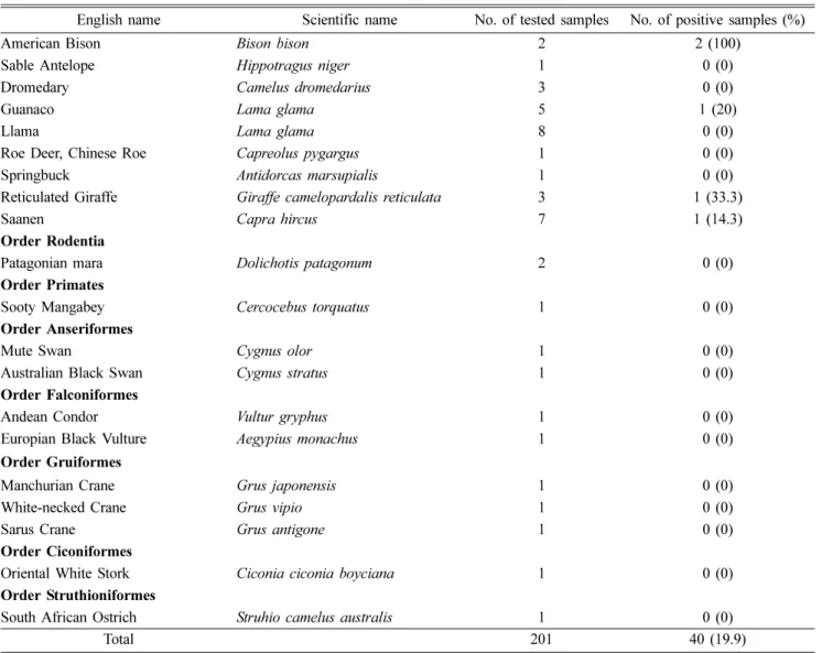
Table 1. continued
English name Scientific name No. of tested samples No. of positive samples (%)
American Bison Bison bison 2 2 (100)
Sable Antelope Hippotragus niger 1 0 (0)
Dromedary Camelus dromedarius 3 0 (0)
Guanaco Lama glama 5 1 (20)
Llama Lama glama 8 0 (0)
Roe Deer, Chinese Roe Capreolus pygargus 1 0 (0)
Springbuck Antidorcas marsupialis 1 0 (0)
Reticulated Giraffe Giraffe camelopardalis reticulata 3 1 (33.3)
Saanen Capra hircus 7 1 (14.3)
Order Rodentia
Patagonian mara Dolichotis patagonum 2 0 (0)
Order Primates
Sooty Mangabey Cercocebus torquatus 1 0 (0)
Order Anseriformes
Mute Swan Cygnus olor 1 0 (0)
Australian Black Swan Cygnus stratus 1 0 (0)
Order Falconiformes
Andean Condor Vultur gryphus 1 0 (0)
Europian Black Vulture Aegypius monachus 1 0 (0)
Order Gruiformes
Manchurian Crane Grus japonensis 1 0 (0)
White-necked Crane Grus vipio 1 0 (0)
Sarus Crane Grus antigone 1 0 (0)
Order Ciconiformes
Oriental White Stork Ciconia ciconia boyciana 1 0 (0)
Order Struthioniformes
South African Ostrich Struhio camelus australis 1 0 (0)
Total 201 40 (19.9)
68 Young-Jo Song, Bo-Sook Kim, Woo-Jung Park, Byung-Joo Park, Seul-Kee Lee, Jong-Il Shin, Nak-Hyung Lee, Joong-Bok Lee, Seung-Yong Park, Chang-Seon Song, Kun-Ho Seo, In-Soo Choi
Acknowledgments
This study was supported by Veterinary Science Research Institute of Konkuk University, Korea.
References
1. Arankalle VA, Joshi MV, Kulkarni AM, Gandhe SS, Chobe LP, Rautmare SS, Mishra AC, Padbidri VS.
Prevalence of anti-hepatitis E virus antibodies in different Indian animal species. J Viral Hepat 2001, 8, 223-227.
2. Bouwknegt M, Teunis PFM, Frankena K, de Jong MCM, de Roda Husman AM. Estimation of the likelihood of fecal-oral HEV transmission among pigs. Risk Anal 2011, 31, 940-950.
3. Choi IS, Kwon HJ, Shin NR, Yoo HS. Identification of swine hepatitis E virus (HEV) and prevalence of anti-HEV antibodies in swine and human populations in Korea. J Clin Microbiol 2003, 41, 3602-3608.
4. Cossaboom CM, Córdoba L, Dryman BA, Meng XJ.
Hepatitis E virus in rabbits, Virginia, USA. Emerg Infect Dis 2011, 17, 2047-2049.
5. Kim YM, Jeong SH, Kim JY, Song JC, Lee JH, Kim JW, Yun H, Kim JS. The first case of genotype 4 hepatitis E related to wild boar in South Korea. J Clin Virol 2011, 50, 253-256.
6. Kuno A, Ido K, Isoda N, Satoh Y, Ono K, Satoh S,
Inamori H, Sugano K, Kanai N, Nishizawa T, Okamoto H. Sporadic acute hepatitis E of a 47-year-old man whose pet cat was positive for antibody to hepatitis E virus.
Hepatol Res 2003, 26, 237-242.
7. Lu L, Li C, Hagedorn CH. Phylogenetic analysis of global hepatitis E virus sequences: genetic diversity, subtypes and zoonosis. Rev Med Virol 2006, 16, 5-36.
8. Meng XJ, Halbur PG, Shapiro MS, Govindarajan S, Bruna JD, Mushahwar IK, Purcell RH, Emerson SU.
Genetic and experimental evidence for cross-species infection by swine hepatitis E virus. J Virol 1998, 72, 9714-9721.
9. Meng XJ. Novel strains of hepatitis E virus identified from humans and other animal species: is hepatitis E a zoonosis?
J Hepatol 2000, 33, 842-845.
10. Pavio N, Meng XJ, Renou C. Zoonotic hepatitis E:
animal reservoirs and emerging risks. Vet Res 2010, 41, 46.
11. Rutjes SA, Lodder-Verschoor F, Lodder WJ, van der Giessen J, Reesink H, Bouwknegt M, de Roda Husman AM. Seroprevalence and molecular detection of hepatitis E virus in wild boar and red deer in The Netherlands. J Virol Methods 2010, 168, 197-206.
12. Song YJ, Jeong HJ, Kim YJ, Lee SW, Lee JB, Park SY, Song CS, Park HM, Choi IS. Analysis of complete genome sequences of swine hepatitis E virus and possible risk factors for transmission of HEV to humans in Korea. J Med Virol 2010, 82, 583-591.