- 151 -
KISEP 대 한 두 경 부 종 양 학 회 지
제 22 권 제 2 호 2006
비인두에 발생한 상피-근상피암종 1예
가톨릭대학교 의과대학 성빈센트병원 내과학교실,* 방사선종양학과교실,***
가톨릭대학교 의과대학 강남성모병원 병리학교실,** 이비인후과학교실****
홍은정*·이연수**·김수지***·김경희*·김민식****·선동일****·심병용*·김훈교*
= Abstract =
A Case of Epithelial-Myoepithelial Carcinoma in the Nasopharynx
Eun Jung Hong, M.D.,* Youn Soo Lee, M.D.,** Su ji Kim M.D.,***
Kyoung Hee Kim, M.D.,* Min Sik Kim, M.D.,**** Dong-Il Sun, M.D.,****
Byoung Yong Shim, M.D.,* Hoon-Kyo Kim, M.D.*
Department of Internal Medicine,* Radiation Oncology,*** St. Vincent’s Hospital, Suwon, Korea Department of Pathology,** Otorhinolaryngology Kangnam,**** St. Mary’s Hospital, Seoul, Korea
Epithelial-myoepithelial carcinoma is rare, low grade malignant tumor of the salivary glands that exhibits a dual composition of epithelial and myoepithelial cells. Most of these tumors arise in the parotid gland, and only few occur in the submandibular gland or minor salivary glands. We describe here a rare case of epithelial- myoepithelial carcinoma arising from a minor salivary gland in the nasopharynx, one of the most unusual locations. The clinical and biological behavior of this tumor is not yet known.
KEY WORDS:Nasopharyngeal neoplasm·Minor salivary gland tumor.
서 론
상피-근상피암종은 모든 타액선 종양의 1% 미만을 차지 하는 아주 드문 종양으로 저등급의 악성 종양이다1). 주 타 액선 특히 이하선에서 가장 흔히 발생하고 그 외에도 악하 선, 소타액선에서 발생하며 드문 발생 장소로 상악동, 구개, 비강, 비인두, 혀의 기저부, 기관지, 폐 등이 보고된 바 있 다2-9). 저자들은 비인두에 발생한 상피-근상피암종을 경 험하였고 국내에서 경험하는 첫 증례로 생각되어 문헌고찰 과 함께 보고하는 바이다.
증 례
70세 남자 환자가 6개월 간 지속된 콧물과 코 막힘을 주
소로 인근 이비인후과 방문하여 비인두강에 종양이 있다는 이야기를 듣고 본원으로 전원 되었다. 환자는 후비루와 간 헐적인 코피, 후각감퇴를 호소하였다.
환자의 과거력과 가족력에서 특이사항 없었으며 환자는 25 갑년의 흡연력이 있었다. 내원 당시 혈압은 120/80mmHg, 맥박은 70회/분, 호흡수 20회/분, 체온은 36.4℃이었다. 이 학적 검사 소견에서 코 중격이 좌측으로 치우쳐 있었고 우 측 후비강을 채우고 있는, 표면이 울퉁불퉁한 흰 색의 종괴 가 관찰되었다. 그 외 다른 소견은 보이지 않았다. 말초혈 액 검사에서 백혈구 8,010/mm3, 혈색소 11.1g/dL, 헤마 토크리트 32.8%, 혈소판 346,000/mm3 이었으며 생화학 검사에서 BUN 10.4mg/dL, creatitine 0.98mg/dL, Na 142 mEq/L, K 4.6 mEq/L, AST 20 IU/L, ALT 25IU/L, 총빌 리루빈 0.57mg/dL, 공복혈당 107mg/dL 이었다. 갑상선 기 능검사, 종양표지자 검사는 정상 범위였다. 안면부 자기공명 영상 소견상 비인두강의 우측 측면과 후면을 침범하는 4×2.3
×4cm 크기의 경계가 불명확한 종괴가 비강의 측면과 연구 개로 뻗어 있었으며 그 외 경부의 림프절은 관찰되지 않았다 (Fig. 1). 세침흡인 세포검사에서 소타액선에서 기원한 저등도 교신저자:심병용, 442-723 수원시 팔달구 지동 93
가톨릭대학교 의과대학 성빈센트병원 내과학교실 전화:(031) 249-8153·전송:(031) 253-8898 E-mail:[email protected]
- 152 - 의 다형성 선종이 의심되는 소견을 보였다. 뼈스캔과 양자 방 출 단층촬영을 시행하였으나 전이성 병변은 관찰되지 않았다.
환자는 비인두의 우측에 위치한 종양을 포함한 선택적 우측 경부곽청술을 시행하였다. 수술 후 병리 조직학적 검사상 비인 두강에 위치한 상피-근상피암종으로 진단되었으며 림프절 전 이는 없었으나 비인두의 외측벽을 침범한 소견이 관찰되었다.
술 후 환자는 비인두에 5,200cGy/ 26Fx의 방사선 치료를 시행하였고 현재 1년째 특별한 재발 없이 추적관찰 중이다.
1. 병리조직학적 검사
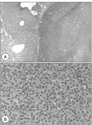
종양은 경계가 비교적 좋았고 다결절성 성장을 하고 있었
다(Fig. 2A). 종양세포는 큰 세포군집이나 판상구조를 만들 고 있었으며 2종류의 세포들로 이루어져 있었는데 일부 관 상구조를 이루는 부분도 있었지만 대부분 고형성 성장을 하 는 다각형 세포들로 이루어져 있었다. 이 세포들은 세포질 이 투명하거나 일부 연한 호산성 세포질을 가지고 있었고 핵 의 비정형은 미약하였으며 유사분열은 10개 고배율 시야에 서 2개 이내로 관찰되었다(Fig. 2B).
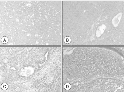
이 판상구조의 투명세포들은 PAS 염색에 양성(Fig. 3) 이 고 diastase 처리 후 모두 소실되어 세포질 내에 glycogen 을 가지고 있는 것이 확인되었다. 또한 이들 세포들은 S100 (Fig. 4A), actin(Fig. 4B)에 대한 면역조직화학염색에 양 성 소견이 관찰되어 근상피세포의 분화를 보이고 있었으며 일부 관상구조를 이루는 종양세포는 cytokeratin 19(Fig.
4C)에 양성소견이 관찰되어 관상피세포의 분화를 나타내고 있었다.
이 소견은 근상피세포의 분화를 보이면서 관상피세포 분 화가 동시에 관찰되는 상피-근상피암종에 합당한 소견이다.
또한 종양세포 Ki-67 labeling index가 40% 정도로 상당 히 높은 증식능을 나타내고 있었다(Fig. 4D).
고 찰
상피-근상피암종이라는 용어는 1972년 Donath 등10)이
Fig. 1. MRI of PNS. There is about 4×2.3×4cm sized soft tissue mass involving the right lateral and posterior wall of the nasopharynx, with anterior extension to the lateral wall of the nasal cavity and soft palate.
Fig. 3. Cytoplasmic periodic acid-Schiff staining indicates signifi- cant glycogen in the clear cells.
Fig. 2. The tumor is well circumscribed and has nodular growth pattern(H&E, ×40)(A). Polygonal tumor cells have uni- form nuclear feature with clear cytoplasm and a mitotic figure(H&E, ×400)(B).
A
B
- 153 - 처음 사용하였으며, 1991년에 WHO 분류에 포함된 드문 타 액선 종양이다. 상피-근상피암종의 호발 연령은 70대, 평 균 연령은 60세이나 27세부터 103세까지 보고된 바 있으 며 여성에서 더 흔히 발생한다1)11). 이하선이 가장 흔한 발생 장소이며 악하선과 구강내의 부선에서도 발생하며 드물게 비 강, 부비동, 기관지, 폐와 같은 구강 외 구조물에서도 보고 된 바 있다2-9). 임상 증상은 비특이적이며 대개 오랫동안 지속되며 진행하는 무통성의 종괴를 특징으로 하고 드물게 통증이나 안면신경 마비와 같은 증상을 동반하기도 한다11).
조직학적으로 2가지 종류의 종양세포를 특징으로 하며, 세관을 형성하는 관상피세포와 그 주위를 둘러싸는 근상피 세포로 구성되며, 이 두 세포의 비율은 종양마다 다르고 한 종양내에서도 부위마다 차이가 있다12)13). 면역조직 화학검 사상 관상피세포는 cytokeratin에, 근상피세포는 S-100단 백, actin에 양성 반응을 보임으로써 진단에 도움이 된다.
타액선 종양의 세포학적 특징은 아주 다양하고 각 종양의 소견들이 서로 중복되기 때문에 항상 정확한 진단이 가능한 것은 아니다. 감별할 질환으로는 관세포, 근상피세포 및 기 질조직으로 혼합 구성된 다형성 선종과 선양낭성암종이 있 으며 투명세포를 갖는 다른 종양과도 감별이 필요한데 여 기에는 피지선종, 점액표피양 암종, 선방세포암종등이 있다.
또한 전이암종, 특히 신세포암은 현미경적 소견이 상피-근 상피암종과 유사하여 감별진단에 고려되어야 한다12). 조직 학적으로 고형성 성장 양식, 핵의 비정형, 핵산의 이수배수 체, 높은 증식능은 일반적으로 공격적 성향과 높은 국소 재 발율 그리고 이하선 주위나 경부림프절로의 전이 등 나쁜 예후와 관련이 있다1)14).
타액선 종양의 원격 전이는 드물지만 일부에서 드물게 원
격 전이의 형태를 보이므로 수술 전 원격 전이를 확인하는 것이 필요하다고 생각되며 수술 시에도 경부림프절의 전이 를 확인 하여야 한다.
상피-근상피암종의 치료는 병변의 광범위 절제가 가장 좋 은 치료법으로 여겨지고 있으며 국소 재발을 막기 위해서 수술 후 방사선 치료가 도움이 된다11)15). 항암치료의 효과 에 대해서는 아직 확립된 바가 없으나 Puri 등6)은 혀의 기 저부에 발생한 상피-근상피암종에 대해 항암치료와 방사 선 치료만 시행하여 완전 관해 된 경우를 보고한 바 있다. 상 피-근상피암종은 저등급의 악성 종양으로 분류되지만 국소 재발이 흔하고 경부림프절로의 전이나 신장, 폐, 두피, 뇌16)17) 등으로의 원격전이도 보고된 바 있어 수술 후에도 전이 여 부를 비롯한 장기간의 추적관찰 기간이 필요하다. 본 증례 의 경우 종양을 포함한 선택적 경부곽청술을 통해 병변이 완전히 제거되었고 술 후 방사선 치료 후 지금까지 1년째 특별한 재발 병변은 보이지 않고 있다.
저자들은 70세 남자 환자의 비인두에 발생한 상피-근상 피암종을 경험하였고 이는 극히 드문 위치에 발생한 종양으 로 국내에서 처음으로 경험하는 증례이기에 문헌고찰과 함 께 보고하는 바이다.
중심 단어:비인두 종양·부타액선 종양.
References
1) Tralongo V, Daniele E: Epithelial-myoepithelial carcinoma of the salivary glands: a review of literature. Anticancer Res. 1998;
18:603-608
2) Li CY, Shirasuna K, Ishibashi H, Nakayama H, Kiyoshima T:
Fig. 4. Immunoreactivity for S100(A), Actin(B) shows positive reaction in the myoepithelial cells. Positive immunoreac-tion for CK-19 indicates ductal differentiation(C). Ki-67 labelling index is 40%(D).
B
D A
C
- 154 - Epithelial-myoepithelial carcinoma arising in pleomorphic ade- noma of the palate. Oral Surg Oral Med Oral Pathol Oral Radiol Endod. 2000;90:460-465
3) Inoue Y, Nomura J, Hashimoto M, Tagawa T: Epithelial-myoe- pithelial carcinoma of the palate: a case report. J Oral Maxil- lofac Surg. 2001;59:1502-1505
4) Lee HM, Kim AR, Lee SH: Epithelial-myoepithelial carcinoma of the nasal cavity. Eur Arch Otorhinolaryngol. 2000;257:376- 378
5) Imate Y, Yamashita H, Endo S, et al: Epithelial-myoepithelial carcinoma of the nasopharynx. ORL J Otorhinolaryngol Relat Spec. 2000;62:282-285
6) Puri T, Singh K, Sharma DN, Khurana N: Epithelial-myoepi- thelial carcinoma of the base of tongue: pathology and manage- ment. Indian J Cancer. 2004;41:138-140
7) Ru K, Srivastava A, Tischler AS: Bronchial epithelial-myoepi- thelial carcinoma. Arch Pathol Lab Med. 2004;128:92-94 8) Doganay L, Bilgi S, Ozdil A, Yoruk Y, Altaner S, Kutlu K: Epi-
thelial-myoepithelial carcinoma of the lung. A case report and review of the literature. Arch Pathol Lab Med. 2003;127:177-180 9) Shanks JH, Hasleton PS, Curry A, Rahman A: Bronchial epi- thelial-myoepithelial carcinoma. Histopathology. 1998;33:90-91 10) Donath K, Seifert G, Schmitz R: Diagnosis and ultrastructure of the tubular carcinoma of salivary gland ducts. Epithelial-myoe- pithelial carcinoma of the intercalated ducts. Virchows Arch A
Pathol Pathol Anat. 1972;356:16-31
11) Simpson RH, Clarke TJ, Sarsfield PT, Gluckman PG: Epithelial- myoepithelial carcinoma of salivary glands. J Clin Pathol. 1991;
44:419-423
12) Deere H, Hore I, McDermott N, Levine T: Epithelial-myoepi- thelial carcinoma of the parotid gland: a case report and review of the cytological and histological features. J Laryngol Otol.
2001;115:434-436
13) Miliauskas JR, Orell SR: Fine-needle aspiration cytological findings in five cases of epithelial-myoepithelial carcinoma of salivary glands. Diagn Cytopathol. 2003;28:163-167
14) Amin KS, McGuff HS, Cashman SW, Newman R: Recurrent epithelial-myoepithelial carcinoma of the parotid with direct in- tracranial extension. Otolaryngol Head Neck Surg. 2002;126:
83-84
15) Senis-Segarra L, Sahuquillo-Arce E, Davo R, Hamad-Arcis P, Floria-Garcia LM, Baquero MC: Salivary Gland Epithelial- Myoepithelial Carcinoma: behaviour, diagnosis and treatment.
Med Oral. 2002;7:391-395
16) Noel S, Brozna JP: Epithelial-myoepithelial carcinoma of Sali- vary gland with metastasis to lung: report of a case and review of the literature. Head Neck. 1992;14:401-406
17) Kwon MS, Lee SS, Koh JS, Chung JH: Fine Needle Aspiration Cytology of Metastatic Epithelial-Myoepithelial Carcinoma of the Scalp: A Case Report. Korean J Cytopathol. 2000;11:93-97.