Archives of Craniofacial Surgery
www.kcpca.or.kr ISSN 2287-1152
76 Copyright © 2012 The Korean Cleft Palate-Craniofacial Association
This is an Open Access article distributed under the terms of the Creative Commons Attribution Non-Commercial License (http://creativecommons.org/
licenses/by-nc/3.0/) which permits unrestricted non-commercial use, distribution, and reproduction in any medium, provided the original work is properly cited.
두피에 발생한 거대 피지샘 상피종 1례
김은연·김선구·김유진·이세일 가천대 길병원 성형외과학교실
Purpose: Sebaceous epithelioma (sebaceoma) is a benign tumor with sebaceous differentiation. It presents primarily as a yellowish papule or nodule on the face and scalp. It must be differentiated from basal cell carcinoma and other appendageal tumors. We report a giant sebaceous epithelioma on the scalp and describe the immunohistochemical character of the cells in sebaceous epithelioma to epithelial membrane antigen (EMA).
Methods: A 55-year-old-man who presented with 5-cm-diameter 2-cm-height, round shape exophytic ulcerated tumor on his head presented for treatment. The patient had noticed the lesion 40 years prior as a small yellowish plaque and 18 months ago, the plaque started to grow progressively larger. We excised the lesion with 1 cm resection margin, considering the possibility of malignancy because this lesion grossly resembled basal cell carcinoma (BCC). The defect was repaired with the use of a split- thickness skin graft.
Results: When we excised the lesion, the margin was clear. Histology showed nodules that consisted of an admixture of basaloid cells and mature adipocytes lacking an organized lobular architecture. Strong expression of EMA on mature adipose cells confirmed the differential diagnosis from BCC with sebaceous differentiation because of the absence of a nuclear palisade pattern and cleft-like spaces on the hematoxylin and eosin (H&E) section.
Conclusion: We treated the giant sebaceous epithelioma on the scalp with surgical excision and a split-thickness skin graft. It is important to know that the diagnosis of sebaceous epithelioma should be made based on the histologic pattern of the H&E section. Immunohistochemistry with EMA can help to confirm the differential diagnosis between sebaceous epithelioma and BCC.
Keywords: Sebaceous epithelioma, Basal cell carcinoma
A Giant Sebaceous Epithelioma on the Scalp: A Case Report
Eun Yeon Kim, Sun Goo Kim, Yu Jin Kim, Se Il Lee
Department of Plastic and Recontructive Surgery, Gachon University Gil Hospital, Incheon, Korea
서 론
피지샘 상피종(sebaceous epithelioma)은 피지샘 분화를 보이는 드문 양성 피부 부속기 종양으로, 임상적으로 기저 세포 상피암의 외양을 지니며, 원발성 또는 피지샘 모반(ne-
vus sebaceus)이나 지루성 각화증(seborrheic keratosis)에서 이차적으로 발생한다. 남성에 비해 여성에서 호발하며 대 개 고령자에서 안면, 두피 등 피지샘이 잘 발달된 부위에 황 색의 구진(papule), 결절(nodule), 또는 판(plaque)의 형태로 나타나고, 크기는 3 mm에서 10 cm까지 다양 하지만 대부 분 직경 1 cm 이하이다. 병리조직학적으로 대부분의 세포 들이 미분화된 기저양세포(basaloid cell)들로 구성되어 있 으며, 일부 성숙된 피지세포와 세포질의 지방성 공동화를 일으키는 세포들이 관찰되지만 성숙된 피지샘 구조를 형 성하지 않는 것이 특징이다.1 간혹 피지선 상피종을 포함한
Arch Craniofac Surg Vol.13 No.1, 76-79 http://dx.doi.org/10.7181/acfs.2012.13.1.76
Correspondence: Yu Jin Kim
Department of Plastic and Recontructive Surgery, Gachon University Gil Hospital, 21 Namdong-daero, 774 beon-gil, Namdong-gu, Incheon 405-760, Korea Tel: +82-1577-2299 / Fax: +82-32-461-2774 / E-mail: [email protected] Received August 19, 2011 / Revised December 29, 2011
Accepted December 29, 2011
Case Report
77
www.kcpca.or.kr
피지선 종양과 내부 장기암이 연관되어 발생되는 경우가 있는데 이를 Muir–Torre 증후군이라고 한다.2 저자들은 원 발성으로 측두부에 발생한 거대 피지샘 상피종을 경험하 여, 드문 예로 생각되어 보고하고자 한다.
증 례
55세 남자 환자가 좌측 측두부에 발생한 가려움증과 삼 출물이 동반된 덩어리를 주소로 내원하였다. 내원 약 40년 전부터 두피에 작은 구진이 발생하였고 서서히 병변이 커져 가던 중, 내원 1년 6개월 전부터 급속히 성장하기 시작하였 다. 외상과 같은 과거력이나 다른 선행된 두피의 피부병변 은 없었다. 진찰 검사에서도 피부 소견 외에 특이 사항은 관 찰되지 않았다. 병변은 직경 5 cm, 높이 2 cm의 적황색의 반 구의 모양을 하고 있으며 부분적으로 삼출물이 흘러나와 굳어있었고 탈모반, 중심부 궤양 소견을 보이고 있었다(Fig.
1). 궤양 주변으로 얇게 표피가 일어나 약한 자극에도 쉽게 손상되었으며, 흘러나온 삼출물에서는 악취가 났다. 일반 혈액 검사와 소변 검사는 정상 범위였다.
수술 전 병변부위에서 시행한 절개생검상 피지샘 분화 를 보이는 종양의 소견이 있었으나, 그 세부 분류에 대해서 는 정확한 감별진단이 이루어지지 않았다. 중심부 궤양과 주변부 미란, 홍반 및 거대 결절 형성 등의 임상소견상 피지 샘 분화가 동반된 결절성 기저세포암의 가능성을 염두에 두고 병변으로부터 1 cm의 여유를 두어 치료적 절제술 후, 연부조직 결손부위에 부분층 피부이식술을 시행하였다.
Fig. 1. Diameter 5 cm sized yellow to orange colored solitary exo- phytic tumor with central ulceration on the left parietal scalp.
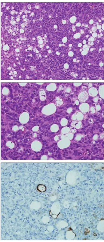
Fig. 2. Histology of sebaceous epithelioma. (Above) Sheets of basa- loid cells area randomly admixed with small mature sebocytes. The basaloid cells have uniform nuclear with small nucleoli (H&E, × 200).
(Center) There is no mitosis (H&E, × 400). (Below) Epithelial mem- brane antigen highlights the mature sebocytes (× 400).
Eun Yeon Kim, et al. A giant sebaceous epithelioma
Archives of Craniofacial Surgery Vol. 13, No. 1, 2012
www.kcpca.or.kr
78
절제한 병변은 조직학적 소견상 기저양세포와 다수의 공포 (vacuole)를 가진 피지세포들로 구성되어 있었고 부분적으 로 표피층과 연결된 피지샘 상피종으로 진단되었다. 면역조 직학적 소견상 S–100 단백, carcinoembryonic antigen 염색 에 음성이었으며, epithelial membrane antigen (EMA)는 성 숙된 공포를 가진 피지세포에만 양성을 보였다(Fig. 2). 내부 장기암과의 연관성을 조사하기 위해 상부위장관 및 결장 의 내시경 검사 또는 조영술을 권유하였으나 아직 시행하 지 않았다. 수술 후 7개월간 추적관찰 중이며 재발은 없는 상태이다(Fig. 3).
고 찰
피지샘 상피종은 1880년 Bock에 의해 처음으로 보고되 었다. 원발성 또는 지루성 각화증이나 피지선 모반에서 이 차적으로 발생하며, 발생연령은 29–87세로 다양한 연령분 포를 보이나 대개 60세 이상에서 발생하고, 남성에 비해 여 성에서 4배정도 더 호발한다.1,3 치료 후 재발이나 전이에 대 해서는 아직 보고된 바 없다.1
피지샘 상피종은 다른 용어들과 혼용되어 사용되어 왔 으며, 피지샘종(sebaceous adenoma)의 변이로 인식되기도 하였고, 피지샘종과 기저세포암의 중간 단계로 불리기도 하였다. 또한, 피지샘 분화가 동반된 기저세포암(basal cell epithelioma with sebaceous differentiation)과 sebomatrico- ma와 같은 의미로 쓰이기도 하였다.4 Troy와 Ackerman3은 이런 문제를 명확히 하고 양성병변이라는 것을 강조하기 위
해 sebaceous epithelioma를 폐기하고, sebaceoma라고 부를 것을 제안하기도 하였다.
피지샘 상피종은 진피층에 중심이 있으며, 표피를 침범 하는 경우는 종종 관찰되고, 아주 드물게 피하조직을 침범 한다. 천천히 자라며 절제술 후 재발은 하지 않는 것으로 알 려져 있다.1,4 피지샘 상피종은 피지세포의 양성 종양으로 기 저세포 상피종이나 미분화 기저양세포들이 우세한 종양과 그 감별이 어려울 때가 많다.피지샘 상피종과 결절성 기저 세포암 모두 소엽(lobule) 모양을 이루고 있고, 경도의 세포 이형성(atypia)과 유사분열(mitotic activity)은 사실 두 종양 에서 모두 나타날 수 있다.
피지샘과 관련된 신생물의 경우 육안적으로는 감별진단 이 어렵기 때문에 조직학적 감별점에 대해서 많이 연구되 어 왔다. Dinneen과 Mehregan5은 21개의 피지샘 상피종의 증례를 통해, 병변의 낭성 변화를 특징적으로 기술하였다.
기저세포암 역시 피지샘의 낭성 변화를 보일 수 있으나, 특 징적인 기저양세포의 울타리배열(palisade of basaloid cells) 이나, 기저양세포가 모여 이룬 섬(islands)들 사이의 간극 (cleft) 등이 나타날 수 있으며, 유사분열이 활발한 것이 특징 이다.1
본 증례는 50대 중반의 비교적 건강한 남성에서 황색를 띤 직경 5 cm의 거대한 덩어리로 두피에 발생하였다. 궤양 을 동반한 성장이 빠른 병변이었기 때문에, 절개생검만으 로는 기저세포암을 배제할 수 없어 안전역(safety margin)을 두고 절제술을 시행하였다. 조직 검사 결과는 대부분 기저 양세포로 이루어져 있었고 공포를 가진 피지세포들이 관 찰되었으며 낭성 공간도 다수 관찰되어 피지샘 상피종의 소견에 부합하였다. 피지샘 상피종에 대한 논문은 국내에 서도 보고된 바 있으나 본 증례의 경우처럼 삼출물을 동반 한 궤양성 결절로 나타난 피지샘 상피종은 드문 예로 생각 된다.6,7
기저세포암과 피지샘 상피종은 그 조직학적 특징이 비슷 하지만, 이 둘을 감별진단하는 것은 환자 평가와 치료법의 선택 및 추적 관찰에 있어서 중요하다. 기저세포암은 국소 적으로 침습적인 피부암으로 정상조직을 포함한 광범위 절 제술이 가장 좋은 치료방법이다. 광범위 절제술에 의해 주 변 조직 특히 입주변이나 코, 눈에 미용적, 기능적으로 변형 을 일으킬 수 있다.
피지샘 상피종의 경우에는 상염색체 우성 유전을 하는 Fig. 3. Postoperative 7 months. Diameter 6 cm sized round shape
slightly depressed area without any signs of marginal recurrence.
79
www.kcpca.or.kr
Muir–Torre 증후군과 관련이 있는 것으로 알려져 있어 조 기에 내장장기의 악성종양에 대한 선별 검사를 환자뿐 아 니라 가족에게도 시행해야 한다. 그래서 Fan 등8은 조직학 적 특성이 비슷하거나 검체의 크기가 너무 작은 경우, EMA 와 같은 특수 염색을 통해 피지샘 상피종과 결절성 기저세 포암을 감별진단 할 수 있다고 하였다. EMA는 성숙한 피지 세포에만 염색이 되기 때문에 EMA에 좀 더 우세하게 염색 되는 것이 피지샘 상피종이다.
본 증례에서와 같이 거대한 크기를 가지며 임상적 양상 에서 피지샘 분화를 보이는 기저세포암과 구별이 힘든 피 지샘 종양의 경우, 절개생검한 검체에 대해서도 hematoxy- lin and eosin 염색과 더불어 EMA 면역조직화학 염색을 동 시에 시행하여 기저세포암과 감별진단을 한다면, 절제범위 와 치료방법을 선택하는데 도움이 될 수 있다고 생각한다.
REFERENCES
1. McKee PH, Calonje E, Granter SR: Pathology of the skin with clinical correlations. 3rd ed, Boston, Elsevier Mosby, 2005, p 157
2. Torre D: Multiple sebaceous tumors. Arch Dermatol 98: 549, 1968 3. Troy JL, Ackerman AB: Sebaceoma. A distinctive benign neoplasm of
adnexal epithelium differentiating toward sebaceous cells. Am J Der- matopathol 6: 7, 1984
4. Barnes L, Eveson J, Reichart P, Sidransky D: Pathology and genetics of head and neck tumors. Lyon, IARC Press, 2005, p 163
5. Dinneen AM, Mehregan DR: Sebaceous epithelioma: a review of twen- ty–one cases. J Am Acad Dermatol 34: 47, 1996
6. Lee UH, Park HS, Choi JC, Chun DK: A case of sebaceous epithelioma.
Korean J Dermatol 42: 904, 2004
7. Mohaghegh F, Asilian A, Taheri S, Rajabi P: A case of giant sebaceoma.
Iran J Dermatol 12: S23, 2009
8. Fan YS, Carr RA, Sanders DS, Smith AP, Lazar AJ, Calonje E: Character- istic Ber–EP4 and EMA expression in sebaceoma is immunohis- tochemically distinct from basal cell carcinoma. Histopathology 51: 80, 2007
Eun Yeon Kim, et al. A giant sebaceous epithelioma