134
with local recurrence occurring in about 25%-35% of pa- tients3.
II. Case Report
This is a case of a 22-year-old female who was previously well till she experienced swelling and pain over her right tem- poral region for 18 months. She had episodes of jaw locking and could hear clicking sounds when chewing or talking. Her hearing was slightly reduced on the right side. She sought medical help but was treated as temporomandibular disorder (TMD) and was given analgesics, physiotherapy and an oc- clusal splint. The pain reduced after the prescribed treatment and the swelling was inconspicuous initially but increased in size and hence further investigations were carried out. Ex- amination revealed a 2×2 cm swelling over the right temporal region which was firm and tender on palpation. Upon open- ing her mouth, her mandible would deviate to the right side.
Facial nerve and trigeminal nerve function was intact. Com- puted tomography (CT) scan showed an aggressive erosive tumor of the squamous temporal bone extending to the right temporomandibular joint (TMJ).(Fig. 1) An ultrasound-guid- ed fine needle aspiration biopsy was performed. The result
I. Introduction
Giant cell tumor (GCT) of bone usually occurs in the epiphyses of long bones like the distal femur, proximal tibia, distal radius and proximal humerus1. Craniofacial bone involvement is rare but has been reported to occur in the mandible, temporal bone, maxilla, occipital and sphenoid2,3. The incidence of GCT varies between regions and is highest amongst the Asian population, especially the Chinese and Japanese where they account for nearly 15% of all primary bone tumors4. GCTs affect women more than men at a ratio of 3:22,5. This tumor is mainly diagnosed during the third to fifth decade of life6. Although generally thought to be benign tumors, GCTs are known to be locally aggressive at times,
CASE REPORT
Jo Ee Sam
Department of Neurosurgery, Hospital Pulau Pinang, Jalan Residensi, 10990 Georgetorn, Pulau Penang, Malaysia
TEL: +60-164990242 FAX: +60-42281737 E-mail: [email protected]
ORCID: http://orcid.org/0000-0002-6279-6433
This is an open-access article distributed under the terms of the Creative Commons Attribution Non-Commercial License (http://creativecommons.org/licenses/by-nc/4.0/), which permits unrestricted non-commercial use, distribution, and reproduction in any medium, provided the original work is properly cited.
CC
Giant cell tumor of temporomandibular joint masquerading as temporomandibular joint pain dysfunction syndrome: a rare case report
Jo Ee Sam1, Rullyandrianto Pan Nuriman Rachmat2, Cri Saiful Jordan Melano3, Nasser Abdul Wahab1 Departments of 1Neurosurgery and 2Oral and Maxillofacial Surgery, Hospital Pulau Pinang,
3Department of Oral and Maxillofacial Surgery, Hospital Seberang Jaya, Pulau Penang, Malaysia
Abstract(J Korean Assoc Oral Maxillofac Surg 2017;43:134-137)
Giant cell tumor (GCT) of the craniofacial bones has been reported but they are not common. This tumor occurs more often in women than in men and predominantly affects patients around the third to fifth decade of life. GCTs are generally benign but can be locally aggressive as well. We report a case of GCT involving the temporomandibular joint (TMJ), which was initially thought to be temporomandibular disorder (TMD). A 22-year-old female presented with swelling and pain over the right temporal region for 18 months associated with jaw locking and clicking sounds. On examination, her jaw deviated to the right during opening and there was a 2×2 cm swelling over the right temporal region. Despite routine treatment for TMD, the swell- ing increased in size. Computed tomography and magnetic resonance imaging of the brain and TMJ revealed an erosive tumor of the temporal bone involving the TMJ which was displacing the temporal lobe. Surgical excision was done and the tumor removed completely. Histopathological exami- nation was consistent with a GCT. No clinical or radiological recurrence was detected 10 months post-surgery.
Key words: Giant cell tumors, Temporomandibular joint, Temporomandibular joint disorders, Temporal bone, Mandible
[paper submitted 2016. 7. 8 / revised 1st 2016. 8. 20, 2nd 2016. 9. 3, 3rd 2016. 9. 17 / accepted 2016. 9. 21]
Copyright Ⓒ 2017 The Korean Association of Oral and Maxillofacial Surgeons. All rights reserved.
https://doi.org/10.5125/jkaoms.2017.43.2.134 pISSN 2234-7550·eISSN 2234-5930
Giant cell tumor of TMJ
135 the ascending ramus of the mandible was used for access to the coronoid process. Intraoperatively, the tumor was seen to invade the temporal bone, mandibular condyle, TMJ and overlying temporalis muscle but did not invade the temporal dura. The tumor was hard and yellowish in color. The patient underwent right partial temporal craniectomy, removal of part of the mandibular condyle and zygomatic arch, excision of the coronoid process, and excision of the TMJ.(Fig. 3) The zygomatic arch was reconstructed with titanium mesh. The patient recovered uneventfully after the surgery. Histopatho- logical examination of the tumor revealed fibrous connective was suggestive of a xanthogranuloma. A magnetic resonance
imaging (MRI) of the brain and TMJ was performed which showed an extra-axial mass at the right middle cranial fossa involving the right TMJ, measuring 4.2×1.6×2.6 cm. The mass was enhanced heterogeneously post-contrast and dis- placed the adjacent temporal lobe.(Fig. 2) During surgery, we used a modified frontotemporal flap for access to the tempo- ral bone and TMJ. An intraoral right sulcular incision along
Fig. 1. Computed tomography scan showing an erosive tumor of squamous temporal bone and temporomandibular joint.
Jo Ee Sam et al: Giant cell tumor of temporomandibular joint masquerading as temporomandibular joint pain dysfunction syndrome: a rare case report. J Korean Assoc Oral Maxillofac Surg 2017
Fig. 2. Magnetic resonance imaging post-gadolinium showing an extra-axial heterogenous mass compressing on the right temporal lobe.
Jo Ee Sam et al: Giant cell tumor of temporomandibular joint masquerading as temporomandibular joint pain dysfunction syndrome: a rare case report. J Korean Assoc Oral Maxillofac Surg 2017
Fig. 3. Intraoperative picture of the right temporal region. Forceps holding part of destroyed temporomandibular joint.
Jo Ee Sam et al: Giant cell tumor of temporomandibular joint masquerading as temporomandibular joint pain dysfunction syndrome: a rare case report. J Korean Assoc Oral Maxillofac Surg 2017
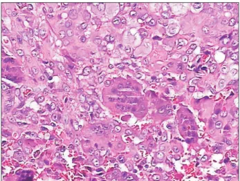
Fig. 4. Numerous multinucleated giant cells (H&E staining, ×200).
Jo Ee Sam et al: Giant cell tumor of temporomandibular joint masquerading as temporomandibular joint pain dysfunction syndrome: a rare case report. J Korean Assoc Oral Maxillofac Surg 2017
J Korean Assoc Oral Maxillofac Surg 2017;43:134-137
136
and it can detect soft tissue and intra-articular extension16. Macroscopically, most GCTs are soft and fleshy and ap- pear grey to light red or dark reddish-brown. There may be areas of cyst, hemorrhage or fibrous septa formation as well.
The margins are usually ill defined, which explains the high percentage of recurrence if only curettage is done4,17.
GCT is a neoplasm of stromal-like neoplastic cells that are able to recruit macrophage and multinucleate osteoclast- like giant cells. Histologically, GCTs are characterized by the finding of large osteoclast-like multinucleated giant cells scattered among a background of plump or spindle shaped mononuclear stromal cells. The stromal cells may be mitoti- cally active but should not have abnormal or atypical mitotic cells. These giant cells have approximately 10-20 nuclei per cell, but may have 100 or more nuclei. There may be reac- tive bone formation usually at the periphery and reactive changes such as reactive fibrosis, necrosis, hemorrhage and xanthogranulomatous inflammation. This is likely the reason the ultrasound-guided fine needle aspiration biopsy result showed a xanthogranuloma. GCTs often have abundance of neovascularization, which explains the hemorrhages that are frequently seen within such tumors4,18.
Important differential diagnoses of GCTs are osseous le- sions that are giant cell rich. It is important to consider le- sions such as giant cell reparative granuloma, hyperparathy- roidism, non-ossifying fibroma, chondroblastoma, solid areas of aneurysmal bone cyst, malignant fibrous histiocytoma and osteogenic sarcoma4.
Surgery with the aim of wide excision is the mainstay of treatment for GCTs, preferably with a wide margin of nor- mal tissue8,16,19. However, total removal of the skull base and cranial GCTs is technically challenging. Radiotherapy is reserved for cases where wide excision cannot be achieved or for patients who are not fit for surgery. Irradiation-induced sarcomatous transformation is a known risk with orthovoltage radiation, but interestingly there is less risk with current use of megavoltage radiation19. Denosumab, a receptor activa- tor of nuclear factor kappa-B ligand (RANKL) inhibitor has been approved for use in recurrent and unresectable GCTs20. High dose dexamethasone therapy had been used effectively to rapidly reduce the size of these tumors but unfortunately, discontinuation of steroids is associated with re-growth in nearly every case4.
Conflict of Interest
No potential conflict of interest relevant to this article was tissue with a few foci of numerous multinucleated giant cells,
histiocytes, neutrophils, lymphocytes and occasional foam cells. There was peripheral bone and adjacent fibrocartilage- neous tissue as well.(Fig. 4) A final diagnosis of benign GCT was made. No recurrence at 10 months post-surgery was de- tected.
II. Discussion
GCTs are usually benign but have been known to be local- ly aggressive and occasionally metastasize, especially to the lung7-9. Very rarely, GCTs may turn into sarcoma4. The usual sites of occurrence are the epiphysis of long bones and less than 2% occur in the head and neck region where the usual sites are the sphenoid and temporal bones10. Less than 30 cas- es of GCT in the TMJ have been reported so far. Portions of the temporal bone form by endochondral ossification, which is the same way epiphyses of long bones are formed and thus it is possible that the temporal bone is more prone to develop GCTs because of this11. Patients usually present with progres- sive pain and swelling over the site. In the temporal region, hearing impairment and facial nerve paralysis can occur due to compression or local invasion from the tumor12. Involve- ment of the TMJ causes jaw locking, deviation of mandibular movement and clicking sounds. These three symptoms and signs are also common with TMD13. We would like to high- light the danger of treating patients as TMD before a precise diagnosis is made. A thorough history and examination should be done and a list of differential diagnoses should be considered, including tumors14. TMD may present with pain and swelling at the temporal area as in this case, but swelling in TMD is different and not common. Some patients may still have temporal swelling but the swelling should be softer on palpation and not persistent in size as compared to tumors.
The swelling seen in TMD may be present during and after chewing and should decrease gradually between meals.
GCTs appear lytic, subarticular, eccentrically located and usually lack a sclerotic rim on radiographs. Local bony de- struction, cortical breakthrough and soft tissue expansion may also be seen. CT will rarely provide information that helps physicians arrive at a diagnosis but may be useful in de- lineating tumor extent, evaluation of cortical integrity and de- termination of tumor recurrence15. On MRI, GCTs have low signal intensity on T1-weighted images, heterogeneous high signal intensity on T2-weighted images and heterogeneous enhancement with gadolinium. MRI is the preferred imaging modality for GCTs, as the diagnostic accuracy of MRI is high
Giant cell tumor of TMJ
137 reported.
ORCID
Jo Ee Sam, http://orcid.org/0000-0002-6279-6433
Rullyandrianto Pan Nuriman Rachmat, http://orcid.
org/0000-0002-6668-5001
Cri Saiful Jordan Melano, http://orcid.org/0000-0001- 8636-7177
Nasser Abdul Wahab, http://orcid.org/0000-0002-5265-3033
References
1. Unni KK. Dahlin's bone tumors: general aspects and data on 11,087 cases. 5th ed. Philadelphia: Lippincott-Raven; 1996:263-85.
2. Schajowicz F. Tumors and tumor-like lesions of bone: pathol- ogy, radiology, and treatment. 2nd ed. Berlin: Springer-Verlag;
1994:257-95.
3. Bertoni F, Unni KK, Beabout JW, Ebersold MJ. Giant cell tumor of the skull. Cancer 1992;70:1124-32.
4. Zheng MH, Robbins P, Xu J, Huang L, Wood DJ, Papadimitriou JM. The histogenesis of giant cell tumour of bone: a model of inter- action between neoplastic cells and osteoclasts. Histol Histopathol 2001;16:297-307.
5. Huvos AG. Bone tumors: diagnosis, treatment and prognosis. Tu- mors of histiocytic or fibrohistiocytic origin. 2nd ed. Philadelphia:
Saunders; 1991:429-67.
6. Robbins SL, Cotran RS, Kumar V. Robbins pathological basis of disease. 5th ed. Philadelphia: WB Saunders; 1994:1245-6.
7. Vanel D, Contesso G, Rebibo G, Zafrani B, Masselot J. Benign giant-cell tumours of bone with pulmonary metastases and favour- able prognosis. Report on two cases and review of the literature.
Skeletal Radiol 1983;10:221-6.
8. Leonard J, Gökden M, Kyriakos M, Derdeyn CP, Rich KM. Malig- nant giant-cell tumor of the parietal bone: case report and review of the literature. Neurosurgery 2001;48:424-9.
9. Min BI, Park YW. A case of metastatic giant cell tumor aris- ing in two-year old girl. J Korean Assoc Oral Maxillofac Surg 1988;14:82-7.
10. Lee HJ, Lum C. Giant-cell tumor of the skull base. Neuroradiology 1999;41:305-7.
11. Morriss-Kay GM. Derivation of the mammalian skull vault. J Anat 2001;199:143-51.
12. Findlay JM, Chiasson D, Hudson AR, Chui M. Giant-cell tumor of the middle cranial fossa. Case report. J Neurosurg 1987;66:924-8.
13. Manfredini D, Guarda-Nardini L, Winocur E, Piccotti F, Ahlberg J, Lobbezoo F. Research diagnostic criteria for temporomandibular disorders: a systematic review of axis I epidemiologic findings.
Oral Surg Oral Med Oral Pathol Oral Radiol Endod 2011;112:453- 14. Lee BK. Evidence-based practice in the treatment of temporoman-62.
dibular disorders. J Korean Assoc Oral Maxillofac Surg 2012;38:
15. Wang CS, Lou JH, Liao JS, Ding XY, Du LJ, Lu Y, et al. Recur-263.
rence in giant cell tumour of bone: imaging features and risk fac- tors. Radiol Med 2013;118:456-64.
16. Purohit S, Pardiwala DN. Imaging of giant cell tumor of bone. In- dian J Orthop 2007;41:91-6.
17. Wysocki RW, Soni E, Virkus WW, Scarborough MT, Leurgans SE, Gitelis S. Is intralesional treatment of giant cell tumor of the distal radius comparable to resection with respect to local control and functional outcome? Clin Orthop Relat Res 2015;473:706-15.
18. Silvers AR, Som PM, Brandwein M, Chong JL, Shah D. The role of imaging in the diagnosis of giant cell tumor of the skull base.
AJNR Am J Neuroradiol 1996;17:1392-5.
19. Chen ZX, Gu DZ, Yu ZH, Qian TN, Huang YR, Hu YH, et al. Ra- diation therapy of giant cell tumor of bone: analysis of 35 patients.
Int J Radiat Oncol Biol Phys 1986;12:329-34.
20. Branstetter DG, Nelson SD, Manivel JC, Blay JY, Chawla S, Thomas DM, et al. Denosumab induces tumor reduction and bone formation in patients with giant-cell tumor of bone. Clin Cancer Res 2012;18:4415-24.