Received: April 30, 2019 Revised: November 29, 2019 Accepted: December 8, 2019
OPEN ACCESS
HORTICULTURAL SCIENCE and TECHNOLOGY 38(1):107-117, 2020
URL: http://www.hst-j.org pISSN : 1226-8763 eISSN : 2465-8588
This is an Open Access article distributed under the terms of the Creative Commons Attribution Non-Commercial License which permits unrestricted non-commercial use, distribution, and reproduction in any medium, provided the original work is properly cited.
Copyrightⓒ2020 Korean Society for Horticultural Science.
This research was supported by the Public Projects of Zhejiang province (LGN18C160005, LGN19C160005); The National Natural Science Foundation of China (31000897, 31301811);
Zhejiang provincial science and technology cooperation project (2019SNLF013); Science Technology Department of Zhejiang Province (No. 2016C02052-1); The earmarked fund for China Agriculture Research System (CARS-27).
Chimerism Evaluation of ‘Hongrou Huyou’,
a Grafted Chimera between Citrus changshan-huyou and Citrus unshiu
Min Zhang 1†* , Zequn Zhang 1† , Qun Wu 2 , Fuzhi Ke 3 , Jianguo Xu 3 , Siqing Zhao 4 , Gang Wang 4 , and Chi Zhang 1
1
State Key Laboratory of Subtropical Silviculture, Zhejiang A& F University, Hangzhou 311300, PR China
2
Quzhou Technical Extension Station for Cash Crops, Quzhou 324000, PR China
3
Zhejiang Citrus Research Institute, Taizhou, Zhejiang 318020, PR China
4
Changshan Huyou Research Institute, Quzhou, Zhejiang 324000, PR China
*Corresponding author: mzhang@zafu.edu.cn
†
These authors contributed equally to the work.
Abstract
Chimeras occur spontaneously or artificially and are valuable in horticultural crop breeding. A new diploid citrus chimera, named ‘Hongrou Huyou’ (abbreviated HH) (Citrus changshan-huyou + C.
unshiu), was found during a bud sports investigation in China. The morphology, flesh carotenoid content, and molecular markers were evaluated in HH and the two grafted donors. The results showed that characteristics derived from L2/L3, such as the fruit size, winged leaf, seed, pollen, and rind aroma, were similar to those of C. changshan-huyou (CH), whereas the juice sac and stomatal characteristics originating from L1 were the same as those of the satsuma mandarin. High-performance liquid chromatography analysis of carotenes from the flesh of HH showed that the content was the same as that of the satsuma mandarin, with β-cryptoxanthin producing the main carotenoid spectrum, whereas lutein and violaxanthin were the main carotenoids in CH. Nuclear simple sequence repeat, chloroplast simple sequence repeat, and mitochondrial simple sequence repeat analyses showed that the leaves, outer pericarp (epidermis and flavedo), segment wall, and juice sac of HH contained the nuclear, chloroplast, and mitochondrial genomes of both donors; however, the albedo of HH only contained the genetic material of CH. Thus, HH is confirmed to be a periclinal chimera that consists of L1 from C. unshiu and L2/L3 from CH.
Additional key words: carotenoid, cpSSR, morphology, mtSSR, periclinal chimera, SSR
Introduction
Citrus is extensively planted in tropical and subtropical regions worldwide and includes five main
economic groups: orange (Citrus sinensis), mandarin (C. reticulata), grapefruit (C. paradisi), pummelo
(C. maxima), and lemon (C. limon). Over the past 30 years, citrus breeding improvement has been
concentrated on seedlessness, ease of peeling, strong flavor and aroma aspects. Bud sport selection
and cross-breeding are the two conventional breeding methods and have been used to develop more
than 100 citrus cultivars (Deng, 2005). Citrus chimera breeding is an emerging breeding strategy that has made great progress in fresh markets.
Generally, the shoot apical meristem (SAM) of plants is composed of cells with two or more different genotypes. In dicotyledon plants, the SAM consists of three cell layers: dermatogen initial (the outermost layer, L1), periblem initial (middle layer, L2), and plerome initial (the innermost layer, L3) (Schmidt, 1924). In citrus fruit, the juice sac and epidermal pericarp develop from L1; the seed, segment wall, hypoderm, and mesocarp of the pericarp are produced by L2;
and the L3 cell layer is often involved in vascular bundle formation (Ohtsu and Kuhara, 1994). Artificial plant chimeras develop spontaneously as intervariety and interspecies chimeras in vitro, although the use of layer-specific mutations to induce phenotypic trait changes is another method to develop a periclinal chimera. Based on previous studies, the research in this field can be divided into two major branches; the first branch seeks to breed new varieties, and the second branch investigates the different genotype cell layer interactions using natural and synthetic plant chimeras. At present, a grafted chimera is an ideal model to investigate stock-scion interactions and different genotype cell layer displacements. Cao et al. (2016) studied the inner mechanism between stock and scion in the artificial plant chimeras Brassica juncea and Brassica oleracea. They reported that short-distance mobile communication via small RNAs could regulate the expression of related genes and lead to different phenotypes. Additionally, DNA methylation and changes in DNA methylation patterns in transposons play a role in graft-induced phenotypic variations. Generation of grafting mutations may be the cause of phenotypic variation through changes in DNA methylation patterns in transposons (Li et al., 2013).
To date, few reports have investigated xylophyta chimeras. Citrus is one of the most commonly described xylophyta types. The occurrence of citrus chimeras usually emerges from an adventitious bud, which is derived from the callus tissue in the graft junction of the stock and scion, and typically the callus is caused by unintentional injury of the scion. As early as 1674, ‘Bizzaria’ was reported as the first chimera that arose from a graft union of C. medica and C. sinensis (Tilney-Bassett, 1986). Later, some citrus-grafted chimeras were successively released (Sugawara et al., 2002; Zhang et al., 2007, 2015).
More studies have investigated artificial synthetic citrus grafting chimeras in Japan, and some new germplasms with high quality and disease resistance have been developed. For example, Kuhara (1989) successfully synthesized the first citrus chimera (C. sinensis + C. natsudaidai), and Ohtsu and Kuhara (1994) also put forward two artificial citrus chimeras named ‘NFF’ and ‘FNN’ from the synthetic donor plants ‘Fukuhara’ sweet orange and ‘Kawano’ natsudaidai.
C. changshan-huyou (abbreviated CH), which is a precious citrus germplasm resource native to Changshan county of Zhejiang province in China, is widely cultivated for its large fruit, high yield, excellent quality, and unique flavor. In the 1990s, CH was used as a scion and was top grafted upon the satsuma mandarin stock. Then, a natural chimera named
‘Hongrou Huyou’ (HH) adventitiously arose at the grafted junction of CH and the satsuma mandarin (C. unshiu), and showed deep-orange color and juicy flesh, strong flavor, and good storage ability.
In this study, to explore the genetic composition of HH, we performed a characteristic evaluation of the morphology, cytology, and molecular markers of this chimera.
Materials and Methods Plant Materials
The chimera HH arose at a graft union zone where a bud of CH was grafted onto a satsuma mandarin in the 1990s.
Recently, HH was discovered in an orchard in Changshan county of Zhejiang province during a bud mutation investigation.
The fruit on the branch with the pericarp was similar to that of CH, and the flesh was similar to that of the satsuma mandarin. The trees of the three varieties were planted in the same orchard with conventional management. All samples, including the fruits and leaves, were picked from the regrafted trees (with Poncirus trifoliata as the rootstock) for decades.
The picked fruits of HH were immediately divided into four parts: outer pericarp (epidermis and flavedo), albedo, segment wall, and juice sac. Then, the fruit parts were quickly frozen in liquid nitrogen and stored at ‑ 80°C. Because CH was extensively used as a high scion to replace the ‘Owari’ satsuma mandarin (OW) in the 1990s, we conjectured that HH was a peripheral chimera formed by grafting; therefore, CH and OW are assumed to be the donor plants of HH.
Morphological Analysis
Both length and width of leaves and fruits of the HH and donor plants were measured. Current spring shoots from the outer layer of the middle part of the canopy were collected in summer, and mature fruits of the same size were selected.
Leaf and fruit samples were taken from three trees, 10 samples each.
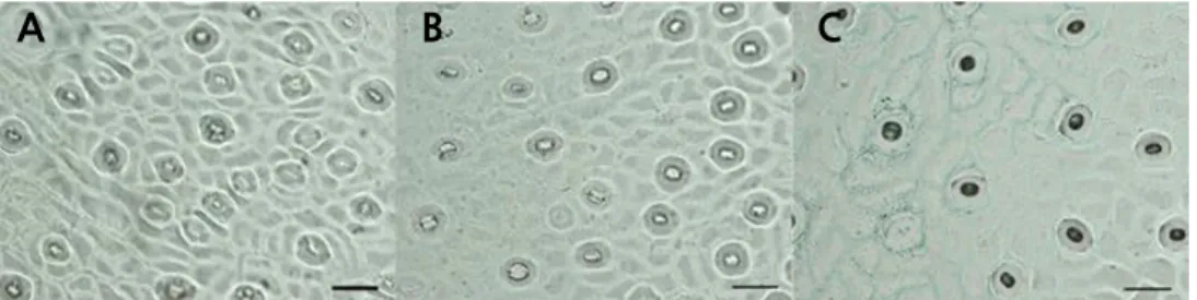
Stomatal Density and Length
The leaves were collected from mature spring shoots, and a thin layer was evenly smeared with nail polish along the two sides of the main vein on the back of the leaf (approximately 5 ‑ 10 mm). After 5 minutes, the imprinted thin layer was torn off with tweezers and observed under an Olympus BX61 universal microscope (Olympus, Japan) directly after pressing with a cover slip. The stomata numbers were counted by observing four visual fields per leaf. The mean value of the stomatal density (per mm
2) was calculated. The guard cell length was measured for 20 stomata per leaf, and the mean stomatal length was calculated.
Observation of the Tissue Structure of the Leaf Cross Section
Paraffin sections were prepared according to the method of Sun et al. (2010). Mature spring shoot leaves were cut into 0.3 × 1-cm pieces with a blade fixed in FAA (ethanol:acetic acid:formaldehyde = 90:5:5), dehydrated in a gradient ethanol series (30 ‑ 100%), and embedded in paraffin. Then, the embedded materials were sliced with a Leica Ultracut R ultrathin slicing machine (Leica, Bensheim, Germany) to 10- µ m thickness. Stained paraffin sections were observed with an Olympus BX61 universal microscope (Olympus, Japan). The upper epidermis, lower epidermis, palisade tissue, and total leaf thickness were measured in the micrographs.
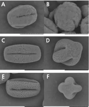
Scanning Electron Microscopy
According to the method of Han et al. (2018), fresh pollen was pretreated on the sample stage and sprayed with the 108 Auto Cressington Sputter Coater (Cressington, UK). The samples were examined with a scanning electron microscope (JSM-6390/LV, NTC, Japan), and representative images were obtained.
Flow Cytometric Analysis
The ploidy level of HH was determined by flow cytometry according to the method of Zhang et al. (2007). Approximately
2 cm
2of leaf was chopped with a razor blade in a Petri dish in 1.5 mL of HR-A nuclei extraction solution (Partec High-Resolution Staining Kit; Partec GmbH, Münster, Germany). After adding 2 mL of HR-B DAPI staining solution (Partec High-Resolution Staining Kit), the mixture was filtered through a 30- µ m nylon filter. Fluorescence of the nuclei was measured with a CA-II flow cytometer (Partec, Germany). Young leaves of CH and OW were used as the controls.
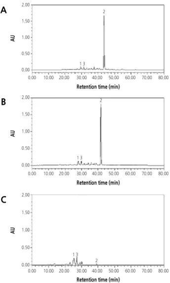
High-Performance Liquid Chromatography Carotenoid Analysis
The carotenoid extraction and saponification processes were performed under the condition of weak or dark light to avoid decomposition. The carotenoid pigments were analyzed by reversed phase high-performance liquid chromatography (HPLC) using the modified binary gradient elution procedure originally developed by Lee et al. (2001). The forms of the carotenoids were identified by the retention time of the standard sample and the characteristic spectral value. Standard samples ( β-cryptoxanthin, violaxanthin, and lutein) were purchased from CaroteNature (Lupsingen, Switzerland).
DNA Extraction and Simple Sequence Repeat Analysis
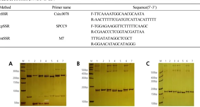
DNA extraction and SSR analysis were conducted based on Cheng et al. (2003a, 2005). PCR products were mixed with an equal volume of formamide loading buffer (98% formamide, 10 mM EDTA pH 8.0, 0.025% Bromophenol Blue, and xylene cyanol). Two milliliters of mixture of the equipartition sample was added to 12% polyacrylamide gel and electrophoresed for 5 hours at 180 V. Bands were displayed after silver staining and recorded on a ScanMaker 3830 (Microtek, Shanghai, China). Primers used for nuclear simple sequence repeat (nSSR) analyses were as described by Xu et al. (2013). Fourteen chloroplast simple sequence repeat (cpSSR) primers (SPCC1 to SPCC14) were utilized to analyze chloroplast genomes according to the report of Cheng et al. (2003b). One mitochondrial simple sequence repeat (mtSSR) primer was used to analyze mitochondria genomes according to the report of Cheng et al. (2003c).
Statistical Analysis
Data are reported as means ± standard error (SE). Statistical analyses were performed with SAS Statistics 9.1 (SAS Institute Inc, NC, USA). Means followed by different letters are significantly different at p = 0.01 according to least significant difference test.
Results
Morphological Characteristics of the Fruit
The fruit index and shape of HH were similar to those of OW and were markedly different from those of CH (Table 1
and Fig. 1A) . However, the fruit width (9.67 cm) and height (8.05 cm) both exceeded those of its donors. The fruit of HH
(384.88 g) was heavier, but the edible rate (67.05%) was less than that of both donors. The flesh weight (257.12 g) of HH
was similar to that of CH. During the experiment, we found that the pericarp of HH was thicker than that of its donor
plants, which might lead to considerable storage quality. The color of the juice sacs in HH (deep orange) was similar to
that of OW (Table 1 and Fig. 1B) , and the peel had a strong aroma similar to that of CH. Additionally, HH was seedy with
an average of 19.82 seeds per fruit, which was similar to that of CH.
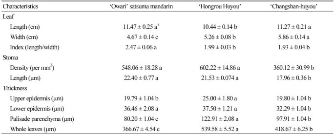
Table 2. Morphological characteristics of leaf between ‘Hongrou Huyou’ and its donor plants
Characteristics ‘Owari’ satsuma mandarin ‘Hongrou Huyou’ ‘Changshan-huyou’
Leaf
Length (cm) 11.47 ± 0.25 a
z10.44 ± 0.14 b 11.27 ± 0.21 a
Width (cm) 4.67 ± 0.14 c 5.26 ± 0.08 b 5.86 ± 0.14 a
Index (length/width) 2.47 ± 0.06 a 1.99 ± 0.03 b 1.93 ± 0.04 b
Stoma
Density (per mm
2) 548.06 ± 18.28 a 602.22 ± 14.86 a 360.12 ± 30.99 b
Length (µm) 22.40 ± 0.77 a 21.53 ± 0.074 a 17.96 ± 0.36 b
Thickness
Upper epidermis (µm) 19.79 ± 1.04 b 25.00 ± 1.80 a 19.80 ± 1.04 b
Lower epidermis (µm) 36.46 ± 2.08 a 37.50 ± 1.21 a 32.29 ± 1.04 b
Palisade parenchyma (µm) 80.20 ± 1.04 c 122.91 ± 2.08 a 97.91 ± 1.04 b
Whole leaves (µm) 366.67 ± 4.54 c 539.58 ± 5.52 a 418.67 ± 6.25 b
z
Means within a line followed by different letters are significantly different at p = 0.01 according to least significant difference test.
Table 1. Morphological characteristics of fruit between ‘Hongrou Huyou’ and its donor plants
Characteristics ‘Owari’ satsuma mandarin ‘Hongrou Huyou’ ‘Changshan-huyou’
Fruit
Width (cm) 7.88 ± 0.19 c
z9.67 ± 0.75 a 8.60 ± 0.21 b
Height (cm) 6.50 ± 0.07 c 8.05 ± 0.14 a 7.63 ± 0.08 b
Size ratio (height/width) 0.83 ± 0.02 b 0.83 ± 0.01 b 0.89 ± 0.01 a
Weight (g) 198.23 ± 9.95 c 384.88 ± 8.28 a 306.43 ± 16.18 b
Flesh weight (g) 150.33 ± 6.38 b 257.12 ± 6.97 a 230.14 ± 12.31 a
Edible rate (%) 77.48 ± 0.07 a 67.05 ± 0.01 b 76.53 ± 0.08 a
Seed number (per fruit) 0 b 19.82 ± 5.58 a 15.58 ± 3.38 a
Color of juice sac Deep orange Deep orange Yellow
Aroma of rind Weak Strong Strong
Shape Oblate Oblate Short Spheroid
z