Ⅰ. 서 론
기질 금속단백분해효소(matrix metalloproteinase, MMP)는 아연 (Zn
++)을 함유하고 있는 세포외 기질단백 분해효소로서 20여종이 있으며, 세포외 기질 중 세포간질이나 기저막의 콜라겐, 단백당,
파이브로넥틴(fibronectin), 라미닌(laminin)과 같은 단백질로 구성 된 물질들을 분해시켜, 태아의 발육, 임신, 성장, 창상의 치유와 같은 결합조직 재형성 과정 및 관절질환, 골다공증, 치주질환, 암 조직의 성장과 침투에 관여하는 것으로 알려져 있다
1).
관절 활액을 이용한 임상 연구에서 측두하악관절 내장증과 관 련해서는 MMP-2와 MMP-9이
2-6), 골관절증과 관련해서는 MMP-1, - 2, -3, -9 이 관련이 있다고
2,3,5,7-9)보고되고 있다. 이 중 MMP-1 (colla- genase 1)은 현재까지 알려진 4 종류의 콜라겐 분해효소 중 하나 로 I, II, III, VII, X형 콜라겐, 젤라틴 등을 분해하고, MMP-2 (gelati- nase A)는 72kDa IV형 콜라겐 분해효소로 젤라틴, I, IV, V, VII, X 형 콜라겐, 엘라스틴(elastin), 파이브로넥틴, 라미닌 등을 분해한 다
10). 측두하악 관절원판 후조직의 세포외 성분은 주로 I형과 III
측두하악 관절원판 후조직의
MMP(matrix metalloproteinase)-1과 MMP-2 mRNA의 발현
허종기∙박광균*∙최민아*∙김형곤
�연세대학교 치과대학 구강악안면외과학교실(용인세브란스병원, 영동세브란스병원
�) 연세대학교 치과대학 구강생물학교실*
Abstract (J. Kor. Oral Maxillofac. Surg. 2003;29:212-218)
김 형 곤
135-720 서울시 강남구 도곡동 146-92
연세대학교 치과대학 구강악안면외과학교실 (영동세브란스 병원) Hyung-Gon Kim
Dept. of Oral and Maxillofacial Surgery, College of Dentistry, Yonsei University 146-92 Dogok-Dong, Gangnam-Gu, Seoul, 135-720, Korea
TEL: +82-2-3497-3560 Fax: +82-2-3463-4052 E-mail : dental@yumc.yonsei.ac.kr
EXPRESSION OF MATRIX METALLOPROTEINASE-1 AND -2 MRNA IN RETRODISCAL TISSUE OF THE TEMPOROMANDIBULAR JOINT
Jong-Ki Huh, Kwang-Kyun Park
*
, Min-Ah Choi*
, Hyung-Gon Kim�Dept. of Oral and Maxillofacial Surgery, College of Dentistry, Yonsei University (Yongin Severance Hospital, Yongdong Severance Hospital
�)
Dept. of Oral Biology, College of Dentistry, Yonsei University *
Matrix metalloproteinases (MMPs) play an important role in the normal morphogenesis, maintenance, and repair of matrix and also have important functions in pathologic conditions characterized by excessive degradation of extracellular matrix, such as rheumatoid arthritis, osteoarthritis, periodontitis and in tumor invasion and metastasis.
In this study, expression of MMP-1 and -2 mRNA in retrodiscal tissue of the temporomandibular joint (TMJ) was examined and com- pared with magnetic resonance imaging (MRI) and surgical findings. MMP mRNAs in the retrodiscal tissue samples were detected by reverse transcription - polymerase chain reaction. TMJ internal derangement (ID) was categorized as normal disc position, disc dis- placement with reduction, early stage of disc displacement without reduction (DDsR) and late stage of DDsR. TMJ osteoarthrosis (OA) was classified with normal, mild and advanced OA. The amount of synovial fluid collection was divided into not detected, small, large and extremely large amount on MR T2-weighted imaging. Perforation and adhesion were examined during open surgery of the TMJ.
Six out of 37 samples were excluded because of little amount of extracted total mRNA. MMP-2 mRNA was detected whole joints, and so the MMP-2 mRNA seems to be expressed normally in retrodiscal tissue. However, MMP-1 mRNA was expressed in 8 of 31 joints.
Frequencies of MMP-1 mRNA expression according to the TMJ IDs, amount of synovial fluid and surgical findings made no significant difference. MMP-1 mRNA was detected more frequently in OA groups (7/16 joints, 43.8%) than in normal bony structure group (1/15 joints, 6.7%).
Expression of MMP-1 mRNA in retrodiscal tissue might be related with OA of the TMJ.
Key words: Matrix metalloproteinase, Collagenase, Gelatinase, Temporomandibular joint, Osteoarthrosis
형 콜라겐 섬유와 탄력 섬유, 그리고 단백당으로 이루어져 있으
므로
11-13), 관절원판 후조직의 기질 변화에 MMP-1과 MMP-2가 관
여할 것이다. 또한 하악과두의 퇴행성 골변화는 하악과두 연골 이나 골조직 자체의 조직 파괴뿐만 아니라, 하악과두가 노출되 어 있는 관절 활액의 조성 변화에도 영향을 받을 것이다. 따라서 관절 활액의 조성 변화에 영향을 끼치는 관절원판 후조직의 기 질 변화가 하악과두의 퇴행성 골변화에도 영향을 끼칠 것으로 추측할 수 있다.
관절 활액은 채취의 용이성으로 인해 많은 연구가 되어 왔으 나, 관절 원판이 전방으로 변위되었을 때, 섬유화 변성, 천공, 충 혈, 유착 등과 같은 다양한 변화과정을 겪게 되는 관절 원판 후조 직에 대한 연구들은 제한적으로 시행되고 있다. 이에 본 연구는 측두하악관절 장애로 인하여 측두하악관절 수술을 받은 환자들 을 대상으로 직접 관절원판 후조직을 채취하여 MMP-1과 MMP-2 mRNA의 발현여부와 자기공명영상 소견 및 수술 소견을 각각 비 교하여 관절원판 후조직에서의 MMP-1과 MMP-2 mRNA의 발현이 측두하악관절을 이루는 구조물들의 변화와 어떠한 연관성을 가 지는지 알아보고자 하였다.
Ⅱ. 연구 대상 및 방법
1. 연구 대상
2002년 3월부터 10월까지 측두하악관절 장애를 주소로 연세대 학교 영동세브란스병원 턱관절 클리닉에 내원하여 임상검사 및 자기공명영상사진 검사 결과 측두하악관절 내장증 또는 골관절 증(osteoarthrosis)으로 진단된 환자들 중 관절원판 성형술(dis- coplasty)이나 절제술(discectomy)을 시행받은 37관절을 대상으로 하였다. 관절 원판 성형술시에는 변위된 관절원판을 정상 위치 로 복원시키기 위해 관절원판의 후외측, 즉 관절원판 후조직의 외측부위를 쐐기 모양으로 제거하였으며, 관절 원판 절제술시에 는 관절원판과 관절원판 후조직을 같이 제거하였다. 관절원판 후조직에서의 MMP-1과 MMP-2 mRNA의 발현을 보기 위해 제거 된 조직에서 관절원판 후조직 부위를 일부 취하여 실험 때까지 RNAlater
TM(Ambion Inc., TX, USA)가 들어있는 폴리프로필렌 튜브
에 담아 -4 ℃에 보관하였다.
2. 연구 방법
가. 리보핵산(RNA)의 추출
폴리프로필렌 튜브에 보관된 조직은 RNAlater
TM를 제거한 후 조 직 분쇄기(tissue homogenizer)에 담아 충분히 으깬 다음, SV Total RNA Isolation System (Promega Co., WI, USA) 을 이용하여 리보핵 산을 추출하였다. 추출된 리보핵산(total RNA) 용액은 역전사 중 합효소 연쇄반응을 시킬 때까지 -70 ℃에 보관하였다.
나. 역전사 중합효소 연쇄반응 (Reverse Transcription Polym- erase Chain Reaction, RT-PCR)
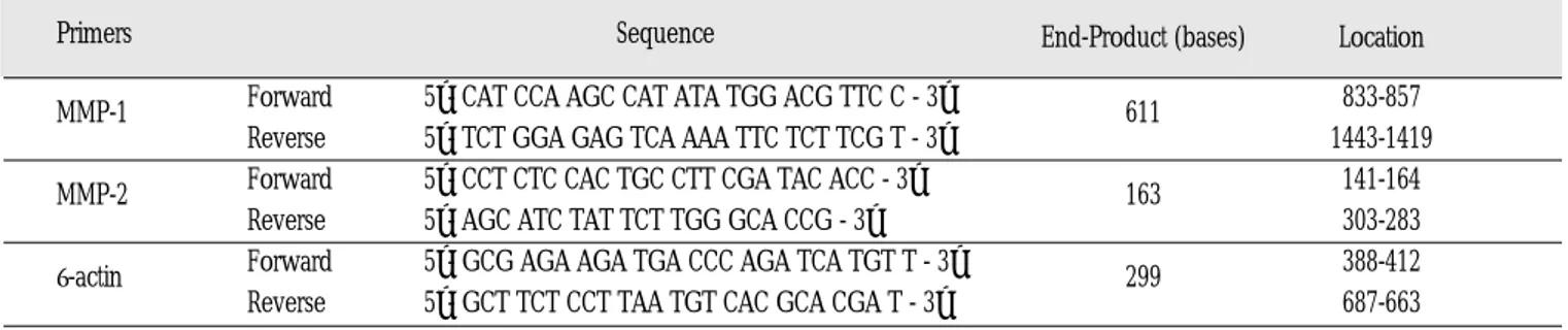
추출된 total RNA는 RNA PCR Kit (Ver. 2.1, TaKaRa Shuzo Co., Japan)을 이용하여 DNA Thermal Cycler 480 (Perkin-Elmer Co., USA)에서 역전사 중합효소 연쇄반응을 시행하였다. 역전사 반응 에는 사용된 키트에서 요구하는 최대용량인 9.5㎕의 total RNA를 사용하였다. 역전사 과정은 55 ℃에서 30분, 99 ℃에서 5분, 5 ℃ 에서 5분 시행하였다. 중합효소 연쇄반응 과정에서 사용된 전향 시발체(primer)와 후향 시발체의 염기배열과 최종산물의 크기는 Table 1과 같으며
14)각각 20 pmole을 사용하였다. 중합효소 연쇄 반응은 초기 denaturation을 94 ℃에서 2분간 1회 시행한 후, denaturation 94 ℃ 45초, annealing 59 ℃ 45초, extension 72 ℃ 1분 의 반응을 35회 반복하고, 최종 extension을 72 ℃에서 5분간 1회 시행하였다.
역전사 중합효소 연쇄반응이 끝난 용액 20㎕를 취하여 4㎕의 6X DNA dye와 잘 혼합하여 ethidium bromide (EtBr)가 든 1.0% 아 가로스 겔(agarose gel)에서 전기영동하고 UV transilluminator에서 결과를 관찰하였다. 원하는 길이의 염기 쌍을 가진 결과물이 얻 어졌는지 확인하기 위해 50bp (base pair) ladder를 사용하였다.
다. 자기공명영상 분석
자기공명 T1 강조영상에서는 관절 원판의 상태 및 복위여부에 따라 정상위치군(normal position), 복위성 변위군(disc displace- ment with reduction), 초기 비복위성 변위군(early stage of disc dis-
Table 1.Sequences of PCR primers in RT-PCR.
Primers Sequence End-Product (bases) Location
MMP-1 Forward 5′- CAT CCA AGC CAT ATA TGG ACG TTC C - 3′ 611 833-857
Reverse 5′- TCT GGA GAG TCA AAA TTC TCT TCG T - 3′ 1443-1419
MMP-2 Forward 5′- CCT CTC CAC TGC CTT CGA TAC ACC - 3′ 163 141-164
Reverse 5′- AGC ATC TAT TCT TGG GCA CCG - 3′ 303-283
β-actin Forward 5′- GCG AGA AGA TGA CCC AGA TCA TGT T - 3′ 299 388-412
Reverse 5′- GCT TCT CCT TAA TGT CAC GCA CGA T - 3′ 687-663
F: forward; R: reverse
placement without reduction), 후기 비복위성 변위군(late stage of disc displacement without reduction)의 네 군으로 나누었으며
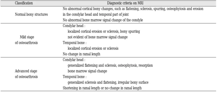
15), 측 두하악관절의 구조를 이루고 있는 관절와나 하악과두의 골변화 정 도 에 따 라 경 도 (mild)와 중 증 도 (advanced) 골 관 절 증 (osteoarthrosis)으로 나누었다(Table 2, Fig. 1, 2). 자기공명 T2 강조 영상에서는 관절 활액의 양을 관찰하였으며, 관절강 내의 고강 도 신호의 관찰 여부 및 범위에 따라, 관찰 안됨(not-observed), 소 량(small amount), 다량(large amount), 과다량(extremely large
amount)의 네 군으로 나누었다
15).
라. 수술 소견
관절원판 후조직의 상태는 상관절강에서 관절원판 또는 관절 원판 후조직이 관절융기나 관절와와 부분적으로 또는 전체가 붙 어있는“유착(adhesion)”, 상관절강과 하관절강이 개통이 되어 있는“천공(perforation)”, 그리고, 유착이나 천공이 관찰되지 않 는“관찰 안됨“으로 구분하여 조사하였다.
Table 2.The classification of status of TMJ with respect to osteoarthrosis.
Classification Diagnostic criteria on MRI
No abnormal cortical bony changes, such as flattening, sclerosis, spurring, osteophytosis and erosion Normal bony structures in the condylar head and temporal part of joint
No abnormal bone marrow signal change of the condyle Condylar head :
localized cortical erosion or sclerosis, bony spurring Mild stage not evident of bone marrow signal change of osteoarthrosis Temporal bone :
localized cortical erosion or sclerosis No change in ramal length
Condylar head :
generalized flattening and sclerosis, osteophytosis, resorption Advanced stage bone marrow signal change
of osteoarthrosis Temporal bone :
generalized sclerosis and flattening, irregular bony surface Shortening in ramal length or no change in ramal length
a. closed mouth b. open mouth
Fig. 1.Mild stage of osteoarthrosis. Erosion of condylar head is observed (a. arrow). No signal change of condylar bone marrow is seen. C: condylar head. E: articular eminence.
a. closed mouth b. open mouth
Fig. 2. Advanced stage of osteoarthrosis. Osteophyte (b.
arrow) and cortical thickening of temporal bone and condylar head are observed. Mild signal change of bone marrow in the condylar head is seen. E: articular eminence.
마. 비교 분석
관절 원판 후조직에서의 MMP-1과 MMP-2 mRNA의 발현 여부 를 자기공명영상 소견 및 수술 소견과 각각 비교하였다. 통계 분 석은 IBM PC용 SAS 통계 분석 프로그램(version 6.04)을 이용하여 카이제곱 검정(기대빈도수가 5이하인 항목이 있을 경우는 피셔 의 정확도 검정(Fisher’s exact test))을 시행하였다.
Ⅲ. 결 과
총 37명 37관절 중 전기영동에서 베타-액틴(β-actin)의 띠가 명 확하게 나타나지 않은 6관절은 제외하였다. 제외된 6관절 중 2관 절은 활액 연골종증(synovial chondromatosis)이었으며, 2관절은
심한 골관절증으로 수술시 관절원판의 천공 및 심한 협착이 관 찰되었고, 2관절은 관절강이 좁아진 상태에서 관절원판의 천공 이 관찰되었다.
연구대상 31명 중 남자는 7명(22.6%)이었고, 여자는 24명 (77.4%)이었으며, 평균나이는 32.1세(17세-62세)였다. 수술은 관절 원판 성형술이 22관절(71.0%)이었고, 나머지는 관절원판 절제술 이었다.
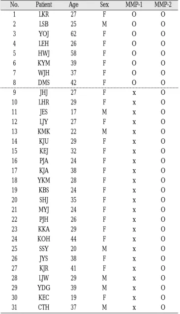
MMP-1 mRNA는 31관절 중 8관절(25.8%)에서 발현이 되었으나 (Fig. 3, Table 3), MMP-2 mRNA는 모든 관절에서 발현이 되었다 (Fig. 4).
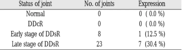
수술환자의 대부분은 비복원성 원판변위 상태였으며, 비복원 성 변위 후기에서 MMP-1 mRNA가 발현된 관절의 빈도(30.4%)는 초기(12.5%)보다 높았으나 통계학적으로 유의한 차이는 없었다 (p=0.642, Table 4).
자기공명영상에서 골관절증이 관찰된 16관절 중 7관절(43.8%) 에서 MMP-1 mRNA가 검출되어, 골관절증이 관찰되지 않은 관절 (6.7%)보다 통계학적으로 유의하게 많은 검출 빈도를 보였다 (p=0.037, Table 5). 비록 관절 수는 작지만 중증도의 골관절증에 서는 5관절 중 3관절(60.0%)에서 MMP-1 mRNA가 검출되어 다른 군에 비해 높은 발현빈도를 보였다.
자기공명 T2 강조영상에 측두하악관절 삼출 소견이 관찰되는 빈도는 MMP-1의 발현여부에 따른 차이를 보이지는 않았다 (Table 6).
Fig. 3. PCR products of MMP-1 mRNA (611bp), β- actin (299bp) and MMP-2 mRNA (163bp) are observed.
MMP-1 -->
β-actin -->
MMP-2 -->
Patient 1 2 3 4 5 6 7 8
Fig. 4. PCR products of MMP-2 mRNA (163bp) and β-actin (299bp) are observed.
β-actin -->
MMP-2 -->
Patient 12 13 14 15 16 17 18
Table 3.Expression of MMP-1 and -2 mRNA in retrodis- cal tissue of the temporomandibular joint.
No. Patient Age Sex MMP-1 MMP-2
1 LKR 27 F O O
2 LSB 25 M O O
3 YOJ 62 F O O
4 LEH 26 F O O
5 HWJ 58 F O O
6 KYM 39 F O O
7 WJH 37 F O O
8 DMS 42 F O O
9 JHJ 27 F x O
10 LHR 29 F x O
11 JES 17 M x O
12 LJY 27 F x O
13 KMK 22 M x O
14 KJU 29 F x O
15 KEJ 32 F x O
16 PJA 24 F x O
17 KJA 38 F x O
18 YKM 28 F x O
19 KBS 24 F x O
20 SHJ 35 F x O
21 MYJ 24 F x O
22 PJH 26 F x O
23 KKA 29 F x O
24 KOH 44 F x O
25 SSY 20 M x O
26 JYS 38 F x O
27 KJR 41 F x O
28 LJW 29 M x O
29 YDG 39 M x O
30 KEC 19 F x O
31 CTH 37 M x O
O: detected, x: not detected
수술시 발견되는 관절원판과 그 주위조직의 변화들 중에서 유 착과 천공은 각각 따로 관찰되기도 하였지만 한 관절에서 같이 관찰되는 경우도 있었다. 유착이 관찰된 총 6관절에서는 MMP-1 mRNA가 2관절(33.3%)에서 검출되었고, 천공이 관찰된 총 14관절 중에서는 5관절(35.7%)에서 MMP-1 mRNA가 검출되어, 유착이나 천공이 관찰되지 않은 관절에서의 MMP-1 mRNA의 발현 빈도 (21.4%)와는 유의한 차이를 보이지 않았다(각각 p=0.613, p=0.678, Table 7).
Ⅳ. 고 찰
측두하악관절 장애는 병태생리학적으로 다양성을 보이기 때 문에, 단층 촬영(tomogram), 관절 조영술, 자기공명영상과 같은 방사선 사진이나, 관절경 또는 수술을 통한 수술적 소견 및 조직 학적 소견을 통해 관절 질환을 규명하려는 많은 노력들이 있었 으며, 분자생물학적으로는 측두하악관절 장애의 다양한 진행 단 계(pathologic stage)에 따른 분자 표식자(molecular marker)를 찾기 위해 관절원판이나 연골 구성물질, 성장 인자, 염증관련 사이토 카인(cytokines), 기질 파괴 효소 등에 대한 연구들이 진행되어 왔 다. 기질 파괴 효소 중 금속단백분해효소(MMP)는 정상 조직의 발육, 성장, 항상성 유지, 재생산과 관련이 있고, 병적 상태에서 는 조직의 파괴 및 섬유성 질환과 관련이 있는 것으로 알려져 있
으며, 최근에는 암의 침투와 전이, 진단과 치료를 위해 많은 관심 을 쏟고 있다
16). 측두하악관절에서 MMP에 대한 연구는 대부분 관절 활액의 분석을 통해 이루어지고 있으며, 측두하악관절을 구성하고 있는 조직에 대한 연구는 거의 없다.
측두하악 관절원판이 전방으로 변위되면 하악과두 상방에 위 치하게 되는 관절원판 후조직은 섬유화 변성, 천공, 충혈, 유착 등과 같은 다양한 변화과정을 겪게 되는 데 이 과정에서 관절원 판 후조직의 주성분인 콜라겐이나 엘라스틴의 변성이 일어날 것 이며, 여기에 콜라겐과 엘라스틴을 분해하는 MMP-1과 MMP-2가 관여할 것이다. 활액을 통한 연구에서 측두하악관절 내장증이나 골관절증이 MMP와 관련이 있다고 보고되고 있으나, 이러한 병 적 상태를 유발하는 부위인 관절원판 후조직에서의 MMP 발현이 나 MMP 조절 기전에 대한 연구는 거의 없으므로, 본 연구는 수 술시 제거한 조직에서의 MMP 발현에 대해 알아보았다. 관절 활액 에서 검출되는 MMP는 활막 세포나 연골세포에서 분비되므로
2,5), 활액에서 검출되는 MMP는 대부분이 관절원판 후조직에서 분비 될 것이다.
본 연구에서 MMP-2 mRNA는 모든 관절에서 검출이 되었다 (Table 3). MMP-2는 관절원판 후조직에 분포하는 엘라스틴을 분 해하여
17)관절원판 후조직의 탄력성을 떨어뜨려 비복원성 원판 변위를 지속시키거나, 심화시킬 것으로 추정하였으나, MMP-2 mRNA는 모든 관절에서 발현이 되었다. 측두하악 관절원판이나
Table 6.Frequency of MMP-1 mRNA expression accord-ing to the amount of synovial fluid collection on MR T2- weighted image.
Amount of synovial
fluid collection No. of joints Expression
Not observed 16 4 (25.0 %)
Small 12 3 (25.0 %)
Large 3 1 (33.3 %)
Extremely large 0 0 ( 0.0 %)
Table 7.Frequency of MMP-1 mRNA expression accord- ing to surgical findings of the retrodiscal tissue.
Surgical findings No. of joints Expression
Not observed 14 3 (21.4 %)
Adhesion 3 0 ( 0.0 %)
Perforation 11 3 (27.3 %)
Adhesion + Perforation 3 2 (66.7 %) Not observed: Adhesion or perforation of the disc apparatus did not observed.
Table 4.Frequency of MMP-1 mRNA expression accord- ing to TMJ internal derangement on MR T1-weighted image.
Status of joint No. of joints Expression
Normal 0 0 ( 0.0 %)
DDcR 0 0 ( 0.0 %)
Early stage of DDsR 8 1 (12.5 %)
Late stage of DDsR 23 7 (30.4 %)
Normal: normal disc position, DDcR: disc displacement with reduction, DDsR: disc displacement without reduction
Table 5.Frequency of MMP-1 mRNA expression accord- ing to TMJ osteoarthrosis.
Stage of OA No. of joints Expression Significance
Normal 15 1 ( 6.7 %)
Mild 11 4 (36.4 %) *
Advanced 5 3 (60.0 %)
OA: osteoarthrosis,
Normal: normal shape of bony structure of TMJ
*: Fisher’s exact test, p-value=0.037
하악과두 연골에서 MMP-2가 분포하는 것
5,18)으로 보아, MMP-2는 조직의 항상성 유지를 위해 항상 유리되어 있으며, 병의 진행 단 계에 따라 성장인자나 사이토카인 또는 다른 MMP 등이 MMP-2 mRNA의 발현정도나 유전자 해석(translation)을 통해 효소로 만들 어진 MMP-2의 작용과 조절에 다양하게 관여할 것으로 보인다
19,20). 대부분의 MMP는 전구형태(pro-form)로 존재하다가 구성물의 일 부를 상실하면서 활성을 나타낸다. 관절원판 후조직에서 정상과 병변이 있는 관절 모두의 활액에서 MMP-2의 전구형과 활성형 (active form)이 같이 검출되나, 방사선 사진상에서 관절 원판이나 연골의 퇴행성 변화를 보이는 관절에서는 정상 소견을 보이는 관절보다 MMP-2의 활성형이 보다 많이 검출되는 것으로 알려져 있다
3). 이는 관절원판 후조직에 주로 존재하는 제 I 형 콜라겐과 엘라스틴이 MMP-2가 주로 분해하는 기질이며
17,21-23), 관절원판 후 조직의 재형성과 파괴를 통한 항상성 유지에 MMP-2가 지속적으 로 관여하기 때문인 것으로 보인다. 하지만, MMP-2가 활성을 나 타내기 위해서는 전구형에서 활성형으로 변해야 하므로, 관절원 판 후조직이 손상을 받기 시작하는 비복원성 원판변위 초기와 적응성 변화를 하여 안정된 상태이거나, 만성적인 조직 파괴가 일어나는 비복원성 원판변위 후기 관절에서의 MMP-2 활성형의 발현 정도는 차이가 있을 것으로 보인다.
MMP-1 mRNA 는 총 31관절 중에서 8관절에서 관찰되었다(Table 3). 활액에서 검출되는 MMP-1에 대해 Kanyama 등
7)은 모든 관절 에서 MMP-1이 검출된다고 하였으나, Ishimaru 등
24)은 통증이 있 는 일부 관절에서 검출이 되었다고 하여 아직 명확히 밝혀져 있 지 않다. 본 연구에서 MMP-2 mRNA가 모든 관절의 원판 후조직 에서 검출된 반면 MMP-1 mRNA는 일부 관절에서 검출된 것으로 보아 MMP-2의 작용이 리보핵산이나 단백효소 단계에서 조절이 되는 것과는 달리 MMP-1은 전사(transcription)나 그 이전 단계에 서 조절될 것으로 추정할 수 있다.
관절원판 후조직에서 가장 많은 성분이 콜라겐이므로 관절원 판의 변위시에 관절원판 후조직에서 MMP-1 mRNA가 검출될 것 이라 추정을 하였다. 특히, 관절원판 후조직의 손상이 많이 일어 나는 관절원판변위 초기에 MMP-1 mRNA가 더 높은 빈도로 관찰 될 것이라고 추정하였으나, 실험결과는 관절원판 변위 정도와는 별 차이가 없었다(Table 4). 이는 MMP-1이 병의 진행단계에서 관 여하는 특정 시기가 있거나, 콜라겐 외에 관절원판 후조직의 주 성분을 이루고 있는 엘라스틴이나 단백당의 파괴가 복합적으로 작용하기 때문일 수도 있겠다.
MMP-1 mRNA가 검출된 8관절 중 7관절에서 골관절증 소견을 보여(Table 5) 관절원판 후조직에서 유리되는 MMP-1이 하악과두 와 하악와의 골과 연골의 파괴와 관련이 있을 것으로 보인다. 이 는 관절 활액에서의 MMP-1, -3, -9의 활성형이 골관절증과 관련성 이 높다는 보고
7)와 비슷한 결과이다. 활막 세포(synoviocyte)와 활 액에서 검출되는 MMP는 동일한 양상을 보이므로
25)관절원판 후 조직에서 검출된 MMP-1은 관절원판 후조직 자체뿐 아니라, 분비 되어 인접 구조물을 이루는 골조직이나 연골조직에 영향을 끼칠 것이라고 추정해 볼 수 있다. 역으로 하악과두 연골의 파괴에 관
여하는 인자들이 활액으로 유리되어 관절원판이나 관절원판 후 조직에 영향을 끼칠 수도 있을 것이다. Tanaka 등
5)은 골관절증을 보이는 하악과두 섬유연골에서 활성형의 MMP-2가 보다 많이 관 찰되므로 활액으로 유리된 활성형의 MMP-2는 골관절증 진단에 유용하게 쓰일 수 있을 것이라고 하였으나, 유리된 MMP-2가 하 악과두의 골관절증을 유발하려면 하관절강에서 작용을 해야 할 것이다. 활액의 MMP에 대한 연구들은“상”관절강에서 채취하였 음에도 하관절강에 드러나 있는 하악과두의 골관절증과의 관련 성에 대해 언급하고 있다
3,6,7). 따라서 하악과두의 골관절증을 유 발하는 인자들은 하악과두 자체뿐만 아니라, 관절 활액 조성변 화에 가장 영향을 많이 미치는 관절원판 후조직에서도 유리된다 고 볼 수 있다. 즉, 상관절강에서 검출된 MMP가 관절원판 후조직 에서 유리되었다면, 이는 관절원판 후조직에서 하관절강으로도 같은 MMP가 유리되어 하악과두의 골관절증을 유발시키는 데 작 용을 할 것이다. 관절와나 관절돌기의 골 파괴 또한, 관절원판 후 조직에서 유리된 골 파괴 인자들에 영향을 받을 것이다.
관절내 삼출 소견은 콜라겐 분해효소의 작용으로 인해 느슨해 진 관절원판 후조직의 세포외 기질을 통해 관절강으로 삼출액이 많이 분비되어 관절원판 변위가 있거나, 원판의 천공이 관찰되 는 관절에서 보다 많이 관찰될 것이라 추정하였으나, MMP-1 mRNA 검출 여부에 따른 삼출소견의 관찰 빈도에는 유의한 차이 가 없었다(Table 6). 연구 대상에서 제외된 2관절은 자기공명 T2 강조영상에서 과다량의 활액이 관찰된 활액 연골종증이었으며, 두 관절 모두에서 MMP-1과 MMP-2 mRNA가 거의 검출되지 않았 다. 이는 연골이나 골조직이 파괴되는 골관절증에서 MMP-1 mRNA가 검출되는 빈도가 높게 나온 것과는(Table 5) 상반된 결 과로써, MMP-1은 연골이나 골조직이 파괴되는 병소에서 주로 검 출되지 않을까 추정해 본다.
관절원판이나 관절원판 후조직의 천공은 관절내의 다양한 변 화에 적응을 하지 못한 경우에 일어나는 가장 심한 형태의 파괴 이므로 천공이 일어난 부위에서는 다양한 기질 파괴효소가 작용 할 것이다. MMP-1과 MMP-2 mRNA 또한 높은 빈도로 검출이 될 것으로 추정하였으나, 천공이 관찰된 관절과 관찰되지 않은 관 절에서의 MMP-1 mRNA의 검출 빈도는 유의한 차이를 보이지 않 았다(Table 7). 유착 소견 또한 천공과 비슷한 결과를 보였다. 따 라서 관절원판 후조직의 조직파괴와 변성에는 MMP-1의 역할이 뚜렷하지 않은 것 같다.
다양한 인자들이 MMP mRNA의 발현을 증가 또는 감소시키는
데 관여할 것이며, MMP mRNA가 검출되었다고 하더라도 이것이
해석이 되어 단백효소로 만들어지는 과정과 단백효소로 만들어
지더라도 비활성형인 전구형태에서 활성을 나타내는 활성형이
되는 과정 사이에 금속단백 분해효소의 조직 억제자(tissue
inhibitors of metalloproteinase; TIMP)와 같은 MMP 억제자들이 작
용하여 MMP의 활성을 제한 또는 조절할 수 있다
7,19,26). 그러나 일
단 MMP mRNA가 검출되었다는 것은 mRNA 단계에서 검출이 되
지 않은 조직보다 MMP 활성형이 만들어질 가능성이 높다고 할
수 있겠다.
Ⅴ. 결 론
본 연구는 수술을 통해 제거된 관절원판 후조직에서 리보핵산 을 추출하여 역전사 중합효소 연쇄반응을 통해 관절원판 후조직 에서의 MMP-1과 MMP-2 mRNA의 발현 양상에 대해 알아보고, 이 들이 관절내의 비정상적인 상태인 내장증, 골관절증, 관절활액 삼출과 어떠한 관련이 있는 지 알아보고자 하였다. MMP-1 mRNA 는 총 31관절 중 8관절에서만 검출되었으나, MMP-2 mRNA는 31 관절 모두에서 검출이 되어 MMP-2 mRNA는 관절원판 후조직에 서 항상 발현되는 것으로 보인다. 자기공명영상에서의 관절원판 변위 정도, 골관절증 유무, 관절활액 삼출 소견과 수술시 관절 원 판 또는 원판 후조직의 천공이나 유착 유무와 비교한 결과, MMP-1 mRNA가 검출된 관절의 관절원판의 내장증 정도와 관절 활액의 삼출 정도 그리고 수술소견은 MMP-1 mRNA가 검출되지 않은 관절과 유의한 차이가 없었으나, 골관절증이 관찰된 관절 에서는 43.8% (7/16 관절)에서 MMP-1 mRNA가 검출되어 골관절 증이 관찰되지 않은 관절의 6.5% (1/15 관절)보다 유의하게 높은 빈도를 보였다. 따라서, 관절원판 후조직에서 발현되는 MMP-1 mRNA는 측두하악관절의 골관절증을 유발하는 데 관여할 것으 로 보이며, MMP-2 mRNA는 관절원판 후조직에 항상 발현이 되어 조직의 항상성 유지에 관여할 것으로 보인다.
참고 문헌
1. Woessner JF Jr. The matrix metalloproteinase family. In: Parks WC, Mecham RP. Matrix metalloproteinases. San Diego, Academic Press, 1998;1-14.
2. Kubota T, Kubota E, Matsumoto A, Kawai Y, Saito H, Mikuni- Takagaki Y, Sato S: Identification of matrix metalloproteinases (MMPs) in synovial fluid from patients with temporomandibular dis- order. Eur J Oral Sci 1998;106:992-998.
3. Mizui T, Ishimaru JI, Miyamoto K, Kurita K: Matrix metallopro- teinase-2 in synovial lavage fluid of patients with disorders of the temporomandibular joint. Br J Oral Maxillofac Surg 2001;39:310-314.
4. Srinivas R, Sorsa T, Tjaderhane L, Niemi E, Raustia A, Pernu H, Teronen O, Salo T: Matrix metalloproteinases in mild and severe temporomandibular joint internal derangement synovial fluid. Oral Surg Oral Med Oral Pathol Oral Radiol Endod 2001;91:517-525.
5. Tanaka A, Kawashiri S, Kumagai S, Takatsuka S, Narinobou M, Nakagawa K, Tanaka S: Expression of matrix metalloproteinase-2 in osteoarthritic fibrocartilage from human mandibular condyle. J Oral Pathol Med 2000;29:314-320.
6. Tanaka A, Kumagai S, Kawashiri S, Takatsuka S, Nakagawa K, Yamamoto E, Matsumoto N: Expression of matrix metalloproteinase- 2 and -9 in synovial fluid of the temporomandibular joint accompa- nied by anterior disc displacement. J Oral Pathol Med 2001;30:59-64.
7. Kanyama M, Kuboki T, Kojima S, Fujisawa T, Hattori T, Takigawa M, Yamashita A: Matrix metalloproteinases and tissue inhibitors of metalloproteinases in synovial fluids of patients with temporo- mandibular joint osteoarthritis. J Orofac Pain 2000;14:20-30.
8. Kubota E, Imamura H, Kubota T, Shibata T, Murakami K:
Interleukin 1 beta and stromelysin (MMP3) activity of synovial fluid as possible markers of osteoarthritis in the temporomandibular joint.
J Oral Maxillofac Surg 1997;55:20-27.
9. Kubota E, Kubota T, Matsumoto J, Shibata T, Murakami KI: Synovial fluid cytokines and proteinases as markers of temporomandibular joint disease. J Oral Maxillofac Surg 1998;56:192-198.
10. Woessner JF, Nagase H. Protein substrates of the MMPs. In:
Woessner JF, Nagase H. Matrix metalloproteinases and TIMPs. New York, Oxford University Press, 2000;87-97.
11. Gage JP, Virdi AS, Triffitt JT, Howlett CR, Francis MJ: Presence of type III collagen in disc attachments of human temporomandibular joints. Arch Oral Biol 1990;35:283-288.
12. Mills DK, Fiandaca DJ, Scapino RP: Morphologic, microscopic, and immunohistochemical investigations into the function of the primate TMJ disc. J Orofac Pain 1994;8:136-154.
13. Nagy NB, Daniel JC: Distribution of elastic fibres in the developing rabbit craniomandibular joint. Arch Oral Biol 1991;36:15-23.
14. Dahan M, Nawrocki B, Elkaim R, Soell M, Bolcato-Bellemin A-L, Birembaut P, Tenenbaum H: Expression of matrix metalloproteinas- es in healthy and diseased human gingiva. J Clin Periodontol 2001;28:128-136.
15. Huh JK, Kim HG, Ko JY: Magnetic resonance imaging of temporo- mandibular joint synovial fluid collection and disk morphology.
Oral Surg Oral Med Oral Pathol Oral Radiol Endod 2003;95:665-671.
16. Vihinen P, Kahari VM: Matrix metalloproteinases in cancer: prognos- tic markers and therapeutic targets. Int J Cancer 2002;99:157-166.
17. Senior RM, Griffin GL, Fliszar CJ, Shapiro SD, Goldberg GI, Welgus HG: Human 92- and 72-kilodalton type IV collagenases are elas- tases. J Biol Chem 1991;266:7870-7875.
18. Breckon JJ, Hembry RM, Reynolds JJ, Meikle MC: Identification of matrix metalloproteinases and their inhibitor in the articular disc of the craniomandibular joint of the rabbit. Arch Oral Biol 1996;41:315- 322.
19. Overall CM, Wrana JL, Sodek J: Independent regulation of collage- nase, 72-kDa progelatinase, and metalloendoproteinase inhibitor expression in human fibroblasts by transforming growth factor-beta.
J Biol Chem 1989;264:1860-1869.
20. Overall CM, Wrana JL, Sodek J: Transcriptional and post-transcrip- tional regulation of 72-kDa gelatinase/type IV collagenase by trans- forming growth factor-beta 1 in human fibroblasts. Comparisons with collagenase and tissue inhibitor of matrix metalloproteinase gene expression. J Biol Chem 1991;266:14064-14071.
21. Aimes RT, Quigley JP: Matrix metalloproteinase-2 is an interstitial collagenase. Inhibitor-free enzyme catalyzes the cleavage of colla- gen fibrils and soluble native type I collagen generating the specific 3/4- and 1/4-length fragments. J Biol Chem 1995;270:5872-5876.
22. Murphy G, Cockett MI, Ward RV, Docherty AJ: Matrix metallopro- teinase degradation of elastin, type IV collagen and proteoglycan. A quantitative comparison of the activities of 95 kDa and 72 kDa gelatinases, stromelysins-1 and -2 and punctuated metalloproteinase (PUMP). Biochem J 1991;277:277-279.
23. Okada Y, Morodomi T, Enghild JJ, Suzuki K, Yasui A, Nakanishi I, Salvesen G, Nagase H: Matrix metalloproteinase 2 from human rheumatoid synovial fibroblasts. Purification and activation of the precursor and enzymic properties. Eur J Biochem 1990;194:721-730.
24. Ishimaru JI, Oguma Y, Goss AN: Matrix metalloproteinase and tissue inhibitor of metalloproteinase in serum and lavage synovial fluid of patients with temporomandibular joint disorders. Br J Oral Maxillofac Surg 2000;38:354-359.
25. Zardeneta G, Milam SB, Lee T, Schmitz JP: Detection and prelimi- nary characterization of matrix metalloproteinase activity in tem- poromandibular joint lavage fluid. Int J Oral Maxillofac Surg 1998;
27:397-403.
26. Sadowski T, Steinmeyer J: Effects of tetracyclines on the production of matrix metalloproteinases and plasminogen activators as well as of their natural inhibitors, tissue inhibitor of metalloproteinases-1 and plasminogen activator inhibitor-1. Inflamm Res 2001;50:175-182.