Introduction
Lung cancer is the most common cause of cancer related death. Alterations of sequence or structure of genes and their expression have an important role in the pathogenesis of lung cancer. Fusion genes and alternative splicing of cancer-related genes have the potential to gain oncogenic activity.
Fusion genes can potentially be used for lung cancer diag- nosis, prognosis, and therapy. EML4-ALK fusion gene gains oncogenic activity by fusing two genes, one that has a role as a dimerization factor and other as a tyrosine kinase, and the oncogenic activity can be prevented by a kinase inhibitor
1. Recent advances in sequencing technology enabled analysis of genetic changes, and there already has been several data
Identification of Alternative Splicing and Fusion Transcripts in Non-Small Cell Lung Cancer by RNA Sequencing
Yoonki Hong, M.D.
1, Woo Jin Kim, M.D.
1, Chi Young Bang, M.D.
1, Jae Cheol Lee, M.D.
2and Yeon-Mok Oh, M.D.
31
Department of Internal Medicine, Kangwon National University School of Medicine, Chuncheon,
2Department of Oncology, Asan Medical Center, University of Ulsan College of Medicine, Seoul,
3Department of Pulmonary and Critical Care Medicine and Clinical Research Center for Chronic Obstructive Airway Diseases, Asan Medical Center, University of Ulsan College of Medicine, Seoul, Korea
Background: Lung cancer is the most common cause of cancer related death. Alterations in gene sequence, structure, and expression have an important role in the pathogenesis of lung cancer. Fusion genes and alternative splicing of cancer-related genes have the potential to be oncogenic. In the current study, we performed RNA-sequencing (RNA-seq) to investigate potential fusion genes and alternative splicing in non-small cell lung cancer.
Methods: RNA was isolated from lung tissues obtained from 86 subjects with lung cancer. The RNA samples from lung cancer and normal tissues were processed with RNA-seq using the HiSeq 2000 system. Fusion genes were evaluated using Defuse and ChimeraScan. Candidate fusion transcripts were validated by Sanger sequencing. Alternative splicing was analyzed using multivariate analysis of transcript sequencing and validated using quantitative real time polymerase chain reaction.
Results: RNA-seq data identified oncogenic fusion genes EML4-ALK and SLC34A2-ROS1 in three of 86 normal-cancer paired samples. Nine distinct fusion transcripts were selected using DeFuse and ChimeraScan; of which, four fusion transcripts were validated by Sanger sequencing. In 33 squamous cell carcinoma, 29 tumor specific skipped exon events and six mutually exclusive exon events were identified. ITGB4 and PYCR1 were top genes that showed significant tumor specific splice variants.
Conclusion: In conclusion, RNA-seq data identified novel potential fusion transcripts and splice variants. Further evaluation of their functional significance in the pathogenesis of lung cancer is required.
Keywords: Sequence Analysis; RNA; Alternative Splicing; Gene Fusion; Lung Neoplasms
Copyright © 2016
The Korean Academy of Tuberculosis and Respiratory Diseases.
All rights reserved.
Address for correspondence: Woo Jin Kim, M.D.
Department of Internal Medicine, Kangwon National University School of Medicine, 1 Gangwondaehak-gil, Chuncheon 24341, Korea
Phone: 82-32-258-9364, Fax: 82-32-258-2404E-mail: [email protected] Received: Jul. 18, 2015 Revised: Nov. 4, 2015 Accepted: Dec. 14, 2015
cc
It is identical to the Creative Commons Attribution Non-Commercial
License (http://creativecommons.org/licenses/by-nc/4.0/).
reported related to lung cancer using the sequencing technol- ogy
2,3.
The recent developments of next-generation sequencing allow for increased base coverage of a DNA sequence, as well as higher sample throughput. This has facilitated the recon- struction of the entire transcriptome by deep RNA sequenc- ing (RNA-seq), even without a reference genome
4. It provides the ability to look at alternative gene spliced transcripts, post- transcriptional modifications, gene fusion, mutations/single- nucleotide polymorphism, and changes in gene expression.
Alternative splicing of cancer-related genes can affect cell cycle control, signal transduction pathway, apoptosis, an- giogenesis, invasion, and metastasis
5. Five different types of alternative splicing affect the resulting translated protein prod- ucts
6. Recent advance in RNA-seq provides the opportunity to quantitatively study alternative splicing
7. Splice isoform can also be a therapeutic target
8.
In the current study, we performed RNA-seq to investigate potential oncogenic alternative splicing and fusion genes in 86 pairs of tissue samples from non-small cell lung cancer and normal lung.
Materials and Methods
1. Preparation of tissue samples
This study included tissues obtained from the Biobank of Asan Medical Center (Seoul, Korea) donated by 88 male smokers who underwent surgery for non-small cell lung car- cinoma (NSCLC) between March 2008 and March 2011. All of the paired NSCLC and adjacent normal tissue specimens used in this study were acquired from surgical specimens.
Cancer and normal tissue specimens were grossly dissected and preserved in liquid nitrogen immediately after surgery.
All protocols were approved by the Institutional Review Board of Asan Medical Center (2011-0711) and Kangwon National University Hospital (2011-04-004).
Resected tumor specimens were evaluated by routine fro- zen section procedures. The study samples were snap-frozen and stored at −80
oC. Tumor and normal lung tissues were selected by a pathologist using manual microdissection under an inverted microscope.
For RNA-Seq, we extracted RNA from tissue using an RNeasy 96 Universal Tissue Kit (Qiagen, Gaithersburg, MD, USA). Total RNA quality and quantity were verified spec- trophotometrically (NanoDrop 1000 Spectrophotometer;
Thermo Scientific, Wilmington, DE, USA) and electropho- retically (Bioanalyzer 2100; Agilent Technologies, Palo Alto, CA, USA). To construct Illumina-compatible libraries, a TruSeq RNA Library Preparation Kit (Illumina, San Diego, CA, USA) was used according to the manufacturer’s instruc- tions. In brief, messenger RNA purified from total RNA using
polyA selection was chemically fragmented and converted into single-stranded cDNA using random hexamer priming.
Double-stranded (ds) cDNA was generated for TruSeq library construction. Short ds-cDNA fragments were joined with se- quencing adapters, and suitable fragments were separated by agarose gel electrophoresis. TruSeq RNA libraries constructed by polymerase chain reaction (PCR) amplification were quan- tified using quantitative PCR (qPCR) according to the qPCR Quantification Protocol Guide, and their quality was assessed electrophoretically (Bioanalyzer 2100; Agilent Technologies).
Sequencing was performed using a HiSeq 2000 platform (Il- lumina).
2. Fusion gene screening and validation
To discover gene fusion from RNA-seq data, we used De- Fuse version 0.4.3 and ChimeraScan version 0.4.5
9,10.
In order to validate fusion transcript by Sanger sequencing, fusion candidate were selected. Fusion transcripts were ob- served only in cancer tissues, and protein coding transcripts were selected. Genes that were reported in cancer gene da- tabase (COSMIC, ChimerDB 2.0) and previous studied were validated.
For Sanger sequencing, 2 g of total RNA was used for cDNA synthesis with an oligo-dT primer and PrimeScript reverse transcription polymerase chain reaction Kit (Takara, Kyoto, Japan) according to the manufacturer’s protocol.
Fusion gene specific primer pairs and TAKARA Ex-Taq polymerase (Takara) were used for the PCR reaction. After purification, PCR products were sequenced with the BigDye Terminator v3.1 Sequencing Kit and a 3730xl automated se- quencer (Applied Biosystems, Foster City, CA, USA). All DNA sequenced comparison alignments were performed using DNAstar SeqMan program (DNAstar, Madison, WI, USA).
3. Alternative splicing detection
To estimate alternative spliced transcripts, the RNA-seq reads were mapped to the human genome using TopHat ver- sion 1.3.3
11. Alternative splicing events were detected using MATS 3.0.6
12. The statistical model calculated the p-value and false discovery rate by the Benjamini-Hochberg method that the difference in the isoform ratio of a gene between two con- ditions.
Results
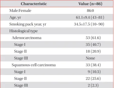
Demographic characteristics of subjects are listed (Table 1).
A total of 86 subjects participated; all were male. Fifty-three
were diagnosed with adenocarcinoma and 33 with squamous
cell carcinoma (SqCC). The average age of subject was 61.1±9.4
and the average of smoking pack-year was 34.5±17.5. All analy-
sis was processed in normal-cancer paired tissue samples.
1. Fusion gene
In the fusion gene analysis, 86 samples were analyzed using DeFuse and 33 SqCC samples were analyzed using both De- Fuse and ChimeraScan. To identify expressed fusion-genes, we used DeFuse and ChimeraScan. From the RNA-seq data, 1,293 and 6,455 fusion transcripts were detected by the two different programs, respectively. Two EML4-ALKs and one SLC34A2-ROS1 fusion gene, already known to be oncogenic,
were detected in analysis result of DeFuse
1,2.
From these results, one EML4-ALK and one SLC34A2- ROS1 fusion genes were validated by Sanger sequencing.
Also according to our procedure, four fusion transcripts were selected (Table 2). The frequencies of the selected fusion tran- scripts were detected in 1%–5% of all samples. The four fusion transcripts were validated by Sanger sequencing (Figure 1).
2. Alternative splicing
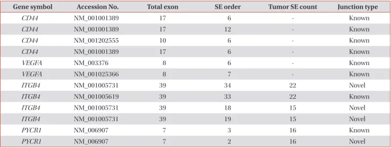
In the alternative splicing analysis, 33 SqCC samples were analyzed among the 86 samples. To identify alternative splic- ing events, total reads and reads aligned of alternative splicing were determined (Table 3).
There were 37 differential skipped exon events and six mu- tually exclusive exon events in the cancer samples compared to the normal samples. Also from these results, we found CD44 and vascular endothelial growth factor A, which were already known as alternative spliced genes, to exist in the can- cer samples, and the most significant gene was CD44 (Table 4)
5,13. As a result of comparing the normal-cancer individual paired samples, there were 12,069 differential skipped exon events. To obtain selective list, 12,069 events were filtered with a condition (normal sample SE count, 0; tumor sample SE count, ≥10; and SE covering sample, ≥30), then 29 differential skipped exon events were selected. In the list of the selected genes, there were ITBG4 and PYCR1 outstanding genes and the most significant gene is ITGB4 (Table 4).
Discussion
In the current study, we have identified candidate fusion genes and alternative splicing in non-small cell lung cancer.
In the present study, EML4-ALK was detected with DeFuse, whereas it was not detected with ChimeraScan. DeFuse is more focused on finding breakposition of fusion candidates and applies various statistical methods and database to filter out fusion candidates, while ChimeraScan concentrates more on finding genes of fusion candidates. Therefore, transcripts from DeFuse were mainly used and those from ChimeraScan were used complementally. One EML4-ALK fusion gene and one SLC34A2-ROS1 fusion gene were detected only in our cancer samples and not detected in the normal samples.
Potential candidate fusion transcripts identified in the pres- ent study are AL137145.2-PFKFB3, C4orf3-KLHL2, TPPP- BRD9, and HNRNPA2B1-SKAP2. AL137145.2-PFKFB3 fusion
Table 2. The list of fusion transcripts from RNA sequencing and validation results of fusion by Sanger sequencing Fusion gene Frequency in RNA sequencing (n=86) Frequency in Sanger sequencing (n=86) p-value
EML4-ALK 2 (2.3) 1 (1.2) 0.560
SLC34A2-ROS1 1 (1.2) 1 (1.2) >0.999
PFKFB3-AL137145.2 4 (4.7) 4 (4.7) >0.999
KLHL2-C4orf3 3 (3.5) 2 (2.3) 0.650
TPPP-BRD9 3 (3.5) 1 (1.2) 0.312
HNRNPA2B1-SKAP2 1 (1.2) 1 (1.2) >0.999
Values are presented as number (%).
The t tests were used to confirm statistical significance between RNA and Sanger sequencing.
Table 1. Baseline characteristics of the subjects Characteristic Value (n=86)
Male:Female 86:0
Age, yr 61.1±9.4 (43–81)
Smoking pack year, yr 34.5±17.5 (10–90) Histological type
Adenocarcinoma 53 (61.6)
Stage I 35 (40.7)
Stage II 18 (20.9)
Stage III None
Squamous cell carcinoma 33 (38.4)
Stage I 9 (10.5)
Stage II 22 (25.6)
Stage III 2 (2.3)
Values are presented as mean±standard deviation (range) or
number (%).
Figure 1. Confirmation by Sanger sequencing of fusion transcript structure according to presence of exon of genes. (A) PFKFB3-AL137145.2.
(B) KLHL2-C4orf3. (C) TPPP-BRD9. (D) HNRNPA2B1-SKAP2. The black arrows indicate orientations.
C
TPPP-BRD9E10 E11 E1
5 E11 E1 3
A
PFKFB3-AL137145.2E11 E12 E13 E14 E2 E3
5 E14 E2 3
B
KLHL2-C4orf3E4 E3 E2 I1
3 E2 I1 5
D
HNRNPA2B1-SKAP2E6 E7 E8 E5
3 E5 E8 5
E6 E7 E8 E5