따른 방사선학적 진단접근
동아대학교 의과대학 진단방사선과 이 기 남
DILD (diffuse infiltrative lung disease); Radiologic Diagnostic Approach According to High-Resolution CT Pattern
Ki-Nam Lee
Department of Diagnostic Radiology, Dong-A University College of Medicine, Pusan, Korea
The introduction of high-resolution CT (HRCT) in recent years has improved the ability of radiologists to detect and characterize the diffuse infiltrative lung disease (DILD).
The detection and diagnosis of diffuse lung disease using HRCT are based on the recognition of specific abnormal findings. In this article, pattern recognition of HRCT findings is reviewed in the differential diagnosis of diffuse infiltrative lung disease.
In general, HRCT findings of lung disease can be classified into four categories based on their appearances. These categories consist of (1) nodules and nodular opacities, (2) linear and reticular opacities, (3) increased lung opacity, and (4) decreased lung opacity, including cystic lesions. (Tuberc Respir Dis 2005; 58:111-119)
Key words : Computed tomography(CT), high-resolution, Lung, cysts, Lung, interstitial disease, Lung, nodule
Address for correspondence : Ki-Nam Lee Department of Diagnostic Radiology, Dong-A University College of Medicine, 3-1-ga,
Dongdaesin-dong, Seo-gu, Pusan, 602-103, Korea Phone : 82-51-240-5375 Fax : 82-51-253-4931 E-mail : [email protected]
서 론
미만성 침윤성 폐질환은 그 원인이 다양할 뿐만 아 니라, 다양한 질병에 의한 병변이 방사선학적 및 병리 적으로 유사한 소견을 보인다는 점에서 진단에 어려 움이 있다.
근래 고해상 CT가 도입됨으로써 폐실질의 상세한 구조물을 파악할 수 있게 되었고, 이에 따라 미만성 침윤성 폐질환의 발견과 진단에 큰 향상이 있었다. 특 히 고해상 CT는 단순흉부촬영이 정상소견을 보일 때 도 이상소견을 발견할 수 있으며, 또한 단순흉부촬영 이 비특이적일 때도 비교적 확신도가 높은 진단이 가 능하다.
고해상 CT를 이용한 미만성 침윤성 폐질환의 감별 진단에는 주된 병변에 대한 형태(pattern)의 인지와 그 분포가 중요하다. 일반적으로 미만성 폐질환의 고
해상 CT상 병변은 크게, 결절 및 결절성 음영(nodules
& nodular opacities), 망상 음영(linear and reticular opacities), 폐음영증가(increased lung opacity), 낭성 병변 및 폐음영감소(decreased lung opacity, cystic lesions)의 네 종류로 나눈다. 또한 병변의 분포가 특징 적이어서 감별진단에 큰 도움이 된다.
본 론
미만성 침윤성 폐질환의 고해상 CT에서 방사선학 적 진단은 주된 병변의 형태와 분포에 따라 분류할 수 있다(Table 1).
1. 병변의 종류 (pattern)
1) 결절 및 결절성 음영 (nodules & nodular opacities)
결절은 모양에 따라 경계가 명확한 간질성 결절과 경계가 불분명한 폐포성 결절로 분류하기도 하지만, 제 2 폐소엽과 연관된 분포에 따라 중심소엽성(centrilobular), 림프관주위(perilymphatic), 임의(random) 분포로 나누는 것이 결절성 폐질환의 진단적 접근에 가장 유용하다1-3.
KN Lee : DILD (diffuse infiltrative lung disease); radiologic diagnostic approach according to high-resolution CT
Table 1. HRCT Pattern of Diffuse Interstitial Lung Disease Predominant pattern Diagnosis
Nodular pattern Centrilobular Silicosis, CWP*,endobronchial spread of tuberculosis, bro
nchopneumonia, hypersensitivity pneumonitis, respiratory bron chiolitis, BOOP†, pulmonary edema
Perilymphatic Sarcoidosis, silicosis, CWP*
Random Miliary tuberculosis, hematogenous metastasis
Reticular pattern Peribronchovascular interstitial thickening
PLC§, pulmonary edema, sarcoidosis
Interface sign Fibrotic lung disease, including IPF‡
Interlobular septal thickening PLC§, pulmonary edema, silicosis, CWP*, IPF‡, sarcoidosis Intralobular interstitial thickening IPF‡, PAP∥, PLC§, pulmonary edema
Honeycombing Late stage of interstitial lung disease, including IPF‡, asbestosis, sarcoidosis, nonspecific interstitial pneumonia
Subpleural line Asbestosis
GGO(Ground-glass opacity) pattern
Subacute or chronic Chronic hypersensitivity pneumonitis, DIP¶, PAP∥, chronic eosinophilic pneumonia, extrinsic lipoid pneumonia, BOOP†, Churg-strauss syndrome, early radiation pnuemonitis
Acute Acute hypersensitivity pneumonitis, pulmonary hemorrhage,
pneumonia(pneumocystis carinii, cytomegalovirus), ARDS**, pulmonary edema, IPF‡
Crazy-paving pattern PAP∥, ARDS**, acute interstitial pneumonia, diffuse alveolar damage superimposed usual interstitial pneumonia, pneumocystis carinii pneumonia
Consolidation pattern Pneumonia(viral), ARDS**, BAC††, PAP∥, extrinsic lipoid pneumonia,
BOOP†, pulmonary hemorrhage, chronic eosinophilic pneumonia, pulmonary edema
Cystic pattern Emphysema Centrilobular, panlobular, paraseptal
Cysts Langerhans cell histiocytosis, lymphangioleiomyomatosis
Mosaic perfusion Air-trapping or poor ventilation, poor perfusion
CWP*, coal-worker’s pneumoconiosis, BOOP†, bronchiolitis obliterans organizing pneumonia, IPF‡, idiopathic pulmonary fibrosis, PLC§, pulmonary lymphagitic carcinomatosis, PAP∥, pulmonary alveolar proteinosis, DIP¶, desquamative interstitial pneumonia, ARDS**, acute respiratory distress syndrome, BAC††, bronchioloalveolar cell carcinoma
중심소엽성(centrilobular) 결절은 소엽 중심부에 1 mm 이상 크기의 결절이 있을 경우이다. 이는 세기관 지 주위의 간질 및 세기관지 자체의 병변을 의미하고4, 자주 보이는 질환에는 규폐증(silicosis), 석탄폐증(coal- worker’s pneumoconiosis), 경기관지 파급의 폐결핵 (endobronchial spread of tuberculosis), 소엽성폐렴 (bronchopneumonia), 과민성폐렴(hypersensitivity pneumonitis), 호흡 세기관지염(respiratory bronchiolitis), 특발성 기질화 폐렴(bronchiolitis obliterans organizing pneumonia; BOOP), 폐부종이 있다. 중심소엽성 결절 은 흉막표면에서 5-10 mm 떨어져서 소혈관을 둘러싸 고 있으며 흉막에 붙어있는 결절은 거의 없다. 중심소
엽성(centrilobular) 결절이 보이는 환자에서 나뭇가지 에 싹이 나는 모양 (tree-in-bud appearance)의 유무 가 감별진단에 도움이 된다. 나뭇가지에 싹이 나는 모 양은 중심소엽내 세기관지의 확장과 감입(impaction) 을 의미하며, 세기관지의 감염이나 점액성 물질의 축 적(mucus retention)을 시사하는 소견이다5(Fig. 1).
림프관주위(perilymphatic) 결절은 흉막하 간질(su
bpleural interstitium), 기관지 및 혈관주위(peribro
nchovascular), 소엽간 중격(interlobular septal), 그 외 중심소엽성 간질을 침범한다. 유육종증은 기관지 및 혈관주위, 흉막하 간질에 우세하게 나타나며, 규폐 증과 석탄진폐증은 흉막하 간질 및 중심소엽성 간질
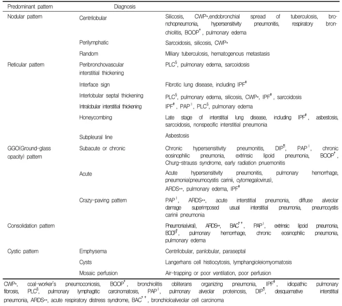
Figure 1. Centrilobular nodules and tree-in-bud app
earance in a patient with endobronchial spread of tuberculosis.
Multiple small nodules occurring in clusters and appearance of tree-in-bud (arrows), being in centrilobular distribution, spare the pleural surfaces. Centrilobular nodules reflect peribronchiolar granuloma formation.
Bronchioles filled with infected material can result in the appearance of tree-in-bud.
Figure 2. Perilymphatic distribution of nodules in a patient with sarcoidosis.
Numerous nodules are predominant in relation to the major fissure (arrow) and perihilar bronchovascular interstitium. Subpleural nodules (arrowheads) are also seen along the costal pleural surfaces.
에, 림프성 폐전이는 기관지 및 혈관주위, 흉막하 간질 에 호발한다6-8(Fig. 2).
임의(random) 분포하는 결절은 제 2 폐소엽의 구조 물에 임의로 분포하고, 흉막하 결절이 빈번하게 나타 나며 특히 엽간열에서 쉽게 인지할 수 있다. 속립성 폐결핵(miliary tuberculosis)과 혈행성 전이(hemato
genous metastasis)가 대표적인 예이다9(Fig. 3).
2) 망상 음영 (linear and reticular opacities) 폐간질을 이루는 조직들이 액성물질, 섬유화조직 혹은 세포 등에 의해서 두꺼워질 때 고해상 CT에서는 선상(linear) 혹은 망상음영으로 나타난다. 이와 같은 망상음영은 기관지및 혈관주위 간질(axial interstitium) 이나 소엽간 중격(interlobular septum, peripheral inter
stitium)이 두터워지거나, 제 2 폐소엽 중심구조물 (core structure), 제 2 폐소엽내 간질(parenchymal inter
stitium)의 비후 및 봉와상폐(honeycombing) 때에 나 타난다.
기관지 및 혈관주위 간질의 비후(peribronchovascular interstitial thickening)는 기관지 및 혈관주위의 축성 간질(axial interstitium)이 두꺼워진 경우로 고해상 CT상 기관지벽 자체가 두꺼워지거나, 혈관의 직경이 국소적으로 두꺼워지거나, 혹은 기관지 및 혈관벽이 우둘투둘하게 나타난다. 기관지 및 혈관주위 간질이 다른 부위보다 선택적으로 두꺼워지는 예로는 림프성 폐전이, 유육종증10 및 울혈성 폐부종11이 대표적이다.
그 중에서 평활한 비후는 림프성 폐전이, 울혈성 폐부 종을 의미하고, 결절성 비후는 유육종증 및 림프성 폐 전이를 시사한다.
계면징후(interface sign)는 기관지 및 혈관의 외벽 이 간질성 섬유화, 염증, 혹은 악성세포 침윤에 의해 불규칙한 비후를 보이는 경우를 일컫는다. 특발성 섬
KN Lee : DILD (diffuse infiltrative lung disease); radiologic diagnostic approach according to high-resolution CT
Figure 3. Miliary nodules in a patient with tuberculosis.
Multiple small, well-defined nodules with involvement of the peripheral pleural surfaces are widely and evenly distributed through the lung. The overall pattern of distribution is random.
Figure 4. Interlobular septal thickening in a patient with hydrostatic pulmonary edema.
Diffuse interlobular septal thickening (arrows) outlines numerous pulmonary lobules in anterior and medias
tinal pleural surfaces of left upper lobe.
유화증의 경우 혈관벽의 불규칙성이 98%, 흉막의 불 규칙성이 94%에서 나타난다12.
소엽간 중격의 비후(thickening of interlobular se
ptum)는 변연부 간질 중 제 2 폐소엽 중격의 비후 로 고해상 CT상 1 cm 이상의 공간을 둘러싸는 다각형 (polygonal) 및 육각형으로 나타난다. 대표적으로 림 프성 폐전이, 울혈성 폐부종에서 보이고, 그 외에 유 육종증, 탄광진폐증, 특발성 폐섬유화증에서도 보인 다(Fig. 4).
제 2 폐소엽 내 간질의 비후 (intralobular interstitial thickening or thickening of parenchymal interstitium) 는 작은 망상 음영으로 종종 섬유화의 초기 징후로 특발성 폐섬유화증, 폐포 단백증(pulmonary alveolar proteinosis), 간질성 폐전이, 폐부종 등에서 나타난 다13.
봉와상폐 (honeycombing)는 미만성 간질성 폐질환 의 말기에 공통적으로 일어나는 변화로 정상 폐구조 의 파괴와 함께 심한 섬유화를 반영하는 소견이다. 특
징적으로 늑막하부 변연부에 다발성의 낭성병변이 형 성되며, 주변 기관지 및 혈관의 분포가 현저하게 변형 되어 나타난다(Fig. 5). 봉와상폐의 낭종(cyst)은 두껍 고 경계가 명확한 섬유화 벽을 가지고, 여러층으로 구 성되며, 주변의 봉와상 낭종과 벽을 공유하고 있는 점 등으로, 벽을 거의 볼 수 없는 폐기종과 구분될 수 있 다14. 그러나 일부는 원위선방형 폐기종(distal acinar emphysema, paraseptal emphysema)와 구별이 힘든 경우도 있다.
늑막하 선상음영(subpleural lines)은 늑막하 1 cm 이내에 늑막면과 평행하게 보이는 얇은 선상의 음영 으로 폐간질의 섬유화를 의미하며, 석면폐증(asbestosis) 또는 봉와상폐의 초기소견으로 기술된 바 있다15. 복 와위나 앙와위에서 형태가 변하지 않으므로 정상인에 서 보일 수 있는 중력방향의 아랫부위에 보이는 음영 (dependent opacity)과는 구별된다. 이들 음영이 병변 과 구별되지 않을 때 환자의 자세를 복와위로 바꾸어 재촬영하면 진단에 도움이 된다.
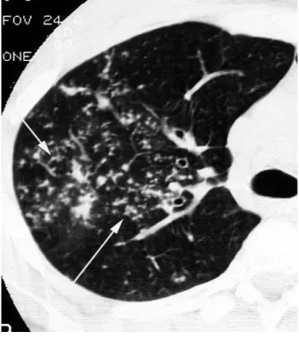
Figure 5. Honeycombing in a patient with IPF.
Honeycombing results in cysts of varying sizes, which have a peripheral predominance.
The cysts with thick and clearly definable walls, sharing with adjacent cysts, show multi-layering.
Figure 6. Ground-glass opacity in a patient with pu
lmonary hemorrhage.
Vessels are visible within patchy areas of ground- glass opacity.
3) 폐음영증가 (increased lung opacity)
간유리음영 (ground-glass opacity)은 폐포강의 부 분적 충만, 또는 폐포벽의 미미한 비후가 있을 경우 고해상 CT가 한 독립된 개체 음영으로 인지해내지 못 하고 뿌옇게 증가된 음영을 보이는 것으로 폐혈관 음 영의 윤곽을 소실시키지 않는 정도의 증가된 음영을 말한다(Fig. 6). 간유리음영 자체는 비특이 소견이지 만, 그 의미는 중요하다. 내부의 불규칙한 선상음영, 견인성 기관지 확장, 열구 또는 해당엽 구조의 비틀림 (fissural or architectural distortion)이 없는 경우 간 유리음영은 급성, 활동성, 가역성 병변이며, 치료가 가 능한 병변임을 60-80%정도에서 시사한다16. 간유리음 영이 보이는 질환을 경과에 따라 크게 두 그룹으로 나누면, 아급성 또는 만성경과를 보이는 군과 급성 경과를 보이는 군으로 볼 수 있다. 전자에 속하는 질 환에는 과민성 폐렴, 박리성 간질성 폐렴(desquamative interstitial pneumonia), 폐포단백증(pulmonary alveolar
proteinosis), 만성 호산구성 폐렴, 외인성 지방폐렴(ex
trinsic lipoid pneumonia; Squalene-induced extrinsic lipoid pneumonia), 특발성 기질화 폐렴(crytogenic or
ganizing pneumonia; COP), 알레르기성 혈관염 및 육 아종증(allergic angiitis and granulomatosis; Churg- Strauss syndrome), 방사선폐렴의 초기 등 이다. 후자 에 속하는 질환에는 급성 과민성 폐렴, 폐출혈, 폐렴 (특히 pneumocystis carinii pneumonia, cytomegalovirus pneumonia), 성인호흡곤란 증후군(adult respiratory distress syndrome; ARDS), 심인성 폐부종, 급성 악 화된 특발성 폐섬유화증(idiopathic pulmonary fibrosis;
IPF)17이 있다.
간유리음영 내에 소엽간 중격의 평활한 비후를 보 일 때 ‘돌조각 보도 모양’(crazy-paving appearance) 이라 부르는데 이는 한때 폐포단백증의 전형적인 소 견으로 알려졌지만, 이외에도 성인호흡곤란 증후군, 급성 간질성 폐렴, 상용 간질성 폐렴에 동반된 급성
KN Lee : DILD (diffuse infiltrative lung disease); radiologic diagnostic approach according to high-resolution CT
Figure 7. Crazy-paving appearance in a patient with acute interstitial pneumonia.
Geographic ground-glass opacities are associated with interlobular septal thickening and intralobular interstitial thickening.
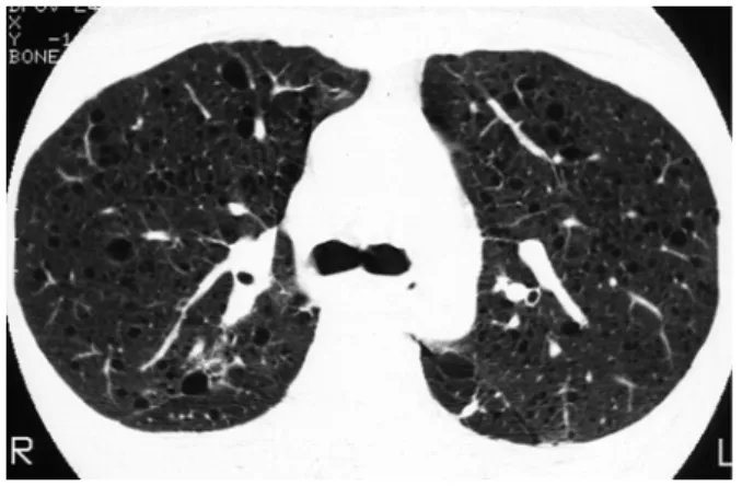
Figure 9. Centrilobular and paraseptal emphysema.
Small lucencies lacking walls in central lung represent centrilobular emphysema (white arrows). Areas of con}
fluent centrilobular emphysema (white arrowheads) are in posterior right lung. Subpleural lucencies reflect associated paraseptal emphysema (black arrows).
Large bullae (black arrowhead) are also subpleural in location.
Figure 8. Consolidations in a patient with bronchiolitis obliterans organizing pneumonia, BOOP.
Airspace consolidations are mainly distributed in peribronchovascular and subpleural regions.
미만성 폐포손상(diffuse alveolar damage superimposed on usual interstitial pneumonia), 약물에 의한 폐손상, 뉴모시스티스 카리니 폐렴(pneumocystis carinii pne
umonia; PCP) 등에서도 보이는 비특이적인 소견이다.
병리적으로는 폐포 충만병변(alveolar filling process), 간질 섬유화병변(interstitial fibrotic process) 그리고 이 두 형태 병변의 혼합병변(combination of interstitital and intraalveolar process)에서도 보인다(Fig. 7).
기강경결 (airspace consolidation)은 폐포강내의 공 기가 체액, 세포 및 단백질로 대부분 혹은 완전히 대 체된 상태를 말하며18, 병변에 의해 폐혈관 윤곽이 소 실되고 공기 기관지조영상(air-bronchogram)이 나타 난다. 폐렴(특히 viral), 성인호흡곤란 증후군, 세기관 지폐포암(bronchioloalveolar carcinoma), 폐포단백증, 외인성 지방폐렴, 특발성 기질화폐염, 폐출혈, 만성 호 산구성폐렴 및 폐부종에서 보인다(Fig. 8).
4) 폐음영감소 및 낭성병변
(decreased lung opacity and cystic lesion)
폐기종은 고해상 CT로 정확하게 진단할 수 있다.
충분히 낮은 감쇄계수(< -600 HU)를 사용하면, 국소적
Figure 10. Langerhans cell histiocytosis.
Multiple, bizarre, variable-sized lung cysts are inter
spersed within normal-appearing lung. These cysts usually spare the costophrenic angles (not shown).
Figure 11. Lymphangioleiomyomatosis.
Cysts are thinner-walled and more regular in size and shape than those seen in patients with Langerhans cell histiocytosis. Cysts are diffusely distributed, and similar in size.
Figure 12. Mosaic perfusion in a patient with post- infectious bronchiolitis obliterans.
Patchy areas of increased or decreased lung attenua
tion, reflects mosaic perfusion are visible. Note that the pulmonary vessels in the lucent-appearing per
ipheral lung (arrows) are smaller than vessels in the denser lungs.
으로 아주 감소된 음영이 주위의 증가된 정상 폐실질 과 대비되어 나타나므로 쉽게 인지 할 수 있다19(Fig. 9).
낭성병변은 1 cm 이상의 직경과 3 mm 이하의 얇은 벽을 갖는 병소로 공기를 함유하고 있으며 그 벽은 여 러 세포로 구성될 수 있으나 보통은 섬유 조직 혹은 상피세포로 구성된다. 고해상 CT상 낭성병변은 망상 병변과 쉽게 구별되고, 대표적인 질환으로는 특발성 폐섬유화증 (IPF), 호산구성 육아종(eosinophilic gra
nuloma), 림프관평활근육종증(lymphangioleiomyomatosis;
LAM)등이 있다.
호산구성 육아종은 원인불명의 육아종성 질환으로, 초기에는 랑게르한스 조직구(Langerhans histiocytes)와 호산구(eosinophils)를 동반한 육아종성 결절이 기관
지 주변에 분포하는 것을 특징으로 하며, 후기에는 폐 섬유화와 낭(cyst) 형성으로 대치된다. 고해상 CT소 견은 다양한 두께의 벽을 가진 1 cm 이하의 크기를 가진 낭성 공간과 5 mm 이하의 결절이 주로 세기관 지주위에 생기며, 폐 상엽과 중엽에 많이 보인다20,21 (Fig. 10).
림프관평활근육종증은 미숙한 평활근(immature smooth muscle)을 닮은 방추세포(spindle cell)의 지 속적인 증식이 폐실질과 림프관을 따라 생기는 질환 이다. 세기관지를 따라 방추세포의 증식에 의해 공기 가둠(air trapping)과 기종(emphysema) 그리고 얇은 벽의 낭(thin-walled cyst)이 형성되고, 이런 낭의 파 열에 의해 기흉이 생길 수 있다. 종격동과 폐문 림프 절 그리고 흉곽외 림프절에 방추세포가 증식하여 폐 내 림프관(intrapulmonary lymphatics)과 흉관(thoracic duct)을 확장시킬 수 있다. 이와 같은 림프관 침범이 유미흉막삼출(chylous pleural effusion)을 유발할 수 있고, 폐동맥의 벽에 방추세포의 증식에 의해 정맥폐 색이 생기고 이로 인하여 폐출혈이 생길 수 있다22 (Fig. 11).
폐음영감소는 낭종성 폐질환이나 폐기종이 아닌 공 기포획이나, 저환기(poor ventilation), 저환류(poor per
fusion)으로 인한 소견으로 나타나면 소엽(lobular),
KN Lee : DILD (diffuse infiltrative lung disease); radiologic diagnostic approach according to high-resolution CT
엽(lobar) 혹은 여러 군데(multifocal) 분포의 소위 ‘모 자이크형 저관류(mosaic perfusion)’를 보인다23 (Fig. 12).
소기도 질환에 의한 모자이크형 저관류를 보이는 경 우 호기 촬영을 하면 더욱 강조되어 보인다24-26. 2. 병변의 분포 (Distribution)
많은 질환들은 병변의 분포가 특징적이어서 감별진 단에 큰 도움이 된다.
폐의 중심부(parahilar)에 주로 병변이 분포하는 질 환으로는 유육종증, 림프성 폐전이 및 중심부 기관지 질환이 있다27. 반대로 폐의 변연부에 주로 분포하는 질환으로는 특발성 폐섬유화증, 교원병(collagen vascular disease), 석면폐증, 및 호산구성 폐렴이 있다.
상엽에 병변의 정도가 심한 질환은 호산구성 육아 종, 유육종증, 규폐증, 소엽중심성 폐기종, 폐결핵이 대표적이고, 하엽에 주로 분포하는 질환은 림프성 및 혈행성 폐전이, 특발성 폐섬유화증(IPF), 교원병, 석면 폐증 등이다.
앞서 기술한 대로 제 2 폐소엽내 구조물과의 관계 에 따라 중심소엽성(centrilobular), 림프관주위(per
ilymphatic), 임의(random) 분포의 3가지로 나눌 수 있다.
폐의 앞쪽에 병변이 주로 분포하는 질환은 기계식 호흡기 치료후의 성인호흡곤란 증후군(adult respiratory distress syndrome: ARDS)만이 알려져 있고 흉막하 섬유화, 봉와상폐, 견인성 기관지확장증 등이 있다28. 반대로 뒤쪽 즉 중력방향에 주로 분포하는 질환에는 석면폐증, 규폐증, 유육종증, 폐부종, 과민성폐렴, 특발 성 폐섬유화증 등이 있다.
편측 분포를 보이는 질환으로는 림프성 폐전이의 약 40%에서, 유육종증의 9-21%에서 볼 수 있다29.
결 론
고해상 CT는 단순흉부촬영에서 정상이거나 비특 이적 소견을 보여도 기질적 변화를 인지해 낼 수 있으 며, 미만성 폐질환의 분포와 양상을 비교적 정확하게 진단 할 수 있다.
이상 본론에서 설명한 고해상 CT에서 보이는 기본 적인 네 가지 폐 병변의 형태를 인지하고 분포를 분석 하며, 임상상을 종합한다면 미만성 침윤성 폐질환의 진단에 그리 어렵지 않게 접근할 수 있을 것으로 생 각된다. 무엇보다도 고해상 CT상 보이는 기본적인 병변의 형태를 숙지하려는 노력이 필요할 것으로 생 각한다.
참 고 문 헌
1. Webb WR, Muller NL, Naidich DP. High-resolution CT of the lung. 3rd ed. Lippincott Williams & Wilkins;
2001. p. 71-171.
2. Gruden JF, Webb RW, Naidich DP, McGuinness G.
Multinodular disease: anatomic localization at high- resolution CTmultireader evaluation of a simple algorithm. Radiology 1999;210:711-20.
3. Lee KS, Kim TS, Han J, Hwang JH, Yoon JH, Kim Y, et al. Diffuse micronodular lung disease: HRCT and pathologic findings. J Comput Assist Tomogr 1999;23:99-106.
4. Gruden JF, Webb WR, Warnock M. Centrilobular opacities on HRCT: diagnostic considerations and pathologic correlation. AJR Am J Roentgenol 1994;
162:569-74.
5. Collins J, Blankenbaker D, Stern EJ. CT patterns of bronchiolar disease: what is "tree-in-bud"? AJR Am J Roentgenol 1998;171:365-70.
6. Lynch DA, Webb WR, Gamsu G, Stulbarg M, Golden J. Computed tomography in pulmonary sarcoidosis.
J Comput Assist Tomogr 1989;13:405-10.
7. Muller NL, Kullnig P, Miller RR. The CT findings of pulmonary sarcoidosis: analysis of 25 patients.
AJR Am J Roentgenol 1989;152:1179-82.
8. Remy-Jardin M, Beuscart R, Sault MC, Marquette CH, Remy J. Subpleural micronodules in diffuse infiltrative lung diseases: evaluation with thin-section CT scans. Radiology 1990;177:133-9.
9. Im JG, Itoh H, Shim YS, Lee JH, Ahn J, Han MC, et al. Pulmonary tuberculosis: CT findings--early active disease and sequential change with antitube
rculous therapy. Radiology 1993;186:653-60.
10. Brauner MW, Grenier P, Mompoint D, Lenoir S, de Cremoux H. Pulmonary sarcoidosis: evaluation with high-resolution CT. Radiology 1989;172:467-71.
11. Storto ML, Kee ST, Golden JA, Webb WR. Hydrostatic pulmonary edema: high-resolution CT findings. AJR Am J Roentgenol 1995;165:817-20.
12. Zerhouni EA, Naidich DP, Stitik FP, Khouri NF,
Siegelman SS. Computed tomography of the pulmo
nary parenchyma: pt 2. interstitial disease. J Thorac Imaging 1985;1:54-64.
13. Webb WR. High-resolution CT of the lung parenchyma.
Radiol Clin North Am 1989;27:1085-97.
14. Primack SL, Hartman TE, Hansell DM, Muller NL.
End-stage lung disease: CT findings in 61 patients.
Radiology 1993;189:681-6.
15. Yoshimura H, Hatakeyama M, Otsuji H, Maeda M, Ohishi H, Uchida H, et al. Pulmonary asbestosis:
CT study of subpleural curvilinear shadow. Radiology 1986;158:653-8.
16. Leung AN, Miller RR, Muller NL. Parenchymal opacification in chronic infiltrative lung diseases:
CT-pathologic correlation. Radiology 1993;188:209-14.
17. Nishimura K, Kitaichi M, Izumi T, Nagai S, Kanaoka M, Itoh H. Usual interstitial pneumonia: histologic correlation with high-resolution CT. Radiology 1992;
182:337-42.
18. Naidich DP, Zerhouni EA, Hutchins GM, Genieser NB, McCauley DI, Siegelman SS. Computed tomo
graphy of the pulmonary parenchyma: pt 1. distal air-space disease. J Thorac Imaging 1985;1:39-53.
19. Klein JS, Gamsu G, Webb WR, Golden JA, Muller NL. High-resolution CT diagnosis of emphysema in symptomatic patients with normal chest radiographs and isolated low diffusing capacity. Radiology 1992;
182:817-21.
20. Moore AD, Godwin JD, Muller NL, Naidich DP, Hammar SP, Buschman DL, et al. Pulmonary histiocytosis X: comparison of radiographic and CT findings. Radiology 1989;172:249-54.
21. Brauner MW, Grenier P, Mouelhi MM, Mompoint
D, Lenoir S. Pulmonary histiocytosis X: evaluation with high-resolution CT. Radiology 1989;172:255-8.
22. Templeton PA, McLoud TC, Muller NL, Shepard JA, Moore EH. Pulmonary lymphangioleiomyomatosis:
CT and pathologic findings. J Comput Assist Tomogr 1989;13:54-7.
23. Lynch DA, Brasch RC, Hardy KA, Webb WR. Pediatric pulmonary disease: assessment with high-resolution ultrafast CT. Radiology 1990;176:243-8.
24. Arakawa H, Webb WR. Expiratory high-resolution CT scan. Radiol Clin North Am 1998;36:189-209.
25. Chen D, Webb WR, Storto ML, Lee KN. Assessment of air trapping using postexpiratory high-resolution computed tomography. J Thorac Imaging 1998;13:
135-43.
26. Worthy SA, Muller NL, Hartman TE, Swensen SJ, Padley SP, Hansell DM. Mosaic attenuation pattern on thin-section CT scans of the lung: differentiation among infiltrative lung, airway, and vascular diseases as a cause. Radiology 1997;205:465-70.
27. Mathieson JR, Mayo JR, Staples CA, Muller NL.
Chronic diffuse infiltrative lung disease: comparison of diagnostic accuracy of CT and chest radiography.
Radiology 1989;171:111-6.
28. Desai SR, Wells AU, Rubens MB, Evans TW, Hansell DM. Acute respiratory distress syndrome: CT abno
rmalities at long-term follow-up. Radiology 1999;210:
29-35.
29. Grinier P, Valeyre D, Cluzel P, Brauner MW, Lenior S, Chastang C. Chronic diffuse interstitial lung disease: diagnostic value of chest radiography and high-resolution CT. Radiology 1991;179:123-32.