• 검색 결과가 없습니다.

Determination of Diastolic Dysfunction Cut-Off Value by Tissue Doppler Imaging in Adults 70 Years of Age or Older: A Comparative Analysis of Pulsed-Wave and Color-Coded Tissue Doppler Imaging

N/A
N/A
Protected

Academic year: 2022

Share "Determination of Diastolic Dysfunction Cut-Off Value by Tissue Doppler Imaging in Adults 70 Years of Age or Older: A Comparative Analysis of Pulsed-Wave and Color-Coded Tissue Doppler Imaging"

Copied!
6
0
0

로드 중.... (전체 텍스트 보기)

전체 글

(1)

137

Open Access

Determination of Diastolic Dysfunction Cut-Off Value

by Tissue Doppler Imaging in Adults 70 Years of Age or Older:

A Comparative Analysis of Pulsed-Wave and Color-Coded Tissue Doppler Imaging

Hee-Kyung Baek, MD, Tae-Ho Park, MD, Sun-Yi Park, MD, Jung-Hwan Kim, MD, Jeong-Min Seo, MD, Woo-Jae Kim, MD, Young-Hee Nam, MD, Moo-Hyun Kim, MD, and Young-Dae Kim, MD

Department of Cardiology, Dong-A University College of Medicine, Busan, Korea

ABSTRACT

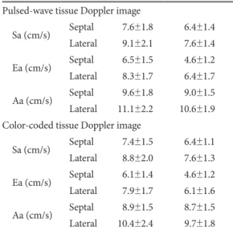
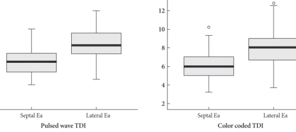
Background and Objectives: The cut-off value of diastolic dysfunction by tissue Doppler imaging (TDI) is affected by ag- ing and modalities used (pulsed-wave vs. color-coded). The purpose of this study was to investigate the diastolic function of healthy elderly people and to determine the appropriate cut-off value of diastolic dysfunction in elderly individuals. Sub- jects and Methods: Healthy volunteers (n=76) and patients with hypertension (n=51) aged ≥70 years underwent 2-dimen- sional and Doppler echocardiography. Mitral annulus velocities of TDI were measured at septal and lateral sites using the pulsed-wave and color-coded modalities. The appropriate cut-off value of diastolic dysfunction for healthy elderly individu- als was defined as the lower limit of the 95% confidence interval for early diastolic mitral annulus velocity (Ea). Results: The mean septal and lateral Ea were 6.5±1.5 and 8.3±1.7 cm/s, respectively, by pulsed-wave TDI, and 6.1±1.4 and 7.9±1.7 cm/s, respectively, by color-coded TDI. The cut-off values for diastolic dysfunction were as follows: septal and lateral Ea were 6.1 and 7.9 cm/s by pulsed-wave TDI, and 5.7 and 7.5 cm/s by color-coded TDI, respectively. When the group was stratified by gen- der, Ea was significantly lower in women than men. Conclusion: When interpreting diastolic function as measured by TDI in elderly subjects, different cut-off values should be considered based on the TDI modality, annulus site, and gender. (Korean Circ J 2011;41:137-142)

KEY WORDS: Heart failure, diastolic; Echocardiography; Aging.

Received: April 3, 2010

Revision Received: June 28, 2010 Accepted: July 30, 2010

Correspondence: Tae-Ho Park, MD, Department of Cardiology, Dong- A University College of Medicine, 1 Dongdaesin-dong 3-ga, Seo-gu, Bu- san 602-715, Korea

Tel: 82-51-240-2964, Fax: 82-51-242-1449 E-mail: [email protected]

• The authors have no financial conflicts of interest.

cc

This is an Open Access article distributed under the terms of the Cre- ative Commons Attribution Non-Commercial License (http://creativecom- mons.org/licenses/by-nc/3.0) which permits unrestricted non-commer- cial use, distribution, and reproduction in any medium, provided the origi- nal work is properly cited.

Introduction

It is important to assess diastolic function in the elderly popu- lation because diastolic dysfunction is responsible for heart failure, especially heart failure with preserved left ventricular

ejection fraction.

1)

The differentiation of pathologic diastolic dysfunction is more difficult in the context of the physiologically decreased diastolic function in elderly subjects because impairment of left ventricular diastolic function is related to the normal ag- ing process as well as pathologic disorders.

2-8)

Hence, appropri- ate cut-off values of diastolic function in the elderly population are needed to discriminate normal healthy diastolic function from true pathologic diastolic dysfunction.

Although there are many reports suggesting a reference

range for tissue Doppler imaging (TDI) velocities of the mi-

tral annulus, the difference between pulsed and color tissue

Doppler-derived velocities for the mitral annulus has not been

systematically investigated. By measuring both pulsed-wave

and color-coded TDI in the same individual, we evaluated the

difference in left ventricular mitral annulus velocity between

the two TDI modalities.

(2)

Subjects and Methods

Subjects

We prospectively enrolled healthy elderly subjects and pa- tients with hypertension who were all aged ≥70 years. The he- althy subjects were recruited from volunteers among relatives of hospital personnel or referrals for consultation in preop- erative evaluation. Inclusion criteria were the presence of nor- mal findings on 2-dimensional echocardiography for cardiac chamber dimension, left ventricular wall thickness, left ven- tricular ejection fraction, and left ventricular wall motion. The presence of hypertension, known congestive heart failure or symptoms, diabetes mellitus, coronary artery disease, cardiac arrhythmia, valvular heart disease, and obesity were exclu- sion criteria for healthy subjects. Fifty-one patients who had an evidence of hypertrophy by electrocardiography while tak- ing concomitant anti-hypertension medication were enrolled in the hypertension group.

Systemic blood pressure, heart rate, height and weight were measured at the time of echocardiographic examination. A 12-lead electrocardiogram was obtained for each subject be- fore or at the time of echocardiographic examination to ex- clude arrhythmia and to detect ST-segment changes, Q waves, or abnormal T waves that represent ischemic heart disease.

Informed consent was obtained from each subject.

Echocardiography

Echocardiography was performed by one experienced in- vestigator using a Hewlett-Packard Sonos 7500 (Hewlett Pa- ckard, Palo Alto, CA, USA) and IE 33 ultrasound system with a 2.5-MHz transducer. The investigator was blinded to the subjects’ clinical information, including blood pressure.

Left ventricular ejection fraction was estimated by the quan- titative biplane Simpson’s method.

9)

Left ventricular mass and left atrial volume were calculated from M-mode and 2-dimen- sional measurements, respectively, as previously described.

10)

Left ventricular internal dimension, interventricular septum, and posterior wall thickness were measured in end-diastole and end-systole according to the recommendations of the Am- erican Society of Echocardiography.

10)

Pulsed-wave Doppler transmitral velocities were recorded from the apical 4-chamber view by placing the sample volume between the leaflet tips in the center of the flow stream. Early diastolic mitral flow velocity (E), late diastolic mitral flow ve- locity (A), the ratio of early to late diastolic mitral flow velo- city (E/A), and deceleration time were measured. From the apical 4-chamber view, the pulmonary venous flow velocities were recorded by placing the sample volume approximately 1 cm into the right upper pulmonary vein. The pulmonary ven- ous peak systolic (PVs) and peak diastolic (PVd) flow veloci- ties, the ratio of peak systolic-to-diastolic flow velocities (PVs/

PVd), and peak reversal flow velocity due to left atrial con-

traction (PVa) were recorded.

All patients underwent repeated echocardiography on the same day to obtain pulsed-wave and color-coded TDI. In puls- ed-wave TDI, velocities were obtained by placing the TDI cursor at the mitral annulus. In color-coded TDI, velocities were measured at the mitral annulus by superimposition of wall motion velocity on the 2-dimensional echocardiogra- phic imaging using velocity color coding. Mitral annulus ve- locities were measured at septal and lateral sites of the mitral annulus. The peak systolic (Sa), early (Ea), and late (Aa) dias- tolic velocities were measured on-line at a sweep speed of 100 mm/s. To obtain the best quality recordings, the filter settings and gains were adjusted at the minimal optimal level to mini- mize noise and to eliminate the signals produced by the trans- mitral flow. The echocardiograms were stored digitally and an- alyzed off-line. A mean of 3 consecutive cycles was used to calculate all echocardiography Doppler parameters.

Statistics

Statistical Package for the Social Sciences (SPSS, version 17, SPSS Inc., Chicago, IL, USA) was used for data management and statistical analysis. Results are presented as mean±SD.

Comparisons of mitral annulus velocities (Sa, Ea, Aa) between pulsed-wave and color-coded TDI were analyzed with the paired t-test. The relationship of transmitral annulus velocities (Sa, Ea, Aa) between pulsed-wave and color-coded TDI were shown using Pearson’s correlation coefficients. Comparisons of mitral annulus velocities according to gender were per- formed by the student t-test.

To determine the appropriate cut-off value of diastolic dys- function for healthy elderly individuals, the lower limit of the 95% confidence interval (CI) for Ea was used. A p of <0.05 was considered as statistically significant. We also evaluated whether these cut-off values were applicable to the detection of hypertensive patients who have diastolic dysfunction.

Results

Clinical and echocardiographic characteristics We enrolled a total 127 subjects (76 healthy volunteers and 51 patients with hypertension). All subjects had adequate echocardiographic images for analysis. Table 1 summarizes demographic data along with 2-dimensional and convention- al Doppler echocardiographic characteristics.

Tissue Doppler echocardiographic characteristics

All values of mitral annulus velocities measured by color-

coded TDI were lower than those measured by pulsed-wave

TDI at both the septal and lateral sites (Table 2). The relation-

ship of each Sa, Ea, and Aa between pulsed-wave and color-

coded TDI showed a positively strong and statistically signifi-

cant correlation (septal Sa, r=0.653; lateral Sa, r=0.682; septal

(3)

Ea, r=0.841; lateral Ea, r=0.826; septal Aa, r=0.612; lateral Aa, r=0.629, all p<0.001).

Cut-off value for diastolic dysfunction in elderly subjects

Median values of septal Ea and lateral Ea measured by puls- ed-wave TDI were 6.5 cm/s (95% CI, 6.1-6.8) and 8.3 cm/s (95% CI, 7.9-8.7), respectively. Median values of septal Ea and lateral Ea measured by color-coded TDI were 6.0 cm/s (95% CI, 5.7-6.4) and 8.0 cm/s (95% CI, 7.5-8.2), respectively (Fig. 1).

Using the lower limit of the 95% CI for Ea, the appropriate cut-off value of diastolic dysfunction in elderly people was determined. In pulsed-wave TDI, the cut-off values of septal Ea and lateral Ea were 6.1 cm/s and 7.9 cm/s, respectively. In

color-coded TDI, the cut-off values of septal Ea and lateral Ea were 5.7 cm/s and 7.5 cm/s, respectively. The sensitivity of cut-off values of septal Ea and lateral Ea by pulsed-wave TDI, and septal Ea and lateral Ea by color-coded TDI were 68%

and 62%, and 59% and 59%. The specificity of cut-off values of septal Ea and lateral Ea by pulsed-wave TDI, and septal Ea and lateral Ea by color-coded TDI were 90% and 79%, and 81% and 83%. Of the 4 cut-off values, septal Ea by pulse-wave TDI (68% sensitivity, 86% specificity, 0.841 area under the curve) was the useful value to diagnose diastolic dysfunction in the current study, but it did not reach statistical signific- ance (Fig. 2).

Transmitral annulus velocities stratified by gender When the subjects were stratified by gender, the values of Sa, Ea, and Aa in men showed a trend toward being higher than those in women. This pattern remained regardless of wh- ether color-coded or pulsed-wave TDI was used or of annulus site measured (Table 3).

Discussion

Diastolic dysfunction in healthy elderly people Heart failure with normal left ventricular ejection fraction is currently accepted as a disease entity, and diastolic dysfunc- tion has a critical role in the clinical presentation of heart fai- lure.

11)

Moreover, asymptomatic diastolic dysfunction is pre- dictive of future occurrence of heart failure and is related to an increase in all-cause mortality.

12)13)

Echocardiography has been used for the noninvasive evalu- ation of diastolic function.

14)

Mitral inflow and pulmonary vein Table 1. Clinical and echocardiographic characteristics of the two

groups

Normal

(n=76) Hypertension

(n=51) p

Clinical baseline characteristics

Age (years) 74±4 73±3 0.083

Male sex (n, %) 34 (44.7) 21 (41.2) 0.691 BMI (kg/m

2

) 22.5±2.8 24.4±3.0 <0.001

SBP (mmHg) 119±11 134±11 <0.001

DBP (mmHg) 76±7 81±6 <0.001

Heart rate (beats/min) 069±13 064±11 0.018 2-dimensional echocardiographic characteristics

LV EF (%) 66±2 65±3 0.258

LVIDd (mm) 44.4±3.8 45.8±4.2 0.050

IVSd (mm) 08.5±1.1 10.2±1.2 <0.001

LVPWd (mm) 08.7±0.9 10.2±1.1 <0.001

LA volume (mL) 045.1±11.6 055.1±16.0 <0.001 LV mass index (g/m

2

) 078.0±14.8 093.3±17.6 <0.001 Conventional Doppler echocardiographic characteristics

Mitral inflow parameters

E (cm/s) 062.5±15.1 057.9±12.7 0.076

A (cm/s) 081.9±17.2 086.6±14.4 0.112

E/A 00.8±0.2 00.7±0.2 0.004

DT (m/s) 224.7±49.2 278.9±64.4 <0.001 Pulmonary vein flow parameters

PVs (cm/s) 063.2±14.5 061.0±13.2 0.394

PVd (cm/s) 038.6±10.2 37.7±9.3 0.604

PVs/d (cm/s) 01.8±1.3 01.7±0.4 0.503

PVa (cm/s) 030.4±10.5 028.0±11.7 0.230

BMI: body mass index, SBP: systolic blood pressure, DBP: diastolic blood pressure, EF: ejection fraction, LVIDd: left ventricular internal diameter in diastole, IVSd: interventricular septal diameter in dias- tole, LVPWd: left ventricular posterior wall diameter in diastole, LA:

left atrium, LV: left ventricular, E: early diastolic mitral flow veloci- ty, A: late diastolic mitral flow velocity, E/A: ratio of early to late di- astolic mitral flow velocity, DT: deceleraton time, PVs: systolic pul- monary vein flow velocity, PVd: diastolic pulmonary vein flow velo- city, PVa: atrial reversal pulmonary vein flow velocity

Table 2. Tissue Doppler echocardiographic characteristics in two groups measured by the pulsed-wave and color-coded modalities

Normal (n=76)

Hypertension

(n = 51) p

Pulsed-wave tissue Doppler image

Sa (cm/s) Septal 07.6±1.8 06.4±1.4 <0.001 Lateral 09.1±2.1 07.6±1.4 <0.001 Ea (cm/s) Septal 06.5±1.5 04.6±1.2 <0.001 Lateral 08.3±1.7 06.4±1.7 <0.001 Aa (cm/s) Septal 09.6±1.8 09.0±1.5 0.098 Lateral 11.1±2.2 10.6±1.9 0.150 Color-coded tissue Doppler image

Sa (cm/s) Septal 07.4±1.5 06.4±1.1 <0.001

Lateral 08.8±2.0 07.6±1.3 <0.001

Ea (cm/s) Septal 06.1±1.4 04.6±1.2 <0.001

Lateral 07.9±1.7 06.1±1.6 <0.001

Aa (cm/s) Septal 08.9±1.5 08.7±1.5 0.566

Lateral 10.4±2.4 9.7±1.80 0.080

Sa: systolic mitral annulus velocity, Ea: early diastolic mitral annulus

velocity, Aa: late diastolic mitral annulus velocity

(4)

flow velocities are representative parameters that provide va- luable information about diastolic function; however, these parameters are affected by the preload condition. In contrast, the TDI-derived mitral annulus velocity is relatively preload- independent and more accurate for the evaluation of diastolic dysfunction.

15-18)

Previous studies have shown that TDI is more sensitive than conventional echocardiography in detect- ing early myocardial dysfunction before development of left ventricular structural changes.

19)20)

Munagala et al.

21)

evaluat- ed the age reference range of Doppler diastolic function pa- rameters and from that study, the reference range for septal Ea was defined as 7 cm/s (5th to 95th percentile value=5-11 cm/s) and for lateral Ea as 8 cm/s (5th to 95th percentile value=5-11 cm/s) in normal adults ≥70 years. Sohn et al.

16)

suggested that the mean septal Ea of the normal elderly group (age range=60-69 years, n=11) was 7.5±1.6 cm/s. In contrast to most previous

studies which presented reference ranges of TDI, we focused on determining a cut-off value for diastolic dysfunction using 2 different TDI modalities and at both sites of the mitral annulus.

Difference between pulsed-wave and color-coded tissue Doppler imaging

In pulsed-wave TDI, the myocardial velocity is obtained from the peak value using the edge of the spectral pulsed Dop- pler envelopes, while in color-coded TDI, the value represents the mean velocity. Therefore, based on previously published data, it appeared that pulsed-wave TDI yielded higher veloci- ties than color-coded TDI.

22)

We performed repeated pulsed- wave and color-coded tissue Doppler echocardiography in the same individual. The results of this study showed a clear dif- ference in mitral annulus velocities according to the TDI mo- dalities. Color-coded TDI derived velocities were lower than Fig. 1. Box plots showing distributions of septal and lateral Ea measured by pulsed-wave TDI and color-coded TDI, respectively. Box heights reflect interquartile ranges (IQRs). Vertical capped lines (whiskers) extend to most extreme data points within 1.5 IQRs of boxes. Data be- yond 1.5 IQRs from boxes are plotted individually. Ea: early diastolic mitral annulus velocity, TDI: tissue Doppler imaging.

14 12 10 8 6 4 2

14 12 10 8 6 4 2

Pulsed wave TDI Color coded TDI

Septal Ea Lateral Ea Septal Ea Lateral Ea

(cm/s) (cm/s)

Fig. 2. Receiver-operator characteristic curve of Ea. The point (●) is the sensitivity and specificity of cut-off value, which is defined by the low- er limit of the 95% CI for Ea to detect hypertensive patients who have diastolic dysfunction. TDI: tissue Doppler imaging

1.0

0.8

0.6

0.4

0.2

0.0

1.0

0.8

0.6

0.4

0.2

0.0 0.2 0.4 0.6 0.8 1.0 0.0 0.0 0.2 0.4 0.6 0.8 1.0 AUC 0.841

AUC 0.793

↓ AUC 0.799

Septal Ea Lateral Ea Reference line

Septal Ea Lateral Ea Reference line

→ AUC 0.783

Se ns iti vi ty Se ns iti vi ty

1-specificity Pulsed wave TDI

1-specificity

Color coded TDI

(5)

pulsed-wave TDI derived velocities with minimum 0.1 to maximum 0.8 cm/s differences. In septal Ea and lateral Ea, color coded TDI values were 0.21 and 0.4 cm/s lower than those of pulsed-wave TDI, respectively. Therefore, the TDI modality must be taken into consideration when interpret- ing results.

Effect of gender on diastolic function

In many previous studies, there were consistent reports that women were more vulnerable to heart failure with preserved left ventricular systolic function when compared with men, which may underlie the premise that diastolic dysfunction could be more prevalent in women than men.

23-25)

In Redfield’s study, for example, women had higher vascular and left ven- tricular systolic and diastolic stiffness than men, which might explain the female predominance in heart failure with pre- served left ventricular systolic function.

26)

Considering this gender characteristic, we stratified the normal healthy group by gender. We did not find significant differences in clinical and 2-dimensional echocardiographic findings between men and women. However, we found that women had lower mi- tral annulus velocities; especially, the Ea of women was signifi- cantly lower than that of men. Although hormonal changes are a possible underlying factor for this difference, the mech- anism underlying the gender difference in diastolic function is unknown. Thus, additional comparative studies are needed to examine the diastolic function difference between women and men.

Limitations of our study

First, although all the normal subjects in our study did not have any signs or symptoms of heart failure, and had normal

findings on electrocardiogram and on 2-dimensional and conventional Doppler echocardiography, we could not com- pletely exclude asymptomatic coronary artery disease. Second, our study had relatively a small population size to determine the cut-off value for diastolic dysfunction.

Conclusion

Although mitral annular velocity is a valuable index of left ventricular diastolic function, before making interpretations of

‘diastolic function’, the normal aging process and gender dif- ferences should be considered, and different cut-off values should be applied to the normal elderly population. Moreover, one should keep in mind that myocardial annulus velocities depend on the TDI modality used, whether pulsed-wave or color-coded.

Acknowledgments

This study was supported by research funds from Dong-A University.

REFERENCES

1) Nishimura RA, Tajik AJ. Evaluation of diastolic filling of left ventricle in health and disease: Doppler echocardiography is the clinician’s Ro- setta Stone. J Am Coll Cardiol 1997;30:8-18.

2) Yamada H, Oki T, Mishiro Y, et al. Effect of aging on diastolic left ven- tricular myocardial velocities measured by pulsed tissue Doppler im- aging in healthy subjects. J Am Soc Echocardiogr 1999;12:574-81.

3) Rodriguez L, Garcia M, Ares M, Griffin BP, Nakatani S, Thomas JD.

Assessment of mitral annular dynamics during diastole by Doppler tis- sue imaging: comparison with mitral Doppler inflow in subjects with- out heart disease and in patients with left ventricular hypertrophy. Am Heart J 1996;131:982-7.

4) Hirota Y. A clinical study of left ventricular relaxation. Circulation 1980;62:756-63.

5) Garcia MJ, Rodriguez L, Ares M, Griffin BP, Thomas JD, Klein AL.

Differentiation of constrictive pericarditis from restrictive cardiomyop- athy: assessment of left ventricular diastolic velocities in longitudinal axis by Doppler tissue imaging. J Am Coll Cardiol 1996;27:108-14.

6) Zarich SW, Kowalchuk GJ, McGuire MP, Benotti PN, Mascioli EA, Nesto RW. Left ventricular filling abnormalities in asymptomatic mor- bid obesity. Am J Cardiol 1991;68:377-81.

7) Wong CY, O’Moore-Sullivan T, Leano R, Byrne N, Beller E, Marwick TH. Alterations of left ventricular myocardial characteristics associ- ated with obesity. Circulation 2004;110:3081-7.

8) Moon CI, Choi JW, Cho YB, Shin WY, Song CS. Assessment of nor- mal mitral annulus velocity using Doppler tissue imaging. Korean Circ J 2001;31:662-9.

9) Schiller NB, Shah PM, Crawford M, et al. Recommendations for quan- titation of the left ventricle by two-dimensional echocardiography. Ame- rican Society of Echocardiography Committee on Standards, Subcom- mittee on Quantitation of Two-Dimensional Echocardiograms. J Am Soc Echocardiogr 1989;2:358-67.

10) Lang RM, Bierig M, Devereux RB, et al. Recommendations for cham- ber quantification. Eur J Echocardiogr 2006;7:79-108.

11) Hunt SA. ACC/AHA 2005 guideline update for the diagnosis and ma- nagement of chronic heart failure in the adult: a report of the American College of Cardiology/American Heart Association Task Force on Prac- tice Guidelines (Writing Committee to Update the 2001 Guidelines for the Evaluation and Management of Heart Failure). J Am Coll Cardiol 2005;46:e1-82.

12) Ren X, Ristow B, Na B, Ali S, Schiller NB, Whooley MA. Preval- ence and prognosis of asymptomatic left ventricular diastolic dysfunc- Table 3. Tissue Doppler echocardiographic characteristics strati-

fied by gender

Men (n=34) Women (n=42) p Pulsed-wave tissue Doppler image

Sa (cm/s) Septal 08.1±2.2 07.2±1.2 0.055

Lateral 09.8±1.8 08.6±2.1 0.010

Ea (cm/s) Septal 07.0±1.6 06.0±1.2 0.003

Lateral 08.8±1.7 07.9±1.5 0.018

Aa (cm/s) Septal 09.9±2.0 09.3±1.7 0.119

Lateral 11.1±2.3 11.1±2.2 0.929

Color-coded tissue Doppler image

Sa (cm/s) Septal 07.9±1.5 06.9±1.5 0.004

Lateral 08.9±1.8 08.6±2.1 0.524

Ea (cm/s) Septal 06.6±1.5 05.6±1.2 0.002

Lateral 08.4±1.8 07.4±1.4 0.006

Aa (cm/s) Septal 09.2±1.7 08.6±1.4 0.073

Lateral 10.4±2.3 10.4±2.5 0.915

Sa: systolic mitral annulus velocity, Ea: early diastolic mitral annulus

velocity, Aa: late diastolic mitral annulus velocity

(6)

tion in ambulatory patients with coronary heart disease. Am J Cardiol 2007;99:1643-7.

13) Ha JW, Oh JK. The pathophysiology and diagnostic approaches for diastolic left ventricular dysfunction: a clinical perspective. Korean Circ J 2005;35:865-76.

14) Redfield MM, Jacobsen SJ, Burnett JC Jr, Mahoney DW, Bailey KR, Rodeheffer RJ. Burden of systolic and diastolic ventricular dysfunc- tion in the community: appreciating the scope of the heart failure epide- mic. JAMA 2003;289:194-202.

15) Nagueh SF. Search for non-invasive load-independent indices of left ventricular relaxation. Clin Sci 2003;105:395-7.

16) Sohn DW, Chai IH, Lee DJ, et al. Assessment of mitral annulus velo- city by Doppler tissue imaging in the evaluation of left ventricular dias- tolic function. J Am Coll Cardiol 1997;30:474-80.

17) Garcia MJ, Thomas JD, Klein AL. New Doppler echocardiographic applications for the study of diastolic function. J Am Coll Cardiol 1998;32:865-75.

18) Nagueh SF, Middleton KJ, Kopelen HA, Zoghbi WA, Quiñones MA.

Doppler tissue imaging: a noninvasive technique for evaluation of left ventricular relaxation and estimation of filling pressures. J Am Coll Cardiol 1997;30:1527-33.

19) Chetboul V, Escriou C, Tessier D, et al. Tissue Doppler imaging de- tects early asymptomatic myocardial abnormalities in a dog model of Duchenne’s cardiomyopathy. Eur Heart J 2004;25:1934-9.

20) Strand A, Kjeldsen SE, Gudmundsdottir H, Os I, Smith G, Bjørnerheim R. Tissue Doppler imaging describes diastolic function in men prone

to develop hypertension over twenty years. Eur J Echocardiogr 2008;

9:34-9.

21) Munagala VK, Jacobsen SJ, Mahoney DW, Rodeheffer RJ, Bailey KR, Redfield MM. Association of newer diastolic function parameters with age in healthy subjects: a population-based study. J Am Soc Echo- cardiogr 2003;16:1049-56.

22) Anderson B. Echocardiography: the normal examination and echo- cardiographic measurements. 2nd ed. Australia: MGA graphics; 2007.

p.323-7.

23) Masoudi FA, Havranek EP, Smith G, et al. Gender, age, and heart fail- ure with preserved left ventricular systolic function. J Am Coll Cardiol 2003;41:217-23.

24) Galvao M, Kalman J, DeMarco T, et al. Gender differences in in-hos- pital management and outcomes in patients with decompensated heart failure: analysis from the Acute Decompensated Heart Failure Na- tional Registry (ADHERE). J Card Fail 2006;12:100-7.

25) Yancy CW, Lopatin M, Stevenson LW, De Marco T, Fonarow GC.

Clinical presentation, management, and in-hospital outcomes of pa- tients admitted with acute decompensated heart failure with preserved systolic function: a report from the Acute Decompensated Heart Fail- ure National Registry (ADHERE) Database. J Am Coll Cardiol 2006;

47:76-84.

26) Redfield MM, Jacobsen SJ, Borlaug BA, Rodeheffer RJ, Kass DA.

Age- and gender-related ventricular-vascular stiffening: a community-

based study. Circulation 2005;112:2254-62.

수치

Table 2. Tissue Doppler echocardiographic characteristics in two  groups measured by the pulsed-wave and color-coded modalities
Fig. 2. Receiver-operator characteristic curve of Ea. The point (●) is the sensitivity and specificity of cut-off value, which is defined by the low- low-er limit of the 95% CI for Ea to detect hyplow-ertensive patients who have diastolic dysfunction

참조

관련 문서

It considers the energy use of the different components that are involved in the distribution and viewing of video content: data centres and content delivery networks

Average of the indexed values of the following data: (1) Total value of the indexed score for disability-adjusted life years (the number of years lost due to illness,

After first field tests, we expect electric passenger drones or eVTOL aircraft (short for electric vertical take-off and landing) to start providing commercial mobility

1 John Owen, Justification by Faith Alone, in The Works of John Owen, ed. John Bolt, trans. Scott Clark, &#34;Do This and Live: Christ's Active Obedience as the

Table 3-4 Design parameters of the superconducting resonance coil according to the number of turns ··· 16.. Table 3-5 Inductance and capacitance value by the number of

(Purpose) Recently early detection of gastric cancer and rate of preoperative staging are increasing due to development of endoscopy and imaging technique,

In addition, the thermal flow analysis and imaging ultrasonic thermography detection method are comparatively analyzed to improve the detection reliability for

A A A A Study Study Study Study Analysis Analysis Analysis Analysis of of of of the the the the J. Bach, a representative composer in Baroque period. Composed