Veterinary Science
DOI: 10.4142/jvs.2010.11.3.185
Received: 12 Oct. 2009, Accepted: 31 Mar. 2010
Original Article
*Corresponding author
Tel: +82-62-530-2831; Fax: +82-62-530-2809
E-mail: [email protected]
†The first two authors contributed equally to this work.
64-Channel multi-detector row CT angiographic evaluation of the micropigs for potential living donor lung transplantation
Woong Yoon
2,†, Jung Min Ryu
1,†, Min Young Lee
1, Yong Ju Moon
2, Sang Hun Lee
1, Jae Hong Park
1, Seung Pil Yun
1, Min Woo Jang
1, Sung Su Park
3, Ho Jae Han
1,*
1
College of Veterinary Medicine, Biotherapy Human Resources Center (BK21), Chonnam National University, Gwangju 500-757, Korea
2
Department of Radiology, Chonnam National University Hospital, Chonnam National University Medical School, Gwangju 501-746, Korea
3
College of Veterinary Medicine, Seoul National University, Seoul 151-742, Korea
Micropigs are the most likely source animals for xenotransplantation. However, an appropriate method for evaluating the lung of micropigs had not been established.
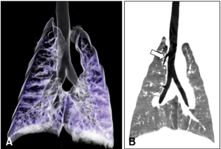
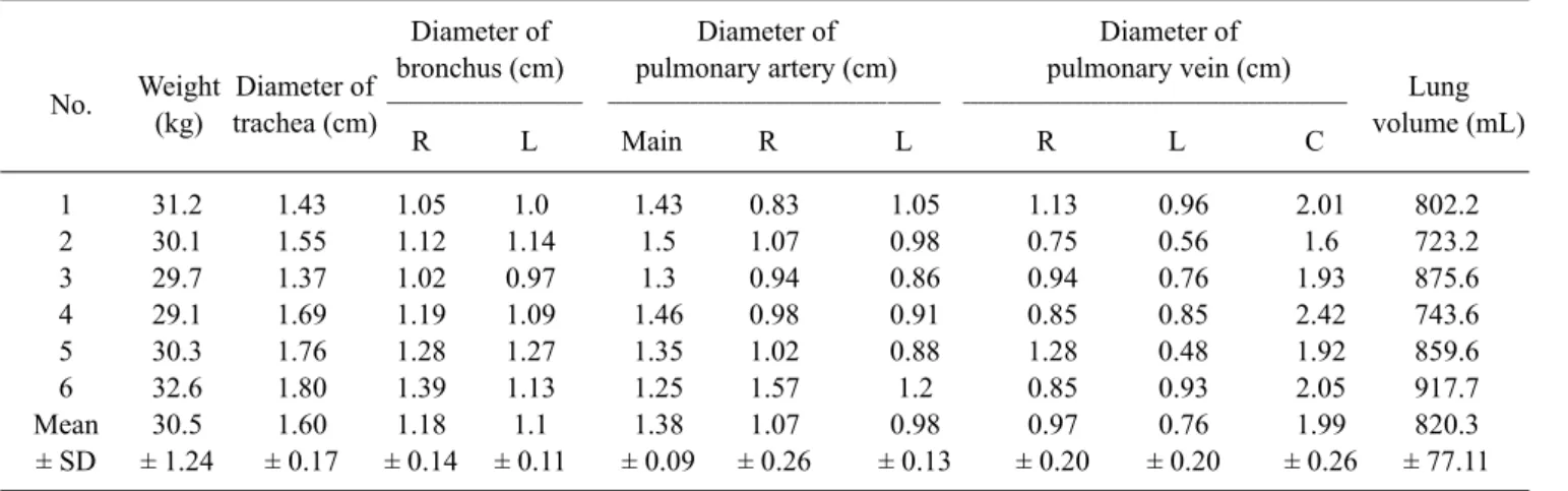
Therefore, this study was performed to evaluate the feasibility of 64-channel multi-detector row computed tomography (MDCT) to measure the diameter of the pulmonary arteries and the lung volume in micropigs. The mean diameters of the trachea, and left and right bronchi were 1.6 ± 0.17, 1.18 ± 0.14, and 1.1 ± 0.11 cm, respectively. The mean diameters of the main, right, and left pulmonary arteries were 1.38 ± 0.09, 1.07
± 0.26, and 0.98 ± 0.13 cm and the diameters of right, left, and common inferior pulmonary veins were 0.97 ± 0.20, 0.76 ± 0.20, and 1.99 ± 0.26 cm, respectively. The mean lung volume was 820.3 ± 77.11 mL. The data presented in this study suggest that the MDCT may be a noninvasive, rapid, and accurate investigational method for pulmonary anatomy in living lung donors.
Keywords: lung, micropig, multidetector row computed tomography (MDCT)
Introduction
Allotransplantation is currently viewed as the preferred solution for the treatment of end-stage organ failure.
However, only a small percentage of those patients who could benefit from this therapy receive it due to a shortage of donor organs [8,21]. This shortage of donors has stimulated interest in the possible use of animal organs for transplantation into humans. Animal-to-human transplantation
(or xenotransplantation) would offer an increasing supply of organs and tissue for transplantation. Xenotransplantation using pig organs could solve the significant increasing shortage of donor organs for allotransplantation due to the anatomical and physiological similarities between pig and human organs [7].
In comparison to other solid organ transplants, there have been few reports regarding lung xenotransplantation between pigs and other non-human primates [4,18]. A previous study reported excellent respiratory function and adequate hemodynamics during the short survival time of xenogeneically perfused porcine lungs [6]. In addition, it has been reported that the transplanted pig lungs into cynomolgus monkeys had adequate function [17]. These results suggest that porcine lungs are physiologically compatible with those of humans, although it would appear that pulmonary xenotransplantation is limited by hyperacute lung injury [25].
In order to transplant swine lungs into humans, physiological
and anatomical comparison and analysis are essential in
assessing the suitability of donor organs for potential
recipients. However, an appropriate method for evaluating
micropig organs has yet to be established. This study
examines the feasibility of evaluating the lung and its related
structures using multi-detector row computed tomography
(MDCT) and establishes standard anatomical reference
values for micropig lungs. Of the major technological
developments in computed tomography in recent years, the
most significant has been the introduction of MDCT, which
has brought about substantial improvements in spatial and
particularly temporal resolution [10,13]. This study is the
first to show MDCT to be a reliable method for noninvasively
assessing the pulmonary anatomy of micropigs.
![Table 2. Comparison of pulmonary diameters between micropigs and humans Micropigs Human Reference (present study) Trachea 16.0 ± 1.78 mm 19.6 ± 2 mm [22] Main bronchus 11.4 ± 1.25 mm 12.6 ± 2 mm [12]](https://thumb-ap.123doks.com/thumbv2/123dokinfo/5446469.652323/4.892.71.431.207.394/comparison-pulmonary-diameters-micropigs-micropigs-reference-trachea-bronchus.webp)
