Candida arthritis is a rare disease which usually oc- curs in immunocompromised conditions such as AIDS, organ transplantation, hematologic malignancy, and dia- betes (1). Among the joints affected by Candida, the knee is the most commonly involved joint and few other joints (elbows and hips) have been reported (2). To our knowledge, there was only one report of Candida parap- silosis arthritis involving the ankle, but it had radi- ographic findings only. We present a case of Candida parapsilosis arthritis involving the ankle in a diabetes pa- tient and describe the MR imaging findings for the first time.
Case Report
A 63-year-old woman with a 13-year history of dia- betes mellitus and hypertension presented at our hospi- tal with right ankle pain and swelling for 4 months. She underwent a trans-sphenoidal adenoidectomy for the pi- tuitary macroadenoma one year previously and had been taking anti-diabetic (metformin) and anti-hyperten- sive drugs, as well as receiving an insulin shot once a day. She had no history of trauma.
Upon physical examination, the lateral aspect of the right ankle was swollen with joint effusion and tender- ness. However, no external wound was evident. Range of motion was nearly full and laboratory findings re- vealed no definite abnormality except for the mild ane- mia (hemoglobin of 10.2 g/dL).
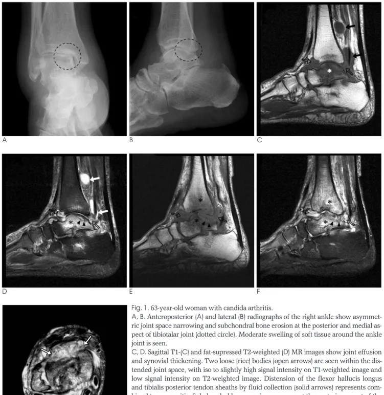
On radiographs of the right ankle (Figs. 1A, B), diffuse periarticular soft tissue swelling around the ankle was noted, with joint space narrowing and subchondral
Candida Parapsilosis Arthritis Involving the Ankle in a Diabetes Patient: A Case Report1
Jinkyeong Sung, M.D., Kyung Ah Chun, M.D.
1
Department of Radiology, The Catholic University of Korea Uijeongbu St.
Mary’s Hospital
Received December 17, 2010 ; Accepted April 7, 2011
Address reprint requests to : Kyung Ah Chun, M.D., Department of Radiology, The Catholic University of Korea Uijeongbu St. Mary’s Hospital, 65-1 Geumo-dong, Uijeongbu-si, Gyeonggi-do 480-130, Korea.
Tel. 82-31-820-3137 Fax. 82-31-846-3080 E-mail: [email protected]
Candida parapsilosis is a rare opportunistic fungal pathogen of the musculoskeletal region. Immune function of almost all patients is severely disturbed. Most reported cases of septic arthritis of joints by Candida involve the knee, especially Candida parap- silosis. To our knowledge, there has been only one case report of Candida parapsilosis involving the ankle presented on only plain radiography. We report a case of Candida parapsilosis arthritis involving the ankle in a diabetes patient which was shown on MR imaging.
Index words : Mycoses Ankle joint Arthritis
Diabetes Mellitus
Magnetic Resonance Imaging
bone erosion at posteromedial aspect of the tibiotalar joint. An initial MR imaging without contrast enhance- ment (Figs. 1C, D) showed diffuse synovial thickening
of the tibiotalar joint with joint effusion. Localized fluid collections were also seen along the posterior tibialis, flexor digitorum longus, flexor hallucis longus, and per-
A B C
D E F
Fig. 1. 63-year-old woman with candida arthritis.
A, B. Anteroposterior (A) and lateral (B) radiographs of the right ankle show asymmet- ric joint space narrowing and subchondral bone erosion at the posterior and medial as- pect of tibiotalar joint (dotted circle). Moderate swelling of soft tissue around the ankle joint is seen.
C, D. Sagittal T1-(C) and fat-supressed T2-weighted (D) MR images show joint effusion
and synovial thickening. Two loose (rice) bodies (open arrows) are seen within the dis-
tended joint space, with iso to slightly high signal intensity on T1-weighted image and
low signal intensity on T2-weighted image. Distension of the flexor hallucis longus
and tibialis posterior tendon sheaths by fluid collection (solid arrows) represents com-
bined tenosynovitis. Subchondral bone erosion was seen at the posterior aspect of the
oneal tendons. Loose bodies were seen at the distended synovial spaces. These were iso- to slighlty hyperintense on T1-weighted images and slightly hypointense on fat- suppressed T2-weighted images. The posterior aspect of the talar dome showed osteochondral lesion. Fluid col- lections and subtle intrasynovial loose bodies at the pos- terior subtalar joint and talonavicular joint were also combined. There was prominent hyperintense bone marrow edema in the talus, distal tibia, and calcaneus on T2-weighted images. Mild periarticular soft tissue edema was combined.
Initially, we presumed that the diagnosis was infec- tious arthritis, such as tuberculous arthritis with associ- ated tenosynovitis and osteomyelitis of the talus. Our differential diagnosis included the pyogenic arthritis or other causes of chronic infectious arthritis including fungal or other bacterial arthritis, neuropathic arthopa- thy, and inflammatory arthritis such as rheumatoid arthritis.
She underwent open tenosynovectomy and arthroto- my of ankle and hypertrophied synovium was found in the tibiotalar joint. Further, the joint space was filled with a pus-like discharge. Intra-articular rice body-like granulation tissues were found and microscopic exami- nations revealed chronic inflammatory changes of the synovial tissue. Foreign body reaction was combined and AFB staining and PCR for tuberculosis were also negative.
During the initial empirical antibiotic (3rd generation cephalosporine) treatment, Candida parapsilosis was cultured. Despite additional systemic infusion of am- photericin B, right ankle pain and swelling did not sub- side. Four months later on a follow up MR imaging (Figs. 1E-G), progression of the synovial thickening and loose bodies within the joint space was noted. Gradual progressive destruction of the tibiotalar joint was com- plicated by the subchondral erosion of the talar dome, and bone marrow abnormality was also noted.
A B
C
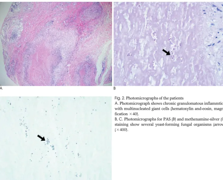
Fig. 2. Photomicrographs of the patients
A. Photomicrograph shows chronic granulomatous inflammtion with multinucleated giant cells (hematoxylin and-eosin, magni- fication ×40).
B, C. Photomicrographs for PAS (B) and methenamine-silver (C)
staining show several yeast-forming fungal organisms (arrow)
(×400).
The patient underwent the operative procedure a sec- ond time. Microscopic examinations showed chronic granulomatous inflammation with multinucleated giant cells. PAS and methenamine-silver staining revealed several yeast-form fungal organisms (Figs. 2A-C).
Despite additional fluconazol treatment, symptoms did not fully subside. Several episodes of pyogenic infection were superimposed, and finally she underwent ankle arthrodesis.
Discussion
Candida infections are very rare in the musculoskele- tal system, especially with immunocompetent patients (3). Among them, the most common cases are septic arthritis by Candida (3). Many clinical settings where patients are immunocompromised are predisposed to the Candidia arthritis. Poor immunity triggers the sys- temic candidema in patients and finally, inoculation at the targeted joint. On the other hand, direct inoculation by iatrogenic or traumatic events induces septic arthritis by the Candida species. Many previous reports showed that variable species of Candida are associated with arthritis and spondylitis. Among them, C. albicans is re- sponsible for 70% of cases, while the most frequent non- albicans species are C. tropicalis, C. parapsilosis, and C.
krusei. Candida parapsilosis is a relatively rare but emerg- ing opportunistic pathogen, especially when associated with arthroplasty (4).
The knee comprises the majority of involved joint of C. parapsilosis throughout the various literature sources.
Prosthetic procedures replacing the knee are thought as the crucial point of infection (4). A report suggests an- other iatrogenic predisposing factor, the intra-articular injection of steroid into a painful knee. Few other joints, such as elbow, hips, and ankle are associated with Candida infections (2).
Candida also involves the spine as a form of spondyli- tis other than synovial joint (5, 6). The sternum is most frequently involved, followed by the vertebrae (5). MR imaging findings of Candida spondylitis are similar to those of pyogenic spondylitis (7). Radiographic findings
patients with Candida arthritis, especially C.
parapsilosis, were profoundly immunocompromised, or underwent invasive procedures at the involved joints.
In fact, there is little information about the MR imag- ing of Candida parapsilosis arthritis. As compared with that of more common fungal pathogens, MR imaging of our case disclosed some similar and non-specific find- ings, including synovial thickening, joint effusion, artic- ular cartilage destruction, subchondral destruction, and marrow edema. However, these findings are not helpful in making a diagnosis. Major differential diagnoses in- clude tuberculosis arthritis and other chronic inflamma- tory or infectious arthritis. Progressive destruction of joint in our patient is similar to that of pyogenic arthritis.
On the other hand, paucity of the cellulitis or sinus tracts around the ankle lowers the probability of pyo- genic arthritis. In addition, an indolent but persistent clinical course also contributes to ruling out pyogenic arthritis.
Loose bodies with low signal intensity on T2-weighted images and slightly high to iso signal intensity on T1- weighted images corresponds to the rice body noted on the surgical and pathologic findings. These rice bodies could represent the chronic granulation tissues, espe- cially the proliferation of synovial tissues. These loose bodies could be helpful in differentiating with pyogenic arthritis, one of the major clinical concerns.
Rheurmatoid arthritis and tuberculous arthritis also have increased synovial inflammation and proliferation, resulting in rice bodies (8). Periarticular cellulitis or ab- scesses, and articular synovitis is uncommon in fungal disease, which is a minor clue to leading our diagnosis with fungal arthritis rather than tuberculosis (9). In our case, there is tenosynovitis around the ankle, which can be seen in rheumatoid arthritis and tuberculous arthri- tis. On the other hand, the finding with a lack of periar- ticular osteopenia on the plain radiography is discrimi- nated from tuberculous arthritis.
Discrimination of the cause of foot pain in the diabetic
patients is critical for a successful treatment, and MR
imaging could be helpful for the challenging clinical set-
ting. Because of diabetes, some differential diagnoses of
cally (10).
Candida arthritis is treated with surgical debridement and antifungal agents. However, Candida parapsilosis arthris usually has poor prognosis, especially in im- munocompromised patients (4). Our patient also has been deteriorated, in spite of aggressive treatment.
In conclusion, we have described a rare case of Candida parapsilosis arthritis involving the ankle in a dia- betes patient with MR imaging. In patients with im- mune suppressed condition, especially diabetes, Candida arthritis should be considered in the differen- tial diagnosis of the cause of ankle pain. Imaging fea- tures of Candida parapsiloisis arthritis are nonspecific with a slow progression. It mimics other chronic infec- tious or inflammatory arthritis on MR imaging; especial- ly tuberculous arthritis.
References
1. Masoud M, Nasser NJ, Karban A, Edelstein S. Candida parapsilo- sis septic arthritis in a renal transplant patient. J Clin Rheumatol
2008;14:56
2. Marmor L, Peter JB. Candida arthritis of the knee joint. Clin Orthop Relat Res 1976:133-135
3. Turgut B, Vural O, Demir M, Kaldir M. Candida arthritis in a pa- tient with chronic myelogenous leukemia (CML) in blastic trans- formation, unresponsive to fluconazole, but treated effectively with liposomal amphotericin B. Ann Hematol 2002;81:529-531 4. Trofa D, Gacser A, Nosanchuk JD. Candida parapsilosis, an
emerging fungal pathogen. Clin Microbiol Rev 2008;21:606-625 5. Miller DJ, Mejicano GC. Vertebral osteomyelitis due to Candida
species: case report and literature review. Clin Infect Dis 2001;33:
523-530
6. Cha JG, Hong HS, Koh YW, Kim HK, Park JM. Candida albicans osteomyelitis of the cervical spine. Skeletal Radiol 2008;37:347-350 7. Munk PL, Lee MJ, Poon PY, O’Connell JX, Coupland DB, Janzen
DL, et al. Candida osteomyelitis and disc space infection of the lumbar spine. Skeletal Radiol 1997;26:42-46
8. Hsu CY, Lu HC, Shih TT. Tuberculous infection of the wrist: MRI features. AJR Am J Roentgenol 2004;183:623-628
9. Parmar H, Shah J, Patkar D, Singrakhia M, Patankar T, Hutchinson C. Tuberculous arthritis of the appendicular skeleton:
MR imaging appearances. Eur J Radiol 2004;52:300-309
10. Karchevsky M, Schweitzer ME, Morrison WB, Parellada JA. MRI findings of septic arthritis and associated osteomyelitis in adults.
AJR Am J Roentgenol 2004;182:119-122
대한영상의학회지 2011;64:587-591
당뇨병 환자의 족관절에서 발생한 Candida Paraspilosis 감염:
증례 보고11