Received: 2013.7.8. Revised: 2013.9.9. Accepted: 2013.9.29.
Corresponding author: Seung-Ho Lee
Department of Obstetrics and Gynecology, Gachon University Gil Medical Center, 21 Namdong-daero 774beon-gil, Namdong-gu, Incheon 405-760, Korea
Tel: +82-32-460-3254 Fax: +82-32-460-3290 E-mail: [email protected]
Articles published in Obstet Gynecol Sci are open-access, distributed under the terms of the Creative Commons Attribution Non-Commercial License (http://creativecommons.
org/licenses/by-nc/3.0/) which permits unrestricted non-commercial use, distribution, and reproduction in any medium, provided the original work is properly cited.
Copyright © 2014 Korean Society of Obstetrics and Gynecology
Introduction
Cervical intraepithelial neoplasia (CIN) diagnoses have in- creased in Korea due to periodic screening and the develop- ment of cervical cancer examination methods including hu- man papillomavirus (HPV) tests. HPV infection is a cause of premalignant and malignant epithelial lesions of the cervix.
Approximately 40 different known HPV genotypes infect the anogenital tract, of which at least 12 genotypes have been linked to the development of CIN [1,2].
The etiological link between HPV genotypes and cervi- cal cancer has been established. Lorincz et al. [3] used the terms low-risk, intermediate-risk, and high-risk to categorize HPV types: low-risk (HPV 6, 11, 42, 43, and 44), which are
Effect of human papillomavirus genotype on severity and prognosis of cervical intraepithelial neoplasia
Chun-Hoe Ku, Seung-Ho Lee, Soon-Pyo Lee
Department of Obstetrics and Gynecology, Gachon University Gil Medical Center, Incheon, Korea
Objective
This study evaluated the effect of the specific human papillomavirus (HPV) genotypes on severity and prognosis in cervical intraepithelial neoplasia (CIN) patients.
Methods
The medical records of 446 patients treated with loop electrosurgical excision procedure (LEEP) were reviewed. The severity of CIN was categorized as CIN1/CIN2 versus CIN3+ including CIN3 and carcinoma in situ (CIS). HPV genotypes were categorized as 1) low risk, 2) intermediate risk, 3) high risk/HPV 16, 4) high risk/HPV 18, and 5) unclassified.
Progression was defined as abnormal cytology, including atypical squamous cells, low-grade squamous intraepithelial lesion and high-grade squamous intraepithelial lesion. The margin status and progression free survival (PFS) by HPV genotypes were analyzed in 355 women with three months or more of post-treatment records.
Results
CIN3+ was the most common CIN type (67.7%), and high risk/HPV 16 (26.9%) was the most common genotype.
Intermediate risk (P < 0.01), high risk/HPV 16 (P < 0.01) and high risk/HPV 18 (P < 0.01) were significantly more common in women with CIN3+ than CIN1/CIN2. Patients with high risk/HPV 18 showed the highest rate of positive margins (P < 0.01). The margin status proved to be the only statistically significant factor affecting PFS.
Conclusion
The proportion of positive margins was significantly different by HPV genotypes and highest in high risk/HPV 18 group.
CIN patients with high risk/HPV 18 need to be more carefully tracked than patients with the other HPV genotypes.
Keywords: Cervical intraepithelial neoplasia; Genotype; Human papillomavirus; Prognosis http://dx.doi.org/10.5468/ogs.2014.57.1.37
pISSN 2287-8572 · eISSN 2287-8580
detected in low-grade CIN but are rare in invasive cancers;
intermediate-risk (HPV 31, 33, 35, 51, 52, and 58), which are detected more frequently in CIN than in invasive can- cers; high risk/HPV 16, which is associated equally with CIN and invasive carcinoma, and high risk/HPV 18 (HPV 18, 45 and 56), which is more prevalent in invasive cancers than in CIN.
CIN lesions do not always progress to invasive lesions.
Spontaneous regression rate in biopsy-proven CIN1 cases is about 60% to 85%. Lesions generally regress, typically within 2 years. CIN2 lesions regress in 40% of cases and persist in 40%. CIN3 lesions regress in 33% of cases but progression to frank carcinoma is about 12% to 22% of cases [4,5]. Hence, it is likely that CIN1/CIN2 represents a less advanced stage of cervical neoplasia than CIN3 including carcinoma in situ (CIS).
CIN1/CIN2 versus CIN3+ (CIN3 and CIS) may therefore serve as a measure of the severity of cervical neoplasia.
The loop electrosurgical excision procedure (LEEP) is the preferred treatment for CIN and has some advantages over other methods. However, residual/recurrent disease after a conization procedure using this technique varies between 5%
and 30%, requiring reassessment and treatment once lesions are identified [6].
The main purpose of observing patients after LEEP due to CIN is the early detection of residual/recurrent cervical disease presenting a risk of progression to invasive carcinoma if an effective treatment is not administered. The status of resection margins is a predictor of residual disease [7]. Current follow- up protocols are mainly based on periodic cytology.
The relative importance of the different HPV genotypes for the development of CIN is not clear, and the effect of specific HPV genotypes on prognosis in CIN patients treated with LEEP is an open question. In this study, we identified the HPV genotypes more common in CIN3+ compared to CIN1/CIN2.
In addition, the effect of specific HPV genotypes on the status of resection margin and prognosis in CIN patients was evalu- ated.
Materials and methods
1. Case selection
From January 2007 to December 2009, 545 women were di- agnosed with CIN and treated by LEEP in our hospital. Ninety- nine women were excluded because they did not have HPV
testing, leaving 446 women who had undergone HPV testing at the time of diagnosis to be included in this study. The his- tological analyses were confirmed with colposcopic-guided biopsy or LEEP. The diagnostic criteria followed the WHO clas- sification of cervical neoplasia.
The HPV genotype was categorized as follows; 1) negative, 2) low-risk (HPV 6, 11, 42, 43, and 44), 3) intermediate-risk (HPV 31, 33, 35, 51, 52, and 58), 4) high risk/HPV 16, 5) high risk/
HPV 18 (HPV 18, 45, and 56), and 6) unclassified. The severity of CIN was categorized as CIN1/CIN2 versus CIN3+. CIN3+
included CIN3 and CIS. The specific HPV genotype according to severity of CIN was analyzed.
At follow-up exams, conventional cytology or liquid based cytology technique (Thin Prep, Cytyc Corporation, Boxbor- ough, MA, USA) was performed. The 2001 Bethesda System was used for terminology for reporting the results of the cervi- cal cytology. Margin status by HPV genotypes and progression free survival (PFS) were analyzed in 355 women with three months or more of follow-up. We excluded 91 patients who did not revisit our hospital after LEEP or patients with less than three months of observation following surgery.
Patients were tracked for a median duration of 11 months (range, 3–54 months). Progression was defined as abnormal cytology including atypical squamous cells (ASC), low-grade squamous intraepithelial lesion (LSIL), and high-grade squa- mous intraepithelial lesion (HSIL).
2. Human papillomavirus testing
For HPV genotyping, a commercially available HPV DNA Chip was purchased from Biomedlab Co. (Seoul, Korea). The manu- facturer’s protocol describes the preparation and testing of specimens, and the genotyping experiment was performed using a procedure provided by Biomedlab Co. The target HPV DNA was amplified by the polymerase chain reaction (PCR) using the primers (HPV and β-globin) and conditions provided by Biomedlab Co. and labeled using Cy5-dUTP (NEN Life Sci- ence Products Inc., Boston, MA, USA). The PCR product was hybridized onto the chip at 40°C for 2 hours and washed with 3× SSPE and with 1× SSPE for 2 minutes each. Hybridized signals were visualized with a DNA Chip Scanner (GSI Lumon- ics, Scanarray lite, Ottawa, ON, Canada).
3. Statistical analysis
The statistical analyses were performed with PASW ver. 18.0
(SPSS Inc., Chicago, IL, USA). The specific HPV genotype by se-
verity of CIN and margin status by HPV genotypes were ana- lyzed with Pearson’s chi-square test or Fisher’s exact test. As independent prognostic factors for PFS, the margin status and HPV genotypes were evaluated by the Kaplan-Meier method with the log-rank test, and multivariate Cox proportional hazard analysis with hazard ratio (HR) and 95% confidence interval (CI). Null hypotheses of no difference were rejected if P-values were less than 0.05, or, equivalently, if the 95% CIs of risk point estimates excluded 1.
4. Ethics
The institutional review board of Gachon University Gil Medi- cal Center, Korea, approved the study (GCIRB2013-91).
Results
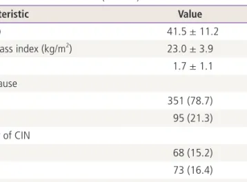
The mean age of all 446 patients was 41.5 ±11.2 years old, and the mean body mass index was 23.0 ± 3.9 kg/m
2. The average parity was 1.7 ±1.1 and 95 patients (21.3%) were menopausal women. By category of CIN severity, CIS was the most common (157 cases, 35.2%) followed by CIN3 (148 cases, 33.2%), CIN2 (73 cases, 15.2%), and CIN1 (68 cases, 15.2%) in sequence (Table 1).
Among the HPV genotypes high risk/HPV 16 was predomi- nant (120 cases, 26.9%), followed by intermediate risk (112 patients, 25.1%), negative (88 cases, 19.7%), unclassified (68 cases, 15.2%), high risk/HPV 18 (53 cases, 11.9%), and low risk (5 cases, 1.1%) in sequence (Table 2). For the analysis of the specific HPV genotype by severity of CIN, each HPV geno- type distribution was compared with all other HPV genotypes aggregated. In the negative group, all 88 patients were diag- nosed as CIN1/CIN2. In the low risk group, all five patients were diagnosed as CIN3+. In the intermediate risk group, all 112 patients were diagnosed as CIN3+. In the high risk/HPV 16 group, all 120 patients were diagnosed as CIN3+. In the high risk/HPV 18 group, all 53 patients were diagnosed as CIN3+. In the unclassified group, 56 patients were diagnoses as CIN1/CIN2 and 12 patients were diagnosed as CIN3+.
Negative (P < 0.01) and unclassified (P < 0.01) group were more common in women with CIN1/CIN2 than CIN3+. Inter- mediate risk ( P < 0.01), high risk/HPV 16 (P < 0.01) and high risk/HPV 18 ( P < 0.01) were significantly more common in women with CIN3+ than CIN1/CIN2 (Table 2).
Margin status by HPV genotypes was analyzed in 355 women with 3 months or more of post-treatment information.
Table 1. Patient characteristics (n = 446)
Characteristic Value
Age (yr) 41.5 ± 11.2
Body mass index (kg/m
2) 23.0 ± 3.9
Parity 1.7 ± 1.1
Menopause
No 351 (78.7)
Yes 95 (21.3)
Severity of CIN
CIN 68 (15.2)
CIN2 73 (16.4)
CIN3 148 (33.2)
CIS 157 (35.2)
Values are presented as mean ± standard deviation or frequency (%).
CIN, cervical intraepithelial neoplasia; CIS, carcinoma in situ.
Table 2. Specific HPV genotype by severity of CIN
HPV genotype CIN1/CIN2 CIN3+
a)Total (%)
b)P-value
c)Negative 88 0 88 (19.7) <0.01
Low risk
d)0 5 5 (1.1) 0.18
Intermediate risk
e)0 112 112 (25.1) <0.01
High risk/HPV 16
f)0 120 120 (26.9) <0.01
High risk/HPV 18
g)0 53 53 (11.9) <0.01
Unclassified 56 12 68 (15.2) <0.01
Total 144 302 446
HPV, human papillomavirus; CIN, cervical intraepithelial neoplasia.
a)
CIN3 and carcinoma in situ;
b)Proportion of the specific HPV genotype in all the patients;
c)Each HPV genotype distribution was compared
with all other HPV genotypes aggregated;
d)Low risk genotypes include HPV 6, 11, 42, 43, and 44;
e)Intermediate risk genotypes include
HPV 31, 33, 35, 51, 52, and 58;
f)High risk/HPV 16 genotype includes HPV 16;
g)High risk/HPV 18 genotypes include HPV 18, 45, and 56.
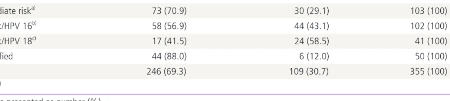
In the negative group, 54 patients (91.5%) had negative mar- gins and 5 patients (8.5%) were positive. In the intermediate risk group, 73 patients (70.9%) had negative margins and 30 patients (29.1%) positive. In the high risk/HPV 16 group, 58 patients (56.9%) were negative and 44 patients (43.1%) had positive margins. In the high risk/HPV 18 group, 17 patients (41.5%) had negative margins while 24 patients (58.5%) were positive. In the unclassified group, 44 patients (88.0%) had negative margin and 6 patients (12.0%) had positive margin. The proportion of the positive margin was significant- ly different (P < 0.01) by HPV genotypes and highest in high risk/HPV 18 group (Table 3).
PFS according to the status of margin and HPV genotype was analyzed in these 355 patients. In univariate analysis, only the status of margin had significant effect (P < 0.01) on
PFS (Fig. 1). PFS was significantly lower in the positive margin group than the negative margin group (Fig. 1A). The slightly lower PFS observed in the high risk/HPV 18 could be ex- plained by chance (Fig. 1B). By multivariate Cox’s proportional hazard analysis, the margin status was the only statistically significant factor affecting PFS (Table 4).
Discussion
HPV 16 and 18 increase the risk of high-grade cervical neo- plasia compared to the HPV genotypes aggregated. The as- sociation between the specific HPV genotype and prognosis in CIN patients treated with LEEP is not clear, so, we designed this study to evaluate the effect of the specific HPV genotypes Table 3. Resection margin status by HPV genotypes in 355 women with three months or more follow-up
Negative margin Positive margin Total
Negative 54 (91.5) 5 (8.5) 59 (100)
Intermediate risk
a)73 (70.9) 30 (29.1) 103 (100)
High risk/HPV 16
b)58 (56.9) 44 (43.1) 102 (100)
High risk/HPV 18
c)17 (41.5) 24 (58.5) 41 (100)
Unclassified 44 (88.0) 6 (12.0) 50 (100)
Total 246 (69.3) 109 (30.7) 355 (100)
P<0.01
d)Values are presented as number (%).
HPV, human papillomavirus.
a)
Intermediate risk genotypes include HPV 31, 33, 35, 51, 52, and 58;
b)High risk/HPV 16 genotype includes HPV 16;
c)High risk/HPV 18 genotypes include HPV 18, 45, and 56;
d)The proportion of the positive margin was signifi cantly different (P < 0.01) by HPV genotypes and highest in high risk/HPV 18 group.
Table 4. Cox proportional hazards analysis for prognostic factors affecting progression free survival
Prognostic factors Hazard ratio 95% Confidence interval P-value
Resection margin
Negative 1 – –
Positive 1.90 1.15–3.13 0.01
HPV genotype
Negative 1 – –
Intermediate risk
a)0.86 0.41–1.83 0.70
High risk/HPV 16
b)0.85 0.39–1.81 0.66
High risk/HPV 18
c)1.45 0.62–3.37 0.38
Unclassified 1.16 0.50–2.68 0.72
HPV, human papillomavirus.
a)
Intermediate risk genotypes include HPV 31, 33, 35, 51, 52, and 58;
b)High risk/HPV 16 genotype includes HPV 16;
c)High risk/HPV 18
genotypes include HPV 18, 45, and 56.
on severity and prognosis in CIN patients. Furthermore, we evaluated the effect of specific HPV genotypes on the status of resection margin, a predictor of residual disease in CIN pa- tients treated with LEEP.
Comparisons among studies of HPV genotype and risk of CIN are difficult, since study designs, grouping of HPV geno- types and outcome measures differ among studies. Lorincz et al. [3] recruited 2,627 women into eight studies analyzing the relationship between HPV infection and cervical neoplasia and defined four categories (intermediate risk, high risk/HPV 16, and high risk/HPV 18). Based on these four categories, we classified HPV genotypes into six groups, adding negative and unclassified groups.
We categorized severity of CIN as CIN1/CIN2 versus CIN3+
because CIN1/CIN2 may represent a less advanced stage of cervical neoplasia than CIN3+. Most CIN1 lesions regress spontaneously if untreated. Generally, women diagnosed with CIN2 are treated with excision. However, there is increasing awareness that not all CIN2 is “precancer” [8]. In fact, a large proportion of CIN2 lesions may resolve without treatment, leading to recommendations not to treat CIN2 immediately in young women [9].
In Korea, HPV prevalence is 11.8% among cytologically normal women [10], 73.2% among those with LSIL [11], and 86.4% among those with HSIL [12]. Based on pooled estimates of 11 published papers, the most common high-risk HPV types in HSIL were HPV 16, 52, 58, 51, and 18. The most
frequent high-risk HPV types among women with LSIL were HPV 16, 52, 51, 56, and 58. HPV 16 remains the most com- mon HPV type across the range of cervical lesions in Korea [13].
In a cross-sectional study in Norway, including 643 women with CIN2, CIN3 and CIS, HPV 16 was the most common HPV genotype, detected in 51.2% of the women, followed by HPV 31, 33, 52, 18, 51, 58, and 45 [14]. In our study, high risk/
HPV 16 was the most common genotype. Intermediate risk, negative, unclassified, high risk/HPV 18 and low risk followed high risk/HPV 16 in order of frequency. We categorized HPV 31, 33, and 52 as intermediate risk.
As for HPV genotypes by severity of CIN, the distribution of HPV types in the CIN1 women and the CIN2/CIN3 women showed a slightly different pattern. In a prospective study in Korea, including 78 CIN patients, the most frequent types in the CIN2/CIN3 women were HPV 16 (29%) and then HPV 58 (13%), 31, 33, and 56. By contrast, the most common HPV types in CIN1 were HPV 58 (18.8%) and HPV 16/35 (18.8%) [15]. In the cross-sectional study in Norway, presence of HPV 16 as a single infection or in combination with another HPV genotype was more common in women with CIN3+ than in women with CIN2. Although less prevalent, HPV 33 was also more common in women with CIN3+ than in women with CIN2 [14]. In a pooled data analysis, including more than 16,000 women from different countries, HPV 16, 18, and 45 were more frequently reported in women with invasive car- cinoma than in women with high-grade squamous intraepi- Fig. 1. Univariate analysis of progression free survival (PFS). (A) PFS was significantly lower (P < 0.01) in the positive margin group than the negative margin group. (B) Human papilloma virus (HPV) genotype had no significant effect on PFS (P = 0.19).
a. Negative b. Unclassified c. High risk/HPV 16 d. Intermediate risk e. High risk/HPV 18 Negative margin
Positive margin
0.00 10.00 20.00 30.00 40.00 50.00 60.00 0.00 10.00 20.00 30.00 40.00 50.00 60.00
P = 0.19
a b
d
e c
P < 0.01
1.0
0.8
0.6
0.4
0.2
0.0 1.0
0.8
0.6
0.4
0.2
0.0