Clinicopathologic characteristics of
human papillomavirus (HPV) infection
and serum anti-HPV 16/18 antibodies
in cervical neoplasia
Doo Byung Chay
Department of Medicine
Clinicopathologic characteristics of
human papillomavirus (HPV) infection
and serum anti-HPV 16/18 antibodies
in cervical neoplasia
Doo Byung Chay
Department of Medicine
Clinicopathologic characteristics of
human papillomavirus (HPV) infection
and serum anti-HPV 16/18 antibodies
in cervical neoplasia
Directed by Professor Jae-Hoon Kim
The Doctoral Dissertation
submitted to the Department of Medicine,
the Graduate School of Yonsei University
in partial fulfillment of the requirements for the degree
of Doctor of Philosophy
Doo Byung Chay
June 2011
This certifies that the Doctoral
Dissertation of Doo Byung Chay is
approved.
---
Thesis Supervisor: Jae-Hoon Kim
---
Hyon-Suk Kim: Thesis Committee Member
---
Nam Hoon Cho: Thesis Committee Member
---
Eunseop Song: Thesis Committee Member
---
Eun-Suk Kang: Thesis Committee Member
The Graduate School
Yonsei University
ACKNOWLEDGEMENTS
I owe my utmost gratitude to my supervisor, Prof.
Jae-Hoon Kim, for his encouragement, guidance and support
from the initial to the final level of this dissertation. It has been
an honor for me to be his student and I will never forget his
sincerity and enthusiasm as a doctor and a professor that
inspired me as an
excellent role model.
One simply could not
wish for a better supervisor.
I would like to express my gratitude to the thesis
committee members: Prof. Hyon-Suk Kim, Prof. Nam Hoon
Cho, Prof. Eunseop Song and Prof. Eun-Suk Kang for their
devoting guidance, advice and insight. I am also heartily
grateful to all my teachers and colleagues in the department of
OB/GY YUMC and Yongin severance hospital who supported
me with their valuable assistance.
Lastly, I would like to thank my parents who raised me
with love and supported me throughout becoming a doctor,
my wife Yeun Jung Shin and my son Hee Geon Chay for all
their love, support and understanding
.
<TABLE OF CONTENTS>
ABSTRACT ··· 1
I. INTRODUCTION ··· 3
II. MATERIALS AND METHODS ··· 5
1. Study subjects ··· 5
2. Cervical HPV DNA testing ··· 6
3. Serum anti-HPV 16/18 antibody ··· 7
4. Statistical analysis ··· 8
III. RESULTS ··· 9
1. Evaluation of cervical HPV infection and serologic HPV detection
with epidemiologic risk factors ··· 9
2. Comparison of cervical HPV infection and serologic HPV detection
according to disease severity ··· 12
3. Evaluation of serologic HPV detection as a prognostic parameter in
cervical cancer patients ··· 16
IV. DISCUSSION ··· 20
V. CONCLUSION ··· 23
REFERENCES ··· 24
LIST OF FIGURES
Figure 1. Comparison of HPV titers according to disease
severity ··· 13
Figure 2. Kaplan-Meier analysis of disease free survival ··· 19
LIST OF TABLES
Table 1. Epidemiologic characteristics according to disease
severity ··· 10
Table 2. Comparison of epidemiologic risk factors with cervical
HPV infection and serologic HPV detection ··· 11
Table 3. Cervical HPV DNA titer and serum anti-HPV 16/18
antibody titer according to disease severity ··· 12
Table 4. Cervical HPV positivity and HPV 16/18 seropositivity
according to disease severity ··· 14
Table 5. Comparison of cervical HPV positivity and HPV 16/18
seropositivity according to disease severity ··· 15
Table 6. Concordance of serologic HPV detection with cervical
HPV infection according to disease severity ··· 16
Table 7. Correlation of serologic HPV detection and prognostic
parameters in cervical cancer ··· 17
Table 8. Multivariate cox proportional hazards analysis on
disease free survival of cervical cancer ··· 18
1
<ABSTRACT>
Clinicopathologic characteristics of human papillomavirus (HPV)
infection and serum anti-HPV 16/18 antibodies in cervical neoplasia
Doo Byung Chay
Department of Medicine
The Graduate School, Yonsei University
(Directed by Professor Jae-Hoon Kim)
Objective: To evaluate the serum anti-HPV 16/18 antibody and cervical
HPV DNA status according to the disease severity of cervical neoplasia
and also to evaluate the prognostic value of serum anti-HPV16/18
antibody in patients with cervical cancer.
Materials and Methods: This study was conducted at the Gangnam
Severance Hospital between July 2002 and December 2010. Data were
analyzed in patients with a histopathological diagnosis of cervical
intraepithelial neoplasia (CIN) 1 (n=64), CIN 2/3 (n=241), cervical
cancer (n=170), and in women with no cervical lesion and with normal
cytology (n=975). Cervical HPV DNA tests were performed by Hybrid
Capture II tests and serum anti-HPV 16/18 antibody was measured by
multiplexed competitive Luminex immunoassay. Results were evaluated
by epidemiological risk factors and compared according to disease
severity. Serum anti-HPV 16/18 antibody was also compared with
histopathologic parameters, and clinical follow-up data to assess
prognostic value in patients with cervical cancer.
2
increased in patients with cervical neoplasia compared to normal
cytology (P <0.001) and although cervical HPV DNA titer was increased
in cervical cancer compared to CIN 1 (P = 0.049), overall there was no
significant difference in cervical HPV DNA titer and positivity according
to the severity of cervical neoplasia. Serum anti-HPV 16 antibody titer
was significantly increased in CIN 2/3 and cervical cancer compared to
CIN 1 and normal cytology (P <0.001), but there was no significant
difference between CIN 1 and normal cytology and between CIN 2/3 and
cervical cancer. HPV 16 seropositivity significantly increased according
to the disease severity up to CIN 2/3 but there was no significant
difference between CIN 2/3 and cervical cancer (P = 0.054). For HPV
type 18, there was no significant difference in the antibody titer and
seropositivity according to the disease severity.
In cervical cancer, HPV
16 seropositivity was significantly associated with longer disease-free
survival (P = 0.044) in the univariate analysis but was not proven in the
multivariate analysis. Kaplan-Meier survival estimates revealed HPV 16
seropositivity was significantly associated with better disease- free
survival (P=0.017).
Conclusions: Cervical HPV DNA and serum anti-HPV 16 antibody
detection maybe useful in cervical screening as an adjuvant test revealing
cervical neoplasia. Serologic detection of anti-HPV 16 antibodies has the
advantage of representing a more advanced cervical neoplasia (more than
CIN 2) and also may have the possibility for a favorable prognostic value
in cervical cancer.
--- Key words: HPV infection, HPV antibody, cervical neoplasia, cervical intraepithelial
3
Clinicopathologic characteristics of human papillomavirus (HPV)
infection and serum anti-HPV 16/18 antibodies in cervical neoplasia
Doo Byung Chay
Department of Medicine
The Graduate School, Yonsei University
(Directed by Professor Jae-Hoon Kim)
I. INTRODUCTION
Cervical cancer is the second most common cancer in women world-wide, with approximately 500,000 new cases and 274,000 deaths reported annually.1 Despite the reduction of cervical cancer incidence and mortality rates which have been achieved through effective screening programs in the developed countries, it is an important public health problem that deserves urgent attention in most of the developing countries, where 80% of the cases occur.2,3
Human papillomavirus (HPV) infection is a common sexually transmitted disease (STD), which causes different diseases such as genital warts, intraepithelial lesions, and even cervical cancer.4 More than 130 HPV genotypes have been identified to date and categorized into high-risk (HR) and low-risk (LR) types on the basis of their epidemiologic association with different disease outcomes. LR types (HPV 6, 11, 40, 42, 43, 44, 54, 61, 70, 72, 81) are mainly found in low grade lesions, genital or skin warts whereas HR types (HPV 16, 18, 31,35, 39, 45, 51, 52, 56, 59, 66, 68, 69 and 73) can cause cervical cancer, anogenital cancers, head and neck cancers.5,6 HPV is associated with nearly all cervical cancers and although there is variability by region, approximately 70% of
4
cervical cancers are associated with HPV 16 and 18. These are followed by subtypes HPV 45, 31, 33, 35, 52, and 58; these variants account for 10% of total cases.7,8 Even though persistent infection of specific types of HR-HPVs is thought to be necessary for most cervical tumorigenesis, HPV infection does not always induce cervical lesion. HPV infection is a transient phenomenon and while most of the HPV infections seem to be cleared spontaneously, some HPV-infected women will develop cervical lesions. Following infection, the virus may persist over time, leading to development of low grade cervical lesions where the virus is present in non-integrated form, progressing in course of time to cancer that takes 15- 20 years.9,10
Considering the epidemiology and the natural history of HPV infection, cervical cancer appears from a defined series of cervical intraepithelial neoplasia (CIN) and models of cervical pathogenesis involve persistent infection caused by HR type HPV infection as the major risk factor, as well as cofactors such as multiparity, cigarette smoking, oral contraceptive use, co-infection with other sexually transmitted diseases whereas the probability of acquiring a HPV-infection is strongly related to age at sexual debut and number of sexual partner.11,12
Due to the close association with persistent HR type HPV infection and cervical cancer, cervical HPV DNA test is clinically applied in primary screening combined with cytology, for the triage of equivocal cervical cytology results, and in the follow-up of patients treated for high-grade cervical intraepithelial neoplasia (CIN).13,14 The cervical HPV DNA test not only identifies women with cervical disease, but also those at risk for developing cervical lesions.15 However, HPV presence in the uterine cervix can only provide data on current infection and cannot fully indicate the persistent exposure to HPV because most HPV infections appear to be transient. Also there are difficulties in cervical HPV DNA testing due to the reluctance and discomfort of unmarried women regarding gynecologic examination. On the other hand, serum antibodies to HPV can provide insights into the natural history of HPV infection and associated diseases by including past
5
infections and cumulative exposure. In fact, it has been reported that 50% to 60% of HPV-infected women are seroconverted 6 to 18 months following HPV-DNA detection and that this seroconversion rarely occurs in women having transitory HPV infections.16,17 Considering these differences represented by cervical HPV and serologic HPV detection, serological assays complementary with cervical HPV DNA detection may be useful in understanding the natural history of HPV infections and the association of cervical tumorigenesis more clearly. Nowadays most serological studies on HPV antibodies including seroprevalence and seroepidemiology studies have been utilized in developing optimal vaccination strategy and assessing the efficacy of HPV vaccination. Despite intensive study with cervical HPV-DNA, studies on serologic antibodies to HPV are relatively insufficient in patients with CIN and cervical cancer.18-20
The aim of this study was to evaluate the serum anti-HPV 16/18 antibody and cervical HPV DNA status according to the disease severity and epidemiological correlates of cervical neoplasia and also to evaluate the prognostic value of serum anti-HPV 16/18 antibody in patients with cervical cancer.
II. MATERIALS AND METHODS
1. Study subjects
The study subjects were composed of 975 women with normal cytology and no cervical lesions, 64 CIN 1 patients, 251 CIN 2/3 patients and 170 cervical cancer patients who visited Gangnam Severance Hospital from July 2002 to December 2010. All patients had a histologically confirmed diagnosis and the cervical cancer patients were clinically staged according to the International Federation of Gynecology and Obstetrics (FIGO) staging system. Biological samples such as vaginal swab specimen and venous blood samples were collected from patients with informed consent at the time of diagnosis before any treatment.
6
Sociodemographic information, histopathologic features and clinical data were obtained by a retrospective review of the medical records. Epidemiological related factors involving cervical pathogenesis such as age, marital status, and age at sexual debut, number of sexual partners, smoking, oral contraceptive use, parity and history of STD were evaluated according to disease severity, cervical HPV DNA status and serum anti-HPV16/18 antibody status. Cervical HPV DNA and serum anti-HPV 16/18 antibody status were also analyzed and compared according to the disease severity of cervical neoplasia. Prognostic outcome were measured by disease-free survival and analyzed with serum anti-HPV16/18 antibody status and other prognostic factors in patients with cervical cancer.
2. Cervical HPV DNA testing
Cervical HPV DNA testing was performed using the Hybrid Capture II assay (Digene, Gaithersburg, MD, USA). To confirm the presence of HR type HPV DNA (HPV subtypes 16, 18, 31, 33, 35, 39, 45, 51, 52, 56, 58, 59, and 68), cells from the uterine cervix were collected with the use of a female swab specimen collection kit (Dacron swab, Digene, Gaithersburg, MD, USA). The cells were placed in a liquid hybridization assay collection kit and then preserved at - 20°C for further analysis. RNA probes of high-risk HPV were hybridized with denatured, single stranded DNA. This reaction mixture was transferred to a tube of which the surface was coated with anti-DNA–RNA hybrid antibodies. The immobilized hybrids were then reacted with an alkaline phosphatase-conjugated antihybrid monoclonal antibody. After rinsing, these reactants were treated with Lumi-Phospho 530 (Lumigen, Inc., Southfield, MI, USA), which reacts to alkaline phosphatase, a dioxetanebased chemiluminescent substrate. The light emitted from the reaction was measured using a luminometer (DML 2000, Digene, Gaithersburg, MD, USA). The intensity was expressed using units that were relative to the reactions. The solutions containing 10 pg HPV 16 DNA per milliliter served as
7
positive controls for high-risk HPV. Relative light unit/cutoff (RLU/CO) ratios were calculated as the ratio of the specimen luminescence to the luminescence of the positive control. A positive cutoff value was set at 1 pg HPV DNA per milliliter in each specimen.
3. Serum anti-HPV16/18 antibody
Blood samples were centrifuged at 1,500×g for 10 minutes for serum separation and stored in prelabeled tubes at -70°C until processing. Frozen samples were thawed and tested for specific neutralizing antibodies to HR HPV subtypes 16 and 18 using a competitive Luminex® immunoassay (Luminex Corporation, Austin, TX, USA). Briefly, the HPV virus-like particles (VLPs) for types 16 and 18 were each covalently conjugated to the free carboxyl groups on xMAP® Multi-Analyte COOH Microspheres (Luminex Corporation, Austin, TX, USA). H16.V5, and H18.J4 monoclonal antibodies (mAbs) were used to chemically couple to phycoerythrin (PE) and used to detect neutralizing epitopes on coupled HPV VLPs 16 and 18. A multiplex reference standard solution was prepared at a concentration of 1000 milli-Merck units per milliliter (mMU/ml) for each HPV type in a prescreened assay matrix of antibody depleted human serum (ADHS) (Human Serum Special Stripped, Valley Biomedical, Winchester, VA, USA). The reference standards were serum samples from African green monkeys hyperimmunized with HPV monovalent L1 VLP types 16 or 18. The multiplex reference standard was diluted in a two-fold serial dilution in ADHS to create a 12-point standard curve with final well concentrations ranging from 0.25 to 500mMU/ml. The assay plate setup contained four controls, ADHS diluents, and high, low, and negative titer controls. All standards, controls, and samples were tested in duplicate. Baseline testing of serum samples was carried out at a 1 : 4 dilution; final assay conditions consisted of 25 ㎕ of each of the following: serum, ADHS, mAb-PE multiplex (0.1 µg/mL for each mAb), and VLP-microspheres
8
(5,000 VLP-microspheres per well per type). The plate was covered with a foil seal and placed on a shaker (600–800 rpm) at room temperature for 16–25 hours. The contents of the assay plate were transferred to a 1.2-㎛ hydrophilic low-protein-binding filter plate (Millipore, Billerica, MA, USA) and washed three times. The microspheres were suspended in 125 ㎕ of assay wash buffer for analysis on a Bio-Plex Suspension Array System (Bio-Rad, Hercules, CA, USA). Fluorescent units were read and averaged, dilution corrected mMU/ml serum values were computed based on four parameter logistics fit of the standard curve on each assay plate. Seropositivity for HPV was defined as having anti-HPV antibodies titers at least 20 mMU/ml for HPV16 and at least 24 mMU/ml for HPV 18 as described by Dias et al in 2005.21
4. Statistical analysis
Comparisons of frequency distributions were analyzed using Chi-squared and Fisher's exact tests. Mantel-Haenszel method was used to analyze ordinal data. Unconditional multiple logistic regression was used to calculate odds ratios (OR) and corresponding 95% confidence intervals (CIs) to assess the associations between cervical HPV DNA status, serologic HPV 16/18 antibody status and the disease severity of cervical neoplasia or selected characteristics. The mean cervical HPV DNA titer and serum anti-HPV16/18 antibody titer was analyzed using ANOVA test after log-transformation of the variables and Bonferroni corrections were used to adjust for multiple comparisons. As measures of prognostic outcome for cervical cancer, Kaplan-Meier survival curves were compared using the log-rank test for disease-free survival and the multivariate Cox proportional hazards model was used to adjust for potential confounding factors. Statistical analyses were performed using the SPSS PASW Statistics 18.0 (SPSS Inc, Chicago, IL, USA). All statistical tests were two-sided and considered to be statistically significant at P < 0.05.
9 III. RESULTS
1. Evaluation of cervical HPV infection and serologic HPV detection with epidemiologic risk factors.
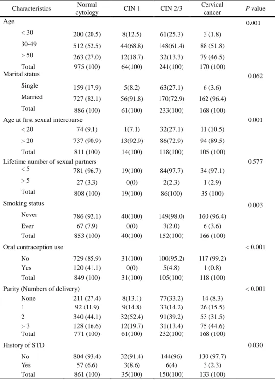
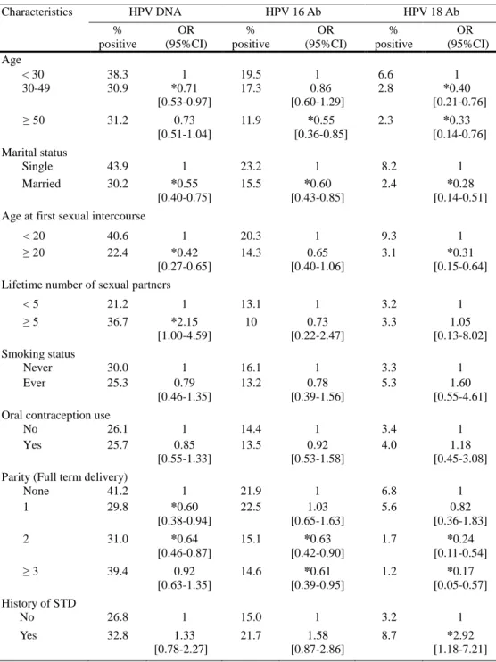
Epidemiological characteristics according to disease severity and the association with cervical HPV DNA and serum anti-HPV 16/18 antibody status are presented in Table 1 and 2. There was a significant increase of cervical HPV DNA positivity in women who were single, in women who had first sexual intercourse at age before 20 years old and in women who had more than 5 sexual partners. Age factor revealed a significant increase of cervical HPV positivity under 30 years old than among age 30 to 49 years old and also showed an increasing tendency than age over 50 years old. Parity status revealed a significant increase of cervical HPV positivity in nulliparous women and then showed a decreasing tendency with increased parity. Considering the serum anti-HPV 16/18 antibody status, women who were single had significantly increased seropositivity to both HPV 16 and 18. Seropositivity to HPV 18 was significantly increased in women who had first sexual intercourse at age before 20 years and in women with a previous history of STD. Seropositivity to HPV 16 and 18 both showed a decreasing tendency to age factor and parity status. There were no significant difference in cervical HPV positivity and HPV 16/18 seropositivity associated with smoking status and oral contraceptive uses. Overall, the results involving sexual behavior of which younger women, with more lifetime partners, with an early sexual debut, who were single and who had a history of STD had an increasing risk of cervical HPV positivity and HPV seropositivity.
10
Table 1. Epidemiologic characteristics according to disease severity
Characteristics Normal
cytology CIN 1 CIN 2/3
Cervical cancer P value Age 0.001 < 30 200 (20.5) 8(12.5) 61(25.3) 3 (1.8) 30-49 512 (52.5) 44(68.8) 148(61.4) 88 (51.8) > 50 263 (27.0) 12(18.7) 32(13.3) 79 (46.5) Total 975 (100) 64(100) 241(100) 170 (100) Marital status 0.062 Single 159 (17.9) 5(8.2) 63(27.1) 6 (3.6) Married 727 (82.1) 56(91.8) 170(72.9) 162 (96.4) Total 886 (100) 61(100) 233(100) 168 (100)
Age at first sexual intercourse 0.001
< 20 74 (9.1) 1(7.1) 32(27.1) 11 (10.5) > 20 737 (90.9) 13(92.9) 86(72.9) 94 (89.5) Total 811 (100) 14(100) 118(100) 105 (100)
Lifetime number of sexual partners 0.577
< 5 781 (96.7) 19(100) 84(97.7) 34 (97.1) > 5 27 (3.3) 0(0) 2(2.3) 1 (2.9) Total 808 (100) 19(100) 86(100) 35 (100) Smoking status 0.003 Never 786 (92.1) 40(100) 149(98.0) 160 (96.4) Ever 67 (7.9) 0(0) 3(2.0) 6 (3.6) Total 853 (100) 40(100) 152(100) 166 (100)
Oral contraception use < 0.001
No 729 (85.9) 31(100) 100(95.2) 117 (99.2) Yes 120 (41.1) 0(0) 5(4.8) 1 (0.8) Total 849 (100) 31(100) 105(100) 118 (100)
Parity (Numbers of delivery) < 0.001
None 211 (27.4) 8(13.1) 77(33.2) 14 (8.3) 1 92 (11.9) 9(14.8) 33(14.2) 26 (15.5) 2 340 (44.1) 32(52.4) 91(39.2) 53 (31.5) > 3 128 (16.6) 12(19.7) 31(13.4) 75 (44.6) Total 771 (100) 61(100) 232(100) 168 (100) History of STD 0.030 No 804 (93.4) 32(91.4) 144(96) 130 (97.7) Yes 57 (6.6) 3(8.6) 6(4) 3 (2.3) Total 861 (100) 35(100) 150(100) 133 (100) Data are presented as number (%).
11
Table 2. Comparison of epidemiologic risk factors with cervical HPV infection and serologic HPV detection
Characteristics HPV DNA HPV 16 Ab HPV 18 Ab % positive OR (95%CI) % positive OR (95%CI) % positive OR (95%CI) Age < 30 38.3 1 19.5 1 6.6 1 30-49 30.9 *0.71 [0.53-0.97] 17.3 0.86 [0.60-1.29] 2.8 *0.40 [0.21-0.76] ≥ 50 31.2 0.73 [0.51-1.04] 11.9 *0.55 [0.36-0.85] 2.3 *0.33 [0.14-0.76] Marital status Single 43.9 1 23.2 1 8.2 1 Married 30.2 *0.55 [0.40-0.75] 15.5 *0.60 [0.43-0.85] 2.4 *0.28 [0.14-0.51] Age at first sexual intercourse
< 20 40.6 1 20.3 1 9.3 1 ≥ 20 22.4 *0.42 [0.27-0.65] 14.3 0.65 [0.40-1.06] 3.1 *0.31 [0.15-0.64] Lifetime number of sexual partners
< 5 21.2 1 13.1 1 3.2 1 ≥ 5 36.7 *2.15 [1.00-4.59] 10 0.73 [0.22-2.47] 3.3 1.05 [0.13-8.02] Smoking status Never 30.0 1 16.1 1 3.3 1 Ever 25.3 0.79 [0.46-1.35] 13.2 0.78 [0.39-1.56] 5.3 1.60 [0.55-4.61] Oral contraception use
No 26.1 1 14.4 1 3.4 1 Yes 25.7 0.85 [0.55-1.33] 13.5 0.92 [0.53-1.58] 4.0 1.18 [0.45-3.08] Parity (Full term delivery)
None 41.2 1 21.9 1 6.8 1 1 29.8 *0.60 [0.38-0.94] 22.5 1.03 [0.65-1.63] 5.6 0.82 [0.36-1.83] 2 31.0 *0.64 [0.46-0.87] 15.1 *0.63 [0.42-0.90] 1.7 *0.24 [0.11-0.54] ≥ 3 39.4 0.92 [0.63-1.35] 14.6 *0.61 [0.39-0.95] 1.2 *0.17 [0.05-0.57] History of STD No 26.8 1 15.0 1 3.2 1 Yes 32.8 1.33 [0.78-2.27] 21.7 1.58 [0.87-2.86] 8.7 *2.92 [1.18-7.21] * P < 0.05
12
2. Comparison of cervical HPV infection and serologic HPV detection according to disease severity
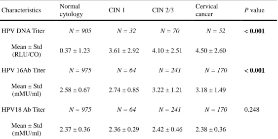
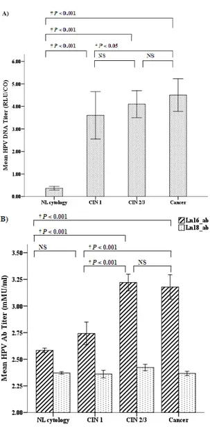
The mean cervical HPV DNA titers and serum anti-HPV 16/18 antibody titers were analyzed according to disease severity and are presented in Table 3. Cervical HPV DNA titer were significantly increased in patients with cervical lesions compared to normal cytology (P <0.001) and although cervical HPV DNA titer was increased in cervical cancer compared to CIN 1 (P = 0.049), overall there was no significant difference according to the severity of cervical neoplasia (Figure 1A). Serum anti-HPV 16 antibody titer was significantly increased in CIN 2/3 and cervical cancer compared with CIN 1 and normal cytology (P <0.001), but there was no significant difference between CIN 1 and normal cytology and between CIN 2/3 and cervical cancer. There was no significant difference in the serum anti-HPV 18 antibody titer according to the disease severity (Figure 1B).
Table 3. Cervical HPV DNA titer and serum anti-HPV 16/18 antibody titer according to disease severity
Characteristics Normal
cytology CIN 1 CIN 2/3
Cervical cancer P value HPV DNA Titer N = 905 N = 32 N = 70 N = 52 < 0.001 Mean ± Std (RLU/CO) 0.37 ± 1.23 3.61 ± 2.92 4.10 ± 2.51 4.50 ± 2.60 HPV 16Ab Titer N = 975 N = 64 N = 241 N = 170 < 0.001 Mean ± Std (mMU/ml) 2.58 ± 0.67 2.74 ± 0.85 3.22 ± 1.21 3.18 ± 1.49 HPV18 Ab Titer N = 975 N = 64 N = 241 N = 170 0.248 Mean ± Std (mMU/ml) 2.37 ± 0.36 2.36 ± 0.29 2.42 ± 0.46 2.38 ± 0.36 Variables were analysed after log-transformation.
13
Figure 1. Comparison of HPV titers according to disease severity. A) Cervical HPV DNA titer. B) Serum anti-HPV 16/18 antibody titer. Bar represents the mean titer and standard error are presented as error bars (I). Bonferroni corrections were used to adjust for multiple comparisons. NS, not significant. Ab, antibody.
14
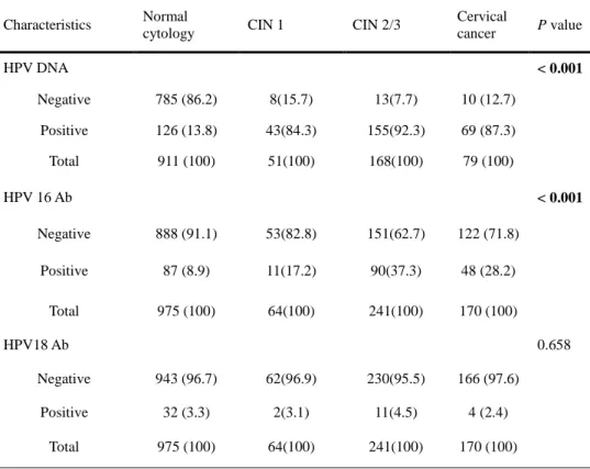
Cervical HPV positivity and HPV 16/18 seropositivity in women with normal cytology, patients with CIN 1, CIN 2/3 and cervical cancer are presented in Table 4. Cervical HPV positivity was 13.8% in normal cytology, 84.3% in CIN 1, 92.3% in CIN 2/3 and 87.3% in cervical cancer. Seropositivity to HPV 16 was 8.9% in normal cytology, 17.2% in CIN 1, 37.3% in CIN 2/3 and 28.2% in cervical cancer. While Cervical HPV positivity and seropositivity to HPV 16 were significantly different among cervical neoplasia (P <0.001) there was no significant difference with seropositivity to HPV 18 (P = 0.658).
Table 4. Cervical HPV positivity and HPV 16/18 seropositivity according to disease severity
Characteristics Normal
cytology CIN 1 CIN 2/3
Cervical cancer P value HPV DNA < 0.001 Negative 785 (86.2) 8(15.7) 13(7.7) 10 (12.7) Positive 126 (13.8) 43(84.3) 155(92.3) 69 (87.3) Total 911 (100) 51(100) 168(100) 79 (100) HPV 16 Ab < 0.001 Negative 888 (91.1) 53(82.8) 151(62.7) 122 (71.8) Positive 87 (8.9) 11(17.2) 90(37.3) 48 (28.2) Total 975 (100) 64(100) 241(100) 170 (100) HPV18 Ab 0.658 Negative 943 (96.7) 62(96.9) 230(95.5) 166 (97.6) Positive 32 (3.3) 2(3.1) 11(4.5) 4 (2.4) Total 975 (100) 64(100) 241(100) 170 (100)
Data are presented as number (%).
15
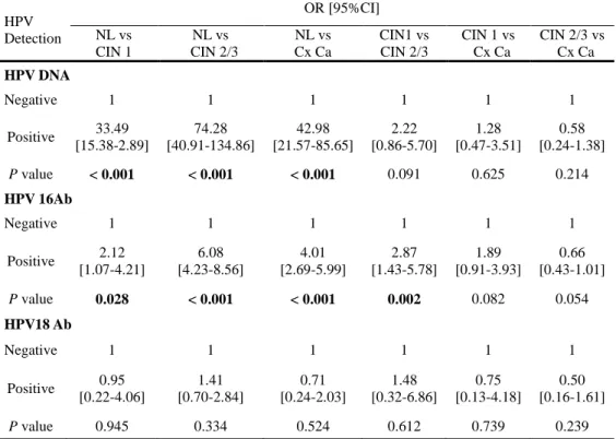
Table 5. Comparison of cervical HPV positivity and HPV 16/18 seropositivity according to disease severity
HPV Detection OR [95%CI] NL vs CIN 1 NL vs CIN 2/3 NL vs Cx Ca CIN1 vs CIN 2/3 CIN 1 vs Cx Ca CIN 2/3 vs Cx Ca HPV DNA Negative 1 1 1 1 1 1 Positive 33.49 [15.38-2.89] 74.28 [40.91-134.86] 42.98 [21.57-85.65] 2.22 [0.86-5.70] 1.28 [0.47-3.51] 0.58 [0.24-1.38] P value < 0.001 < 0.001 < 0.001 0.091 0.625 0.214 HPV 16Ab Negative 1 1 1 1 1 1 Positive 2.12 [1.07-4.21] 6.08 [4.23-8.56] 4.01 [2.69-5.99] 2.87 [1.43-5.78] 1.89 [0.91-3.93] 0.66 [0.43-1.01] P value 0.028 < 0.001 < 0.001 0.002 0.082 0.054 HPV18 Ab Negative 1 1 1 1 1 1 Positive 0.95 [0.22-4.06] 1.41 [0.70-2.84] 0.71 [0.24-2.03] 1.48 [0.32-6.86] 0.75 [0.13-4.18] 0.50 [0.16-1.61] P value 0.945 0.334 0.524 0.612 0.739 0.239
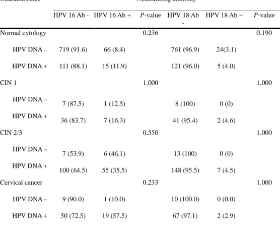
In the analysis according to disease severity, cervical HPV positivity and HPV 16 seropositivity was significantly higher in the presence of cervical lesions compared to normal cytology (P <0.001). Cervical HPV positivity showed no significance between the severity of cervical neoplasia, whereas HPV 16 seropositivity was significantly increased in CIN 2/3 compared with CIN 1 (P = 0.002) but showed no significant differences between CIN 2/3 and cervical cancer (Table 5). The concordance between cervical HPV positivity and seropositivity to HPV 16 was 11.9% in normal cytology, 16.3% in CIN 1, 35.5% in CIN 2/3 and 57.5% in cervical cancer. For seropositivity to HPV 18, the concordance with cervical HPV positivity was 4.0% in normal cytology, 4.6% in CIN 1, 4.5% in CIN 2/3 and 2.9% in cervical cancer (Table 6).
16
Table 6. Concordance of serologic HPV detection with cervical HPV infection according to disease severity
Characteristics Neutralizing antibody
HPV 16 Ab - HPV 16 Ab + P-value HPV 18 Ab - HPV 18 Ab + P-value Normal cytology 0.236 0.190 HPV DNA – 719 (91.6) 66 (8.4) 761 (96.9) 24(3.1) HPV DNA + 111 (88.1) 15 (11.9) 121 (96.0) 5 (4.0) CIN 1 1.000 1.000 HPV DNA – 7 (87.5) 1 (12.5) 8 (100) 0 (0) HPV DNA + 36 (83.7) 7 (16.3) 41 (95.4) 2 (4.6) CIN 2/3 0.550 1.000 HPV DNA – 7 (53.9) 6 (46.1) 13 (100) 0 (0) HPV DNA + 100 (64.5) 55 (35.5) 148 (95.5) 7 (4.5) Cervical cancer 0.233 1.000 HPV DNA – 9 (90.0) 1 (10.0) 10 (100.0) 0 (0.0) HPV DNA + 50 (72.5) 19 (57.5) 67 (97.1) 2 (2.9)
Data are presented as number (%). P values were analyzed by Fisher’s test.
3. Evaluation of serologic HPV detection as a prognostic parameter in cervical cancer patients
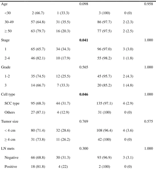
Correlation of serologic HPV detection and prognostic parameters of cervical cancer are shown in Table 7. Patients with Stage 1 cervical cancer had significantly higher HPV 16 seropositivity compared to stage 2 to 4 cervical cancer patient. Seropositivity to HPV 16 was also significantly higher in Squamous Cell Carcinoma (SCC) type compared to other cell types.
17
Table 7. Correlation of serologic HPV detection and prognostic parameters in cervical cancer
Characteristics HPV 16 Ab - HPV 16 Ab + P-value HPV 18 Ab - HPV 18 Ab + P-value
Age 0.098 0.958 <30 2 (66.7) 1 (33.3) 3 (100) 0 (0) 30-49 57 (64.8) 31 (35.5) 86 (97.7) 2 (2.3) ≥ 50 63 (79.7) 16 (20.3) 77 (97.5) 2 (2.5) Stage 0.041 1.000 1 65 (65.7) 34 (34.3) 96 (97.0) 3 (3.0) 2-4 46 (82.1) 10 (17.9) 55 (98.2) 1 (1.8) Grade 0.565 1.000 1-2 35 (74.5) 12 (25.5) 45 (95.7) 2 (4.3) 3 14 (66.7) 7 (33.3) 20 (85.2) 1 (4.8) Cell type 0.046 1.000 SCC type 95 (68.3) 44 (31.7) 135 (97.1) 4 (2.9) Others 27 (87.1) 4 (12.9) 31 (100) 0 (0) Tumor size 0.769 0.575 < 4 cm 80 (71.4) 32 (28.6) 108 (96.4) 4 (3.6) ≥ 4 cm 31 (73.8) 11 (26.2) 42 (100) 0 (0) LN mets 0.300 1.000 Negative 66 (68.8) 30 (31.3) 93 (96.9) 3 (3.1) Positive 18 (81.8) 4 (22) 2 (100) 0 (0)
Data are presented as number (%).
Cut off value: HPV 16 Ab > 20 mMU/ml, HPV 18 Ab > 24 mMU/ml.
In the univariate survival analysis, seropositivity to HPV 16, FIGO stage, tumor size and lymph-node status were found to be significantly associated with
18
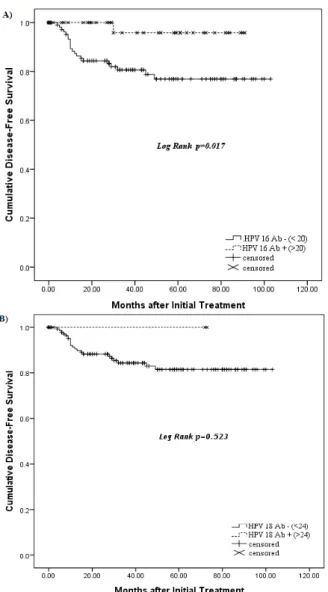
longer disease-free survival. However among the significant parameters, only advanced stage 2 or more patients and lymph node metastasis status were found to be significantly correlated with survival and other factors showed no significant differences in the multivariate analysis (Table 8). Kaplan-Meier survival estimates revealed that seropositivity to HPV 16 was significantly associated with better disease-specific survival (P = .017) (Figure 2A) and seropositivity to HPV 18 showed a trend (P = .523, not significant) for better disease-free survival (Figure 2B).
Table 8. Multivariate cox proportional hazards analysis on disease free survival of cervical cancer Disease-Free Survival Univariate Multivariate Age NS HPV 16 Ab positive 0.12 [0.01-0.94], 0.044 NS HPV 18 Ab positive NS Stage 1 1 1 2-4 10.02 [3.37-29.81], < 0.001 8.14 [2.01-32.94], 0.003 Grade 1-2 1 3 NS Cell type NS Tumor size (> 4 cm) 2.98 [1.26-7.04], 0.013 NS LN mets 6.25 [2.09-18.69], 0.001 3.63 [1.14-11.52],0.029 NS, not significant.
19
Figure 2. Kaplan-Meier analysis of disease free survival. A) Disease free survival according to HPV 16 seropositivity. B) Disease free survival according to HPV 18.
20 IV. DISCUSSION
This study investigated serum anti-HPV 16/18 antibody and cervical HPV DNA status in women with cervical neoplasia, revealing the association according to
the disease severity,
with epidemiological risk factors in cervical carcinogenesis and with prognostic factors of cervical cancer. The overall findings in this study showed that cervical HPV DNA indicated current infection status and distinguished cervical neoplasia with normal cytology, while serum antibodies to HPV 16/18, revealing cumulative status of sustained HPV exposure including past infections, distinguished CIN 1 with advanced cervical neoplasia (more than CIN 2). The
findings were more positively associated with seropositivity to HPV 16 and this may be due to the lower power for the HPV-18 analysis since the seropositivity to HPV 18 was low in patients with CIN 1 (3.1%), CIN 2/3 (4.5%) and cervical cancer (2.4%) and the mean titers were low compared to HPV 16. The limitations of this study was mainly due to the cross sectional nature representing a single time point during the disease process and a lack of cervical HPV DNA type-specific information since HCII test detects 13 high-risk types of HPV at a time and provides the overall current status of HR-HPV infection.Regarding the association with epidemiological risk factors in cervical carcinogenesis the results involving sexual behavior of which younger women, with more lifetime partners, with an early sexual debut, who were single and
who had a history of STD had an increasing risk of cervical HPV DNA
positivity and HPV16/18 seropositivity. On the other hand the results on parity, which has been recognized as a risk factor for cervical carcinogenesis decreased
with the number of full term delivery in this study. Hormonal influences and
long maintenance of the transformation zone on the exocervix is suggested for the explanation of the risk in multiparity, and the lack of this influence in this study can be presumed, but still a feasible explanation is needed for the
21
decreased risk with parity.22,23
The main interest of this study was the evaluation of serum anti-HPV 16/18 antibody and cervical HPV DNA status according to disease severity since it has been reported in many studies that there was a strong association between HPV infection and advanced cervical neopasia.24-26 Persistent detection of cervical HPV DNA was an evident predictor of cervical neoplasia, and serum HPV antibodies were associated with persistent DNA detection indicating life time exposure to HPV.27,28 In accordance with the findings previously reported, cervical HPV positivity was 13.8% in normal cytology, 84.3% in CIN 1, 92.3% in CIN 2/3 and 87.3% in cervical cancer.29-31 Cervical HPV DNA titer were significantly increased in patients with cervical neoplasia compared to normal cytology (P <0.001). Seropositivity to HPV 16 was higher in patients with CIN 1 (17.2%), CIN 2/3 (37.3%) and cervical cancer patients (28.2%) compared with normal cytology (8.9%). The trend for a high percentage of HPV16 seropositivity with advanced cervical neoplasia suggests that HPV antibodies may correlate with the inability of a patient to clear their HPV infection. Thus, continued exposure to HPV increases the likelihood of seroconversion.32,33 The findings that HPV 16 seropositivity were significantly higher with CIN 2/3 than CIN 1 and normal cytology, reveals a strong association between advanced cervical neoplasia. Also serum anti-HPV 16 antibody titers which were significantly higher with CIN 2/3 and cervical cancer compared with CIN 1 and normal cytology supports the hypothesis that serum HPV antibodies correlates with the presence of advanced cervical neoplasia and suggest that serum HPV antibody assay may be useful as an additional tool in combination with cervical HPV DNA testing for the identification of women at risk for advanced cervical neoplasia. The concordance of cervical HPV positivity and HPV 16 seropositivity was 11.9% in normal cytology, 16.3% in CIN 1, 35.5% in CIN 2/3 and 57.5% in cervical cancer but there showed no statistical significance (P > 0.05, Fisher’s test). Concordance can be effected by several factors such as
22
lag time required for seroconversion, waning of detectable antibodies, and variable persistence of type-specific antibodies especially in women with normal cytology. Interestingly the discordance showed a tendency to decrease with disease severity and this may be suggestive, but was not statistically significant, support for the utilization of adjuvant serological assays in detecting advanced cervical lesions.
Since positive antibody responses indicates a prolonged exposure to
replicating virus and antibodies to HPV16/18 are a highly type-specific markers
for HPV infections associated with developing cervical cancer, the correlation of serum anti-HPV 16/18 antibody with prognostic parameters and survival were investigated in patients with cervical cancer. Among the prognostic parameters, squamous cell carcinoma (SCC) cell type and early FIGO stage1
were significantly related with higher HPV 16 seropositivity and detection of
antibodies to HPV-16 was associated with a better disease-free survival in patients with cervical cancer in this study. Higher HPV 16 seropositivity in SCC cell type can be explained since HPV 16 contributes to more than 70% of squamous cell carcinoma worldwide, but the association in early FIGO stage1
and with a better survival prognosis is unclear.34 In previous reported studies,
Heim et al. have proposed an explanation of the observed correlation between prognosis and HPV serostatus.35Within benign HPV lesions, the HPV genome is episomal, whereas in malignant lesions HPV DNA integration into the host
chromosome is frequently observed. In advanced CIN, a subset of lesions can
be identified in which the viral genome has integrated and there is a greater risk of malignant progression.36 In invasive cancer, HPV capsid protein expression is rare, but persistent episomal HPV DNA was detected in 38% of invasive cervical carcinomas. Interestingly, tumors containing episomal DNA were associated with longer survival than tumors with only integrated DNA. The presence of antibodies directed against capsid proteins could therefore indicate the persistence of episomal HPV DNA and more prolonged expression of late
23
genes. In tumors with a lower rate of integration, therefore, the viral genome must remain extra chromosomally which would provide a mechanism for
expression of the late viral genes.37,38 Thus, a relationship between the presence
of capsid antibodies and the time elapsing before integration of HPV DNA in different subsets of tumors may explain the association between HPV serology and prognosis. Another similar study involving 150 cervical cancer patients by Skiba et al. reported that anti-HPV16 seropositivity correlated with prolonged, progression-free and overall survival in the FIGO stage 1 and 2 patients. They hypothesized that the seronegative patients had reduced HPV-specific immune competence, so that the virus might have been able to escape both the humoral and the cellular defense mechanisms, resulting in an impaired ability of the immune system to control the HPV-induced tumor.39,40 The results in this study also revealed a correlation with HPV 16 seropositivity and a better disease free survival supporting the above hypothesis.
V. CONCLUSION
The result of this study reveals the features of serum anti-HPV 16/18 antibody and cervical HPV DNA in women with cervical neoplasia. Among the epidemiologic correlates, sexual behavioral factors were more at risk with both cervical and serological HPV positivity. While serum anti-HPV 18 antibody demonstrated no significant difference according to disease severity, cervical HPV DNA detection and serum anti-HPV 16 antibody detection maybe useful in cervical screening as an adjuvant test revealing cervical neoplasia. Although the low prevalence of serum anti-HPV 16 antibody suggest insufficient provocation of immunogenic response in cervical neoplasia, serologic detection of anti-HPV 16 antibodies has the advantage of representing a more advanced cervical neoplasia (more than CIN 2) and also may have the possibility for a favorable prognostic value in cervical cancer.
24 REFERENCES
1. Hakim, AA. Dinh, TA. Worldwide impact of the human papillomavirus vaccine. Curr Treat Options Oncol 2009;10:44–53.
2. Marais DJ, Constant D, Allan B, Carrara H, Hoffman M, Shapiro S, et al. Cervical human papillomavirus (HPV) infection and HPV type 16 antibodies in south african women. J Clin Microbiol 2008;46(2):732–9. 3. Achour M, Kahla S, Zeghal D, Kochbati L, Mongi M, Zouari F, et al.
Analysis of antibody response to HPV 16 and HPV 18 antigens in Tunisian patients.Viral Immunol 2009;22:7-15.
4. Clifford GM, Smith JS, Plummer M, Munoz N, Franceschi S. Human papillomavirus types in invasive cervical cancer worldwide:a meta-analysis. Br J Cancer 2003;88:63–73.
5. de Villiers EM, Fauquet C, Broker TR, Bernard HU, zur Hausen H. Classification of papillomaviruses. Virology 2004;324:17–27.
6. Shukla S, Bharti AC, Mahata S, Hussain S, Kumar R, Hedau S, et al. Infection of human papillomaviruses in cancers of different human organ sites. Indian J Med Res 2009;130:222-33.
7. Munoz N, Bosch FX, de Sanjose S, Herrero R, Castellsague X, Shah KV, et al. Epidemiologic classification of human papillomavirus types associated with cervical cancer. N Engl J Med 2003;348:518–27.
8. zur Hausen H. Papillomaviruses in the causation of human cancers - a brief historical account. Virology 2009;384:260-5.
9. Carter JJ, Koutsky LA, Hughes JP, Lee SK, Kuypers J, Kiviat N, et al. Comparison of human papillomavirus types 16, 18, and 6 capsid antibody responses following incident infection. J Infect Dis 2000;181:1911–9. 10. Snijders P, Steenbergen R, Heideman D, Meijer C. HPV-mediated cervical
25 2006;208:152–64.
11. Hildesheim A, Herrero R, Castle PE, Wacholder S, Bratti MC, Sherman ME, et al. HPV co-factors related to the development of cervical cancer: results from a population-based study in Costa Rica. Br J Cancer 2001;84(9):1219–26.
12. Stone KM, Karem KL, Sternberg MR, et al. Seroprevalence of human papillomavirus type 16 infection in the United States. J Infect Dis 2002; 186:1396–402.
13. Brummer O, Hollwitz B, Bohmer G, Kühnle H, Petryet KU. Human papillomavirus-type persistence patterns predict the clinical outcome of cervical intraepithelial neoplasia. Gynecol Oncol 2006;102:517-22.
14. Coutlee F, Mayrand MH, Roger M, Franco c EL. Detection and typing of human papillomavirus nucleic acids in biological fluids. Public Health Genomics 2009;12:308–18.
15. Pereira CRN, Rosa MLG, Vasconcelos GALBM, Faria PCP, Cavalcanti SMB, Oliveira LHS. Human papillomavirus prevalence and predictors for cervical cancer among high-risk women from Rio de Janeiro, Brazil. Int J Gynecol Cancer 2007;17:651–660.
16. Ho GY, Studentsov YY, Bierman R, Burk RD. Natural history of human papillomavirus type 16 virus-like particle antibodies in young women. Cancer Epidemiol Biomarkers Prev 2004;13:110–6.
17. Insinga RP, Dasbach E, Elbasha EH, Liaw KL, Barr E. Incidence and duration of cervical human papillomavirus 6, 11, 16 and 18 infections in young women: an evaluation from multiple analytic perspectives. Cancer Epidemiol Biomarkers Prev 2007; 16:709–15.
18. Bosch FX, de Sanjosé S. The epidemiology of human papillomavirus infection and cervical cancer. Dis Markers 2007;23(4):213–27.
19. Markowitz LE, Sternberg M, Dunne EF, McQuillan G, Unger ER. Seroprevalence of human papillomavirus types 6, 11, 16, and 18 in the
26
United States: national health and nutrition examination survey 2003–2004. J Infect Dis 2009;200:1059–67.
20. Jit M, Vyse1 A, Borrow R, Pebody R, Soldan R, Miller E. Prevalence of human papillomavirus antibodies in young female subjects in England.Br J Cancer 2007:97:989 – 91.
21. Dias D, Van Doren J, Schlottmann S, Kelly S, Puchalski D, Ruiz W, et al. Optimization and validation of a multiplexed luminex assay to quantify antibodies to neutralizing epitopes on human papillomaviruses 6, 11, 16, and 18. Clin Diagn Lab Immunol 2005;12:959–69.
22. Wang SS, Zuna RE, Wentzensen N, Dunn ST, Sherman ME, Gold MA, et al. Human papillomavirus cofactors and disease progression and human papillomavirus types in the Study to understand cervical cancer early endpoints and determinants. Cancer Epidemiol Biomarkers Prev 2009;18(1):113-20.
23. Porras C, Bennett C, Safaeian M, Coseo S, Rodríguez AC, González P, et al. Determinants of seropositivity among HPV-16/18 DNA positive young women. BMC Infect Dis 2010;10:238-47.
24. Saunier M, Monnier-Benoit S, Mauny F, Dalstein V, Briolat J, Riethmuller D, et al. Analysis of human papillomavirus type 16 (HPV16) DNA load and physical state for identification of HPV16-infected women with high-grade lesions or cervical carcinoma.J Clin Microbiol 2008;46:3678-85.
25. Cricca M, Morselli-Labate AM, Venturoli S, Ambretti S, Gentilomi GA, Gallinella G, et al. Viral DNA load, physical status and E2/E6 ratio as markers to grade HPV16 positive women for high-grade cervical lesions. Gynecol Oncol 2007;106:549-57.
26. van Doorn LJ, Molijn A, Kleter B, Quint W, Colau B. Highly effective detection of human papillomavirus 16 and 18 DNA by a testing algorithm combining broad-spectrum and type-specific PCR. J Clin Microbiol 2006;44(9):3292–98.
27
27. Einstein MH, Studentsov YY, Ho GY, et al. Combined human papilloma virus DNA and human papillomavirus-like particle serologic assay to identify women at risk for high-grade cervical intraepithelial neoplasia. Int J Cancer 2007; 120:55–59.
28. Leon S, Sanchez R, Patarroyo MA, Camargo M, Mejia A, Urquiza M, et al. Prevalence of HPV-DNA and anti-HPV antibodies in women from Girardot, Colombia. Sex Transm Dis 2009;36(5):290-6.
29. Panotopoulou E, Tserkezoglou A, Kouvousi M, Tsiaousi I, Chatzieleftheriou G, Daskalopoulou D, et al. Prevalence of human papillomavirus types 6, 11, 16, 18, 31, and 33 in a cohort of Greek women. J Med Virol 2007;79:1898-905.
30. Shin HR, Lee DH, Herrero R, Smith JS, Vaccarella S, Hong SH, et al. Prevalence of human papillomavirus infection in women in Busan, South Korea. Int J Cancer 2003;103:413-21.
31. Skjeldestad FE, Mehta V, Sings HL, Ovreness T, Turpin J, Su L, et al. Seroprevalence and genital DNA prevalence of HPV types 6, 11, 16 and 18 in a cohort of young Norwegian women: study design and cohort characteristics. Acta Obstet Gynecol Scand 2008;87:81-8.
32. Wang SS, Schiffman M, Herrero R, et al. Determinants of human papilloma-virus 16 serological conversion and persistence in a population-based cohort of 10 000 women in Costa Rica. Br J Cancer. 2004;91(7):1269–74.
33. Monsonego J, Bosch FX, Coursaget P, Cox JT, Franco E, FrazerI, Sankaranarayanan R,et al. Cervical cancer control, properties and new directions. Int J Cancer 2004;108: 329-33.
34. Grm HS, Bergant M, Banks L. Human papillomavirus infection, cancer & therapy. Indian J Med Res 2009 Sep;130: 277-85.
35. Heim K, Widschwendter A, Pirschner G, Wieland U, Awerkiew S, Christensen ND, et al. Antibodies to human papillomavirus 16 L1 virus-like particles as an independent prognostic marker in cervical cancer. Am J
28 Obstet Gynecol 2002;186:705-11.
36. Pirami L, Giache V, Becciolini A. Analysis of HPV16,18, 31, and 35 DNA in pre-invasive and invasive lesions of the uterine cervix. J Clin Pathol 1997;50: 600-04.
37. Wanram S, Limpaiboon T, Leelayuwat C, Yuenyao P, Guiney DG, Lulitanond V, et al. The use of viral load as a surrogate marker in predicting disease progression for patients with early invasive cervical cancer with integrated human papillomavirus type 16. Am J Obstet Gynecol 2002;186:705- 11.
38. Jeong NH, Lee NW, Woo MK, Kim HJ. Serologic response to human papillomavirus type 16 virus-like particles in Korean women with cervical precancerous and cancerous lesions. Arch Pharm Res 2009;32(3):383-9. 39. Ohlschlager P, Osen W, Dell K, Faath S, Garcea RL, Jochmus I, et al.
Human papillomavirus type 16 L1 capsomeres induce L1-specific cytotoxic T lymphocytes and tumor regression in C57BL/6 mice. J Virol 2003;77: 4635-45.
40. Skiba D, Mehlhorn G, Fasching PA, Beckmann MW, Ackermann, S. Prognostic significance of serum antibodies to HPV-16 L1 virus-like particles in patients with invasive cervical cancer. Anticancer Res 2006;26:4921-26.
29
< ABSTRACT (IN KOREAN)>
자궁경부 종양에서 자궁경부 인유두종 바이러스 (HPV) 감염 및
혈청 HPV 16/18 항체의 임상병리학적 특성
<지도교수 김 재 훈>
연세대학교 대학원 의학과
채 두 병
목적: 본 연구는 자궁경부 종양에서 질병의 진행에 따른 자궁경
부 인유주종 바이러스 (HPV) 감염 및 혈청 HPV 16/18 항체를
평가하고 또한 자궁경부암 환자의 예후에 대한 혈청 HPV
16/18 항체의 가치를 평가하기 위하여 시행하였다.
재료 및 방법: 본 연구는 2002년 7월부터 2010년 12월까지
강남 세브란스 병원에서 조직병리학적으로 확인된 64명의 저위
자궁경부 상피내종양 (CIN 1), 241명의 고위 자궁경부 상피내종
양 (CIN 2/3), 170 명의 자궁경부암 환자 및 자궁경부에 병변이
없고 세포학적으로 정상인 975명의 여성을 대상으로 시행하였
다. 자궁경부 HPV 감염은 Hybrid Capture II test로 검사하였으
며 혈청 HPV 16/18 항체는 multiplexed competitive Luminex
immunoassay 방법을 이용해 측정하였다. 자궁경부 HPV 감염
과 혈청 HPV 16/18 항체는 역학적 위험인자들 및 자궁경부종
양의 질병 진행에 따라 비교 분석 하였으며, 자궁경부암에서 위
험인자 및 임상적 추적 관찰 자료를 분석하여 자궁경부암의 예
후에 대한 혈청 HPV 16/18 항체의 가치를 평가하였다.
결과: 자궁경부 HPV DNA 수치 및 양성빈도 분석에서 자궁경부
30
![Table 8. Multivariate cox proportional hazards analysis on disease free survival of cervical cancer Disease-Free Survival Univariate Multivariate Age NS HPV 16 Ab positive 0.12 [0.01-0.94], 0.044 NS HPV 18 Ab positive NS Stage 1 1 1 2-4](https://thumb-ap.123doks.com/thumbv2/123dokinfo/5071644.71709/25.786.124.681.448.880/multivariate-proportional-analysis-survival-survival-univariate-multivariate-positive.webp)