Cancer Medicine. 2018;7:5655–5664. wileyonlinelibrary.com/journal/cam4
|
5655 O R I G I N A L R E S E A R C HProfiling of serum antibodies against human papillomavirus
antigens in Korean women with cervical intraepithelial neoplasia
and cervical cancer
Yingji Jin
1|
Jae Woong Choi
1|
Hyoung Jin Kim
1|
Jamel Eddouzi
1|
Seung Cheol Kim
2|
Woong Ju
2|
Yun Hwan Kim
2|
Hong‐Jin Kim
1This is an open access article under the terms of the Creative Commons Attribution License, which permits use, distribution and reproduction in any medium, provided the original work is properly cited.
© 2018 The Authors. Cancer Medicine published by John Wiley & Sons Ltd. Yingji Jin and Jae Woong Choi contributed equally to this work.
1Laboratory of Virology, College of
Pharmacy, Chung‐Ang University, Seoul, South Korea
2Department of Obstetrics and
Gynecology, Ewha Womans University College of Medicine, Seoul, South Korea Correspondence
Hong‐Jin Kim, Laboratory of Virology, College of Pharmacy, Chung‐Ang University, Seoul, South Korea. Email: [email protected] Funding information
The present study was supported by Basic Science Research Program through the National Research Foundation of Korea (NRF) funded by the Ministry of Education (NRF‐2015R1D1A1A01057370).
Abstract
Sero‐epidemiological studies of human papillomavirus (HPV) have been undertaken over the last two decades. In this study, the prevalences of nine serum antibodies (anti‐E6, E7 and L1 antibodies of HPV types 16, 18, and 58) were evaluated in nor-mal (control) Korean women and women with cervical intraepithelial neoplasia (CIN) I, CIN II, CIN III, and cervical cancer. The frequencies of all types of anti‐ HPV antibodies were higher in the CIN stages and cervical cancer than in normal women, and those of anti‐HPV16 E6 and E7, anti‐HPV18 E6 and E7, and anti‐ HPV58 E7 antibodies were higher in the cervical cancer group than in the CIN stages. The frequencies of antibodies against HPV16, 18, and 58 E7 tended to in-crease with increasing severity of cervical lesions. However, there were few differ-ences in the frequencies of antibodies against the L1 antigens of HPV16, 18 and 58 in cervical cancer versus CIN stages. The anti‐HPV antibodies were detected in 26.5% of normal, 46.3% of CIN I, 62.5% of CIN II, 51.6% of CIN III, and 75% of cancers when any of the nine antigens was used as a criterion. Correlations between HPV DNA positivity and seropositivity for anti‐HPV E6, E7, or L1 antibodies were found only in HPV16 DNA‐positive cervical cancers for anti‐HPV16 E6 and L1 antibodies. In addition, strong positive correlations in seropositivity were found be-tween anti‐HPV16 E7 and anti‐HPV58 E7 antibodies, and bebe-tween anti‐HPV18 E6 and anti‐HPV58 E6 antibodies. These findings should advance global profiling of the seroprevalences of antibodies against HPV antigens.
K E Y W O R D S
1
|
INTRODUCTION
Human papillomavirus (HPV) is a nonenveloped double‐ stranded DNA virus found in 99.7% of patients with cervical cancer.1 More than 170 types of HPV have been identified, and they are subdivided into those with a high risk of causing cervical cancer (types 16, 18, 31, 33, 45, 52, and 58) and wart‐ causing types (types 6 and 11) with a low risk of causing cer-vical cancer.2,3 Invasive cervical cancer develops from cervical intraepithelial neoplasia (CIN), which comprises precancerous stages during which infection with a high‐risk HPV persists.4-6
Humoral immune responses to HPV L1, E6, and E7 an-tigens have been targets for studying the natural history of cervical carcinogenesis.7,8 Various types of approaches such
as enzyme‐linked immunosorbent assay (ELISA), proteome microarray, and radioimmune precipitation assay were ap-plied for profiling the anti‐HPV antibody responses so far.
7-11 It was suggested that the antibody responses to early and
late HPV antigens occur at different times or phases of HPV pathogenesis.9 Meanwhile, the low sensitivities of anti‐HPV
antibody‐based markers for detecting the cervical lesions were drawbacks indicated.7
There is evidence that geographical distribution affects HPV prevalences.6,12 On the basis of HPV DNA analysis,
HPV16 and 18 are the most common and second most com-mon HPV types, respectively, in cervical cancer, and together are responsible for about 70% of cervical cancers world-wide.13 HPV58 is present in 3.3% of cervical cancers globally
and is the fifth most common type (after HPVs 16, 18, 45, and 33) worldwide.14 However, it is the third most frequent
type in cervical cancers in East Asia.15,16 In fact, according to one report, it is actually the second most frequent type (after HPV16) in Korea, present in 16% of cervical cancers.17 This prevalence rate of HPV58 in Korea (16%) is significantly higher than that in Europe (1%).18
In this study, the seroprevalences of antibodies against nine types of HPV antigen (E6, E7, and L1 of HPV16, 18, and 58) were evaluated in Korean women with CIN I, CIN II, CIN III, and cervical cancer, and in normal controls.
2
|
MATERIALS AND METHODS
2.1
|
Study population
This study was carried out with the approval of the Ewha Womans University Mokdong Hospital Institutional Review Board (approval No. ECT 13‐15A‐28), and all samples were obtained from the same hospital. Samples were collected in a prospective manner after obtaining written informed con-sent from participants. A total of 249 serum samples were collected from women with normal cytology (n = 49), CIN I (n = 41), CIN II (n = 39), CIN III (n = 64), and cervical cancer (n = 56). Participants were screened by liquid‐based
cytology prior to biopsies. Sera from the normal group were collected after examining hysterectomy specimens. Individuals with negative results in the examination of hema-toxylin and eosin‐stained sections of hysterectomy specimens were classified as a normal group. Sera from the CIN I group were collected immediately after punch biopsy, and those from the CIN II and CIN III groups were collected before large loop excision of the transformation zone. Sera from cer-vical cancer patients were collected before surgery. Women over age 20 who have resulted with an abnormality in the cervix from the cytology examination and are designed for biopsy or surgery under suspicion of CINs or cervical cancer were included. Immunocompressed individuals (infection with human immunodeficiency virus, transplant operation, or immunocompressive medications) or individual who has record of another type of cancer was excluded.
Each cervical lesion was graded by hematoxylin and eosin review of sections cut from formalin‐fixed and paraffin‐em-bedded tissue blocks. Cervical cancer was graded according to the International Federation of Gynecology and Obstetrics (FIGO) staging system.
2.2
|
HPV DNA testing
HPV DNA was detected as described previously, with modi-fications.19 Samples for the HPV DNA testing were obtained
from the liquid‐based cytology above. Polymerase chain reac-tions for HPV types 16, 18, and 58 were carried out in 20 µL volumes containing: 1× Go Taq reaction buffer (1.5 mM MgCl2), 10 mM dNTP Mix, 5U Go Taq DNA polymerase
(Promega, USA), 0.1‐1.0 µM of each primer, and an extract of cervix cell lysate. Primers were as follows: for HPV16 E6 (target size 120 bp) forward 5′‐tcaaaagccactgtgtcctga‐3′ and reverse 5′‐cgtgttcttgatgatctgcaa‐3′; for HPV18 E6 (tar-get size 202 bp) forward 5′‐cgacaggaacgactccaacga‐3′ and reverse 5′‐gctggtaaatgttgatgattaact‐3′; for HPV58 E7 (target size 109 bp) forward 5′‐cgaggatgaaataggcttgg‐3′ and reverse 5′‐acacaaacgaaccgtggtgc‐3′.
2.3
|
Enzyme‐linked immunosorbent assays
Enzyme‐linked immunosorbent assays (ELISAs) were used to detect serum antibodies against nine HPV antigens (E6, E7, and L1 of HPV types 16, 18, and 58). Glutathione S transferase‐fused E6 proteins were expressed in Escherichia coli and purified as described previously.20 6× histidine‐
tagged E7 proteins were expressed in Escherichia coli and purified by nickel‐nitrilotriacetic acid chromatography, and L1 proteins were expressed in Saccharomyces cerevisiae and purified as described previously.21 96‐well ELISA plates
(Greiner Bio‐One, Kremsmünster, Australia) were coated overnight with 100 ng of each viral protein at 4°C. The plates were blocked with 5% skim milk (Bioworld, Dublin, Ohio,
USA) in phosphate‐buffered saline (PBS) containing 0.05% Tween 20 (PBS‐T) at room temperature (RT) for 2 hours. Then, 1:200 dilutions of sera in PBS‐T with 0.5% skim milk were incubated in the wells at RT for 2 hours Serum antibod-ies bound to the immobilized HPV antigens were detected with horseradish peroxidase‐conjugated goat anti‐human IgG antibody (Sigma, St. Louis, MO, USA, #A8667). The plates were washed three times with PBS‐T between reactions and five times before substrate reactions. Color was developed with o‐phenylenediamine dihydrochloride (Sigma) and measured at 492 nm with a Flexstation 3 multi‐mode micro-plate reader (Molecular Devices, San Jose, CA, USA).
2.4
|
Statistical analysis
Age differences between groups were analyzed by Student’s t test. Differences in levels of antibodies between groups were evaluated using the Mann‐Whitney U test. Bonferroni correc-tions were performed for multiple comparisons. To identify seropositivity, cutoff values were set at the 95th percentile of the normal group. Differences between groups in the propor-tions of seropositivity were analyzed by Fisher’s exact test with Bonferroni corrections for multiple comparisons. Chi‐square tests for trends were used to evaluate whether changes of an-tibody prevalence were significant. P < 0.05 was considered
statistically significant in all tests. Relationships between an-tibody prevalences were analyzed with Spearman’s rank cor-relation coefficient. All tests were conducted with GraphPad program version 6 (Graphpad software Inc, La Jolla, CA, USA).
3
|
RESULTS
3.1
|
Clinicopathological characteristics of
normal, CIN I, CIN II, CIN III, and cervical
cancer groups
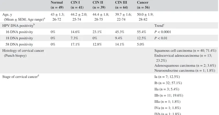
The clinicopathological characteristics of cervical lesions are presented in Table 1. A total of 249 samples was collected from normal (n = 49), CIN I (n = 41), CIN II (n = 39), CIN III (n = 64), and cervical cancer (n = 56) groups. Mean ages of the normal, CIN I, CIN II, CIN III, and cervical cancer groups were 43, 44.2, 44.4, 39.7, and 50.0, respectively. The proportions of squamous cell carcinoma and adenocar-cinoma among the cervical cancers were 71.4% and 23.2%, respectively, similar to those found generally (squamous cell carcinoma, 80%; adenocarcinoma, 20%).22 The frequencies
of HPV DNAs in the cervical cancers were, in descending order, HPV16, 18, and 58, while in the CINs it was HPV16, 58, and 18 (Table 1). The frequencies of HPV16 and 18 DNAs showed increasing trends as the severity of cervical
TABLE 1 Clinicopathological characteristics of normal, CIN I, CIN II, CIN III, and cancer groups Normal
(n = 49) CIN I (n = 41) CIN II (n = 39) CIN III (n = 64) Cancer (n = 56) Age, y
(Mean ± SEM; Age range)a 43 ± 1.3; 26‐72 44.2 ± 2.0; 25‐74 44.4 ± 1.8; 28‐75 39.7 ± 1.6; 22‐74 50.0 ± 1.9; 28‐82
HPV DNA positivityb Trendc
16 DNA positivity 0% 14.6% 23.1% 45.3% 55.4% P < 0.0001
18 DNA positivity 0% 7.3% 0% 9.4% 12.5% P < 0.01
58 DNA positivity 0% 17.1% 12.8% 14.1% 5.0%
Histology of cervical cancer
(Punch biopsy) Squamous cell carcinoma (n = 40; 71.4%)Endocervical adenocarcinoma (n = 13;
23.2%)
Adenosquamous carcinoma (n = 2; 3.6%) Neuroendocrine carcinoma (n = 1; 1.8%)
Stage of cervical cancerd Ia (n = 7; 12.5%)
Ib (n = 32; 57.1%) IIa (n = 3; 5.4%) IIb (n = 11; 19.6%) IIIa (n = 1; 1.8%) IVa (n = 1; 1.8%) IVb (n = 1; 1.8%)
aMean age of cancer group was higher than that of normal, CIN I, CIN II, or CIN III group. Comparison of age between groups was calculated by Student’s t test: cancer
vs normal, P < 0.01; cancer vs CIN I, P < 0.05; cancer vs CIN II, P < 0.05; cancer vs CIN III, P < 0.0001.
bHPV DNA positivity = number of HPV DNA presence sample × 100/total sample. cTrends of HPV DNA positivity were analyzed by chi‐square for trend test. dStage of cervical cancer was classified by FIGO clinical staging system.
lesion increased (Table 1). No HPV DNA positives were found in normal group. HPV16, 18, and 58 DNA positives in CIN I and CIN II group were 14.6%, 7.3%, and 17.1% and 23.1%, 0%, and 12.8%, respectively, while those in CIN III and cervical cancer group were 45.3%, 9.4%, and 14.1% and 55.4%, 12.5%, and 5%, respectively.
3.2
|
Comparison of seroprevalences of
antibodies to nine HPV antigens in normal,
CIN I, CIN II, CIN III, and cervical
cancer groups
To compare the prevalences of serum antibodies against the nine HPV antigens in each cervical lesion group, antibody levels were measured by ELISA (Figure S1). The linearities and reproducibilities of the ELISAs were found to be excel-lent (Figure S2 and Table S1).
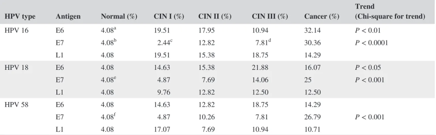
As shown in Table 2, the frequencies of the nine antibod-ies in all the cervical lesions (CIN I, CIN II, CIN III, and cancer) were generally higher than in the normal group. Antibodies against HPV16, 18, and 58 E7 antigens tended to increase in frequency with increasing stage of cervical lesion (P < 0.001 for each antibody; Table 2). Antibodies to HPV16 and HPV18 E6 also tended to be more common with increas-ing stage of cervical lesion, but the trends were weaker than for the antibodies against HPV16, 18, and 58 E7 (anti‐HPV16 E6, P < 0.01; anti‐HPV18 E6, P < 0.05; Table 2).
Serum anti‐HPV16 E6 and E7, anti‐HPV18 E6 and E7, and anti‐HPV58 E7 antibodies were more common in cervical
cancer than in the CIN stages, whereas the frequencies of anti‐HPV16 L1, anti‐HPV18 L1, and anti‐HPV58 E6 and L1 antibodies differed little between cervical cancer and CIN stages (Table 2). Serum antibody frequencies against HPV16, 18, and 58 L1 increased from the CIN I stage and tended to be maintained up to the cancer stage itself (Table 2).
In conclusion, the seroprevalences of antibodies against E7 antigens (of HPV16, HPV18, and HPV58) appear to be the best indicators of the severity of cervical lesions.
3.3
|
Seroprevalences of antibodies
against the nine HPV antigens as a function of
HPV DNA prevalence
The question whether the seroprevalences of antibodies against HPV antigens are influenced by the status of HPV infection was investigated (Table 3). There were no major changes in antibody prevalence associated with HPV DNA prevalence. However, the frequencies of antibodies against HPV16 E6 and L1 were higher in HPV16 DNA‐positive cervical cancers than in HPV16 DNA‐negative ones. Thus, there is little cor-relation between anti‐HPV antibody seroprevalence and HPV DNA positivity, except in the case of HPV16 DNA.
3.4
|
Correlations between antibodies
against the nine HPV antigens
The correlations between antibodies against the nine HPV antigen in normal, CIN I, CIN II, CIN III, and cervical cancer
TABLE 2 Comparison of seroprevalence in the level of antibodies against HPV16/18/58 E6, E7, and L1 antigen in normal, CIN I, CIN II, CIN III, and cervical cancer groups
HPV type Antigen Normal (%) CIN I (%) CIN II (%) CIN III (%) Cancer (%) Trend (Chi‐square for trend)
HPV 16 E6 4.08a 19.51 17.95 10.94 32.14 P < 0.01 E7 4.08b 2.44c 12.82 7.81d 30.36 P < 0.0001 L1 4.08 19.51 15.38 18.75 14.29 HPV 18 E6 4.08 14.63 15.38 21.88 16.07 P < 0.05 E7 4.08e 4.87 7.69 14.06 25 P < 0.001 L1 4.08 9.76 12.82 12.50 12.50 HPV 58 E6 4.08 14.63 12.82 18.75 14.29 E7 4.08f 4.87 10.26 7.81 26.79 P < 0.001 L1 4.08 17.07 7.69 10.94 10.71
Cutoff of the seroprevalence was set at the 95th percentile of normal group. Fisher’s exact test was used to compare the seroprevalence of the anti‐HPV antigen IgGs between groups. P value was adjusted by Bonferroni correction. Chi‐square for trend was used to evaluate seroprevalence trends of the IgGs with increasing stage of cervical lesions. P < 0.05 was considered statistically significant.
aNormal vs Cancer; P < 0.01 (Fisher’s exact test with Bonferroni correction). bNormal vs Cancer; P < 0.01 (Fisher’s exact test with Bonferroni correction). cCIN I vs Cancer; P < 0.01 (Fisher’s exact test with Bonferroni correction). dCIN III vs Cancer; P < 0.05 (Fisher’s exact test with Bonferroni correction). eNormal vs Cancer; P < 0.05 (Fisher’s exact test with Bonferroni correction). fNormal vs Cancer; P < 0.05 (Fisher’s exact test with Bonferroni correction).
TABLE 3
Seroprevalence of HPV DNA‐negative and HPV DNA‐positive individuals to nine types of HPV antigens Normal (n =
49) (%) CIN I (n = 41) (%) CIN II (n = 39) (%) CIN III (n = 64) (%) Cancer (n = 56) (%) HPV16 HPV16 DNA Negative (n = 35) HPV16 DNA Positive (n = 6) Total (n = 41) HPV16 DNA Negative (n = 30) HPV16 DNA Positive (n = 9) Total (n = 39) HPV16 DNA Negative (n = 35) HPV16 DNA Positive (n = 29) Total (n = 64) HPV16 DNA Negative (n = 25) HPV16 DNA Positive (n = 31) Total (n = 56) E6 4.1 22.9 0 19.5 20 11.1 18.0 11.4 10.3 10.9 12 48.4 a 32.2 E7 4.1 2.9 0 2.4 16.7 0 12.8 8.57 6.9 7.8 32 29.0 30.4 L1 4.1 20.0 16.7 19.5 10 33.3 15.4 14.29 24.1 18.7 0 25.8 b 14.3 HPV18 HPV18 DNA Negative (n = 38) HPV18 DNA Positive (n = 3) Total (n = 41) HPV18 DNA Negative (n = 39) HPV18 DNA Positive (n = 0) Total (n = 39) HPV18 DNA Negative (n = 58) HPV18 DNA Positive (n = 6) Total (n = 64) HPV18 DNA Negative (n = 49) HPV18 DNA Positive (n = 7) Total (n = 56) E6 4.1 15.8 0 14.6 15.4 0 15.4 20.7 33.3 21.8 18.4 0 16.1 E7 4.1 5.3 0 4.9 7.7 0 7.7 15.5 0 14.1 20.4 57.1 25 L1 4.1 10.5 0 9.8 12.8 0 12.8 13.8 0 12.5 14.3 0 12.5 HPV58 HPV58 DNA Negative (n = 34) HPV58 DNA Positive (n = 7) Total (n = 41) HPV58 DNA Negative (n = 34) HPV58 DNA Positive (n = 5) Total (n = 39) HPV58 DNA Negative (n = 55) HPV58 DNA Positive (n = 9) Total (n = 64) HPV58 DNA Negative (n = 53) HPV58 DNA Positive (n = 3) Total (n = 56) E6 4.1 14.7 14.3 14.6 14.7 0 12.82 18.2 22.2 18.75 13.21 33.3 14.3 E7 4.1 5.9 0 4.9 11.8 0 10.3 5.5 22.2 7.8 28.3 0 26.8 L1 4.1 20.6 0 17.1 5.8 20 7.5 10.9 11.1 11 11.3 0 10.7
Cutoff of seroprevalence was set at the 95th percentile of normal group.
P value was analyzed by the Fisher’s exact test to compare the seroprevalence of the IgGs between groups. Bolds are significant
difference between DNA
negative and positive. aDNA positive vs DNA negative (anti‐HPV16 E6 antibody seroprevalence in cancer group):
P
<
0.01, Fisher’s exact test was applied.
bDNA positive vs DNA negative (anti‐HPV16 L1 antibody seroprevalence in cancer group):
P
<
are presented in Table 4. R > 0.8 was considered a strong positive correlation. There were strong positive correlations between anti‐HPV16 E7 and anti‐HPV58 E7 antibodies, and between anti‐HPV18 E6 and anti‐HPV58 E6 antibodies in the normal, CIN III, and cancer groups (Table 4). A corre-lation between HPV18 E6 and HPV58 E6 was also found in the CIN I group. Thus, sera reactive with HPV16 E7 and HPV18 E6 tended to react with HPV58 E7 and HPV58 E6, respectively. We suggest that immune responses to HPV58 should be noted together with those to HPV16 and 18, when examining the development of cervical cancer.
4
|
DISCUSSION
4.1
|
Practical use of ELISA‐based serology
assay
In summary, our results imply that the seroprevalence of anti‐HPV E7 may be the best indicator of the severity of cervical lesions: The seroprevalences of antibodies against HPV16, 18, and 58 E7 appeared to be 30%, 25%, and 27% in cervical cancer group, respectively (Table 2). Meanwhile, current HPV DNA testing (Hybrid Capture II) provides over 90% sensitivity for detecting CIN II or worse (CIN II+).23
Therefore, it is thought that the sensitivities of the ELISA‐ based serology assays are too low to consider practical use of primary screening of cervical lesions.
4.2
|
Correlation between
expression of HPV antigens and prevalence of
anti‐HPV antibodies
When HPV infection in the cervix persists, integration of HPV DNA into host chromosomes can occur, and integration rates increase with stage of cervical lesion.24-26 As a result,
the expression of E6 and E7 proteins increases with stage of cervical lesion.25,26 In this study, the prevalence of serum
antibodies against HPV E6 and HPV E7 oncoproteins also increased with increasing stage of cervical lesion (Table 2). On the other hand, the expression of L1 (HPV capsid pro-tein), unlike that of E6 and E7 antigens, is known to increase preferentially in CIN I stage and to decline with increasing severity of the cervical lesions,26 and we found, in agreement
with this, that the prevalence of anti‐HPV L1 antibodies in-creased in CIN I and did not increase further with stage of cervical lesion (Table 2). Therefore, it seems that the limited expression of L1 protein in high‐grade CIN and cervical can-cer limits the immune response to it.
4.3
|
Comparison with previous studies
Overall, as shown in Table S2, our findings for anti‐HPV E6, E7, and L1 antibodies are consistent with previous
observations in terms of increased seroprevalence in cervi-cal lesions.27-34 Our results also show that the frequencies of
antibodies against HPV E6 and E7 (in types 16 and 18) are higher in cervical cancer than in CIN stages. Similar trends in the seroprevalence of antibodies against HPV E6 and E7 antigen in Korean women have been reported previously.35
Meanwhile, we noted little difference in the frequencies of anti‐HPV16 or 18 L1 antibodies between CIN stages and cervical cancer proper (Table 2 and Table S2). In contrast, an increased prevalence of anti‐HPV16 L1 antibody in cervical cancer compared to CIN stages was found in Mexico.36
4.4
|
Correlation between HPV DNA
prevalence and anti‐HPV antibody prevalence
It is thought that persistent infection with HPV can induce the production of antibodies that eventually lead to serocon-version.37 In this study, we enquired whether the presence
of antibodies against antigens of a given type of HPV was associated with the retention of HPV DNA corresponding to that HPV type. Overall, the presence of HPV DNA was not associated with increased seroprevalence, except in the case of HPV16 DNA‐positive cervical cancer where higher anti‐HPV16 E6 and L1 antibody frequencies were found in HPV16 DNA‐positive cervical cancer than in HPV16 DNA‐ negative cervical cancer (Table 3). Jean‐Damien et al38
simi-larly found that HPV16 DNA‐positive individuals had higher levels of anti‐HPV16 E6 and anti‐HPV16 L1 antibodies than HPV16 DNA‐negative individuals, and Chee et al35 found a
similar prevalence trend in HPV16 DNA‐positive individu-als. However, we did not observe the same correlation in HPV16 DNA‐positive CIN III (Table 3).
4.5
|
Correlation between antibodies against
different HPV antigens
We found a strong positive correlation between anti‐ HPV16 E7 and anti‐HPV58 E7 antibodies, and between anti‐HPV18 E6 and anti‐HPV58 E6 antibodies (Table 4). There is considerable amino acid sequence conservation within the same species of HPV, and this allows cross‐reac-tivity between anti‐HPV antibodies.39,40 HPV 16 and 58
be-long to the A9 species while HPV18 bebe-longs to A7,34,35 and
there is also some amino acid sequence conservation be-tween species. Previous research in Algeria and South India found a strong correlation between the seroprevalences of anti‐HPV16 E7 and anti‐HPV58 E7 antibodies.38 This is
consistent with our findings, but a correlation between anti‐ HPV18 E6 and anti‐HPV58 E6 antibodies was also noted,38
and these do not accord with our findings. This discrepancy may be due to differences in major histocompatibility com-plex (MHC) restriction of the production of antibodies in different ethnic groups.
TABLE 4 Correlation between the seroprevalences of antibodies against nine types of HPV antigens HPV16 E6 HPV16 E7 HPV16 L1 HPV18 E6 HPV18 E7 HPV18 L1 HPV58 E6 HPV58 E7 HPV58 L1 Normal HPV16 E6 0.38 −0.28 0.28 0.11 0.09 0.28 0.40 −0.14 HPV16 E7 −0.06 0.69 0.75 0.31 0.66 0.81 0.06 HPV16 L1 −0.09 0.02 0.26 0.01 0.05 0.64 HPV18 E6 0.55 0.27 0.86 0.66 0.00 HPV18 E7 0.26 0.41 0.62 0.29 HPV18 L1 0.34 0.39 0.35 HPV58 E6 0.65 −0.02 HPV58 E7 0.10 HPV58 L1 HPV16 E6 HPV16 E7 HPV16 L1 HPV18 E6 HPV18 E7 HPV18 L1 HPV58 E6 HPV58 E7 HPV58 L1 CIN I HPV16 E6 0.53 0.43 0.74 0.21 0.07 0.75 0.52 0.13 HPV16 E7 0.47 0.61 0.58 0.14 0.63 0.66 0.11 HPV16 L1 0.60 0.37 0.30 0.59 0.34 0.53 HPV18 E6 0.30 0.18 0.97 0.63 0.36 HPV18 E7 0.11 0.34 0.57 0.14 HPV18 L1 0.16 −0.06 0.36 HPV58 E6 0.64 0.24 HPV58 E7 0.09 HPV58 L1 HPV16 E6 HPV16 E7 HPV16 L1 HPV18 E6 HPV18 E7 HPV18 L1 HPV58 E6 HPV58 E7 HPV58 L1 CIN II HPV16 E6 0.42 −0.07 0.46 0.25 0.12 0.45 0.36 0.03 HPV16 E7 −0.21 0.44 0.73 0.35 0.55 0.66 −0.14 HPV16 L1 −0.08 −0.13 −0.27 −0.12 0.04 0.57 HPV18 E6 0.44 0.23 0.74 0.45 0.07 HPV18 E7 0.19 0.56 0.54 0.05 HPV18 L1 0.13 0.21 −0.28 HPV58 E6 0.53 0.04 HPV58 E7 0.12 HPV58 L1 HPV16 E6 HPV16 E7 HPV16 L1 HPV18 E6 HPV18 E7 HPV18 L1 HPV58 E6 HPV58 E7 HPV58 L1 CIN III HPV16 E6 0.49 −0.09 0.60 0.36 0.17 0.47 0.44 −0.04 HPV16 E7 0.15 0.71 0.77 0.03 0.64 0.81 −0.03 HPV16 L1 0.08 0.16 0.42 0.16 0.14 0.61 HPV18 E6 0.40 0.13 0.93 0.74 0.00 HPV18 E7 0.00 0.33 0.62 0.14 HPV18 L1 0.13 0.03 0.23 HPV58 E6 0.72 0.04 HPV58 E7 0.12 HPV58 L1 (Continues)
4.6
|
HPV type 58: a unique causative of
cervical cancer in East Asia
HPV58 DNA positivity is found in 3.3% of cervical cancers and is the fifth most frequent type worldwide.14 Meanwhile,
HPV58 is the third most common type found in cervical can-cers in East Asia, after HPV16 and HPV18.15,16 High rates of
HPV58 DNA prevalence of 26%, 8%, and 16% were reported in cancer patients in Shanghai (China), Japan, and Korea, respec-tively.17,41,42 Moreover, HPV58 was found in 17.2% of high‐
grade CIN groups in East Asia.43 In our study cases, HPV58
was more common than HPV18 in CIN stages (Table 1). These findings indicate that HPV58 is an important causative agent of cervical cancer in East Asia. Our results confirm that Korean women not only have high levels of HPV58 DNA positivity but also high levels of antibody seropositivities to HPV58 antigens in CINs (Tables 1 and 2). Therefore, we suggest that HPV58 must be taken into account in the prevention, treatment, and diagnosis of cervical cancer in the East Asian region.
4.7
|
Limitations of this study
In the present study, only three types of HPVs (HPV16, 18, and 58) were considered to investigate the anti‐HPV anti-body seroprevalences and HPV DNA positivities, and limited number of serum samples per group were used (n = 39‐64). Also, folding property or antigenicity of the HPV E6 or E7 produced in E. coli may be different from that of na-tive antigen is a considerable factor in interpretation of the seroprevalences.
4.8
|
Use of antibodies against HPV
antigens as biomarkers, and future directions
for their use
Antibodies against nine types of HPV antigens were as-sessed as biomarkers for diagnosing cervical lesions,
and we found that rates of detection of cervical lesions increased significantly when the seroprevalence factors were used in parallel (Table S3). When “any of nine anti-gens” was used as criterion, CIN II, CIN III, and cervical cancer were detected (CIN II, 61.5%; CIN III, 51.6%; cer-vical cancer, 75%). However, 26.5% of normal individu-als individu-also registered as seropositive (Table S3). Meanwhile, considerable parts of cervical lesions (CIN II, 38.5%; CIN III, 48.4%; cervical cancer, 25%) were not detectable when screening protocol using “any of nine antigens” was applied. It seems that the serology assays have lim-ited usefulness as primary screening system. In the serial assay strategy, the detection rate was zero in all groups when the “all of nine antigens” combination strategy was used (Table S3). In the “all of E7 (HPVs16, 18, and 58)” combination strategy, no false positives were found in the normal or CIN I group, and seroprevalence displayed an increasing trend as the severity of cervical lesion in-creased. However, the rates of detection of CIN II, CIN III, and cervical cancer were too low for this strategy to be applied in practice (CIN II, 2.6%; CIN III, 3.6; cervical cancer, 12.5%).
All in all, the antibodies against E7 proteins appeared to have the highest potential of the anti‐HPV antibodies tested as markers for detecting cervical lesions (Table 2), and fur-ther studies are needed to identify peptide regions of the E7 proteins whose antibody levels reflect most accurately the se-verity of cervical lesions.
We believe that our results will contribute to global pro-filing of the prevalence of serum antibodies against HPV an-tigens. The accumulated results of such efforts are expected to provide the fundamental basis for monitoring and treating cervical lesions.
CONFLICT OF INTEREST
None of the authors have conflict of interests to disclose.
HPV16 E6 HPV16 E7 HPV16 L1 HPV18 E6 HPV18 E7 HPV18 L1 HPV58 E6 HPV58 E7 HPV58 L1 Cancer HPV16 E6 0.30 0.11 0.05 0.12 −0.11 0.09 0.35 0.15 HPV16 E7 0.01 0.30 0.58 −0.20 0.29 0.80 0.01 HPV16 L1 −0.44 −0.07 −0.22 −0.48 0.04 0.55 HPV18 E6 0.28 0.23 0.92 0.34 −0.21 HPV18 E7 0.01 0.29 0.38 −0.09 HPV18 L1 0.21 −0.08 −0.20 HPV58 E6 0.32 −0.21 HPV58 E7 −0.01 HPV58 L1
Correlations were analyzed by Spearman correlation. Bolds are cases when the R value is over 0.8 (high positive correlation) between two types of antibodies.
ORCID
Hong‐Jin Kim http://orcid.org/0000-0002-2539-7134 REFERENCES
1. Walboomers JM, Jacobs MV, Manos MM, et al. Human papillo-mavirus is a necessary cause of invasive cervical cancer world-wide. J Pathol. 1999;189:12‐19.
2. de Villiers EM. Cross‐roads in the classification of papillomavi-ruses. Virology. 2013;445:2‐10.
3. Ghittoni R, Accardi R, Chiocca S, et al. Role of human papilloma-viruses in carcinogenesis. Ecancermedicalscience. 2015;9:526. 4. Burd EM. Human papillomavirus and cervical cancer. Clin
Microbiol Reviews. 2003;16:1‐17.
5. Snijders PJ, Steenbergen RD, Heideman DA, et al. HPV‐medi-ated cervical carcinogenesis: concepts and clinical implications. J Pathol. 2006;208:152‐164.
6. Bosch FX, Burchell AN, Schiffman M, et al. Epidemiology and natural history of human papillomavirus infections and type‐spe-cific implications in cervical neoplasia. Vaccine. 2008;26(Suppl 10):K1–K16.
7. Lehtinen M, Pawlita M, Zumbach K, et al. Evaluation of antibody response to human papillomavirus early proteins in women in whom cervical cancer developed 1 to 20 years later. Am J Obstet
Gynecol. 2003;188:49‐55.
8. Gutierrez‐Xicotencatl L, Salazar‐Pina DA, Pedroza‐Saavedra A, et al. Humoral immune response against human papillomavirus as source of biomarkers for the prediction and detection of cervical cancer. Viral Immunol. 2016;29:83‐94.
9. Stanley M. Antibody reactivity to HPV E6 and E7 oncoproteins and early diagnosis of invasive cervical cancer. Am J Obstet
Gynecol. 2003;188:3‐4.
10. Ewaisha R, Panicker G, Maranian P, et al. Serum immune pro-filing for early detection of cervical disease. Theranostics. 2017;7:3814‐3823.
11. Luevano M, Bernard HU, Barrera‐Saldana HA, et al. High‐ throughput profiling of the humoral immune responses against thirteen human papillomavirus types by proteome microarrays.
Virology. 2010;405:31‐40.
12. Orozco‐Colin A, Carrillo‐Garcia A, Mendez‐Tenorio A, et al. Geographical variation in human papillomavirus prevalence in Mexican women with normal cytology. Int J Infect Dis. 2010;14:e1082–e1087.
13. Wardak S. Human papillomavirus (HPV) and cervical cancer.
Med Dosw Mikrobiol. 2016;68:73‐84.
14. Smith JS, Lindsay L, Hoots B, et al. Human papillomavirus type distribution in invasive cervical cancer and high‐grade cervical lesions: a meta‐analysis update. Int J Cancer. 2007;121:621‐632. 15. Chan PK. Human papillomavirus type 58: the unique role in
cer-vical cancers in East Asia. Cell Biosci. 2012;2:17.
16. Lin QQ, Yu SZ, Qu W, et al. Human papillomavirus types 52 and 58. Int J Cancer. 1998;75:484‐485.
17. Hwang T. Detection and typing of human papillomavirus DNA by PCR using consensus primers in various cervical lesions of Korean women. J Korean Med Science. 1999;14:593‐599. 18. de Sanjose S, Quint WG, Alemany L, et al. Human
papilloma-virus genotype attribution in invasive cervical cancer: a ret-rospective cross‐sectional worldwide study. Lancet Oncol. 2010;11:1048‐1056.
19. Karlsen F, Kalantari M, Jenkins A, et al. Use of multiple PCR primer sets for optimal detection of human papillomavirus. J Clin
Microbiol. 1996;34:2095‐2100.
20. Xu ML, Kim SC, Kim HJ, et al. Two‐step chromatographic purifi-cation of glutathione S‐transferase‐tagged human papillomavirus type 16 E6 protein and its application for serology. Protein Expr
Purif. 2017;132:19‐26.
21. Kim HJ, Jin Y, Kim H‐J. The concentration of carbon source in the medium affects the quality of virus‐like particles of human papillomavirus type 16 produced in Saccharomyces cerevisiae.
PLoS One. 2014;9:e94467.
22. Lee MY, Shen MR. Epithelial‐mesenchymal transition in cervical carcinoma. Am J Transl Res. 2012;4:1‐13.
23. Soderlund‐Strand A, Rymark P, Andersson P, et al. Comparison between the hybrid capture II test and a PCR‐based human pap-illomavirus detection method for diagnosis and posttreatment follow‐up of cervical intraepithelial neoplasia. J Clin Microbiol. 2005;43:3260‐3266.
24. Li W, Wang W, Si M, et al. The physical state of HPV16 infection and its clinical significance in cancer precursor lesion and cervi-cal carcinoma. J Cancer Res Clin Oncol. 2008;134:1355‐1361. 25. Narisawa‐Saito M, Kiyono T. Basic mechanisms of high‐risk
human papillomavirus‐induced carcinogenesis: roles of E6 and E7 proteins. Cancer Sci. 2007;98:1505‐1511.
26. Doorbar J, Quint W, Banks L, et al. The biology and life‐cycle of human papillomaviruses. Vaccine. 2012;30(Suppl 5):F55–F70. 27. Jeong NH, Lee NW, Woo MK, et al. Serologic response to human
papillomavirus type 16 virus‐like particles in Korean women with cervical precancerous and cancerous lesions. Arch Pharm Res. 2009;32:383‐389.
28. Matsumoto K, Yoshikawa H, Yasugi T, et al. IgG antibodies to human papillomavirus 16, 52, 58, and 6 L1 capsids: Case‐control study of cervical intraepithelial neoplasia in Japan. J Med Virol. 2003;69:441‐446.
29. Matsumoto K, Yoshikawa H, Taketani Y, et al. Antibodies to human papillomavirus 16, 18, 58, and 6b major capsid proteins among Japanese females. Jpn J Cancer Res. 1997;88:369‐375. 30. Sun YP, Eluf‐Neto J, Bosch FX, et al. Serum antibodies to human
papillomavirus 16 proteins in women from Brazil with invasive cervical carcinoma. Cancer Epidem Biomar. 1999;8:935‐940. 31. Zumbach K, Hoffmann M, Kahn T, et al. Antibodies against
on-coproteins E6 and E7 of human papillomavirus types 16 and 18 in patients with head‐and‐neck squamous‐cell carcinoma. Int J
Cancer. 2000;85:815‐818.
32. Meschede W, Zumbach K, Braspenning J, et al. Antibodies against early proteins of human papillomaviruses as diagnostic markers for invasive cervical cancer. J Clin Microbiol. 1998;36:475‐480. 33. Paez CG, Yaegashi N, Sato S, et al. Prevalence of serum Igg
antibodies for the E7 and L2 proteins of human papillomavirus type‐16 in cervical‐cancer patients and controls. Tohoku J Exp
Med. 1993;170:113‐121.
34. Combita AL, Bravo MM, Touze A, et al. Serologic response to human oncogenic papillomavirus types 16, 18, 31, 33, 39, 58 and 59 virus‐like particles in Colombian women with invasive cervi-cal cancer. Int J Cancer. 2002;97:796‐803.
35. Chee YH, Namkoong SE, Kim DH, et al. Immunologic diagnosis and monitoring of cervical cancers using in vitro translated HPV proteins. Gynecol Oncol. 1995;57:226‐231.
36. Salazar‐Pina DA, Pedroza‐Saavedra A, Cruz‐Valdez A, et al. Validation of serological antibody profiles against human
papillomavirus Type 16 antigens as markers for early detection of cervical cancer. Medicine. 2016;95:e2769.
37. Clifford GM, Shin HR, Oh JK, et al. Serologic response to onco-genic human papillomavirus types in male and female university students in Busan, South Korea. Cancer Epidemiol Biomarkers
Prev. 2007;16:1874‐1879.
38. Combes JD, Pawlita M, Waterboer T, et al. Antibodies against high‐risk human papillomavirus proteins as markers for invasive cervical cancer. Int J Cancer. 2014;135:2453‐2461.
39. Park JS, Park DC, Kim CJ, et al. HPV‐16‐related proteins as the serologic markers in cervical neoplasia. Gynecol Oncol. 1998;69:47‐55.
40. Wang SS, Schiffman M, Shields TS, et al. Seroprevalence of human papillomavirus‐16, ‐18, ‐31, and ‐45 in a population‐ based cohort of 10000 women in Costa Rica. Br J Cancer. 2003;89:1248‐1254.
41. Huang S, Afonina I, Miller BA, et al. Human papillomavirus types 52 and 58 are prevalent in cervical cancers from Chinese women. Int J Cancer. 1997;70:408‐411.
42. Asato T, Maehama T, Nagai Y, et al. A large case‐control study of cervical cancer risk associated with human papillomavirus
infection in Japan, by nucleotide sequencing‐based genotyping. J Infect Diss. 2004;189:1829‐1832.
43. Chan PK, Cheung TH, Li WH, et al. Attribution of human papil-lomavirus types to cervical intraepithelial neoplasia and invasive cancers in Southern China. Int J Cancer. 2012;131:692‐705. SUPPORTING INFORMATION
Additional supporting information may be found online in the Supporting Information section at the end of the article.
How to cite this article: Jin Y, Choi JW, Kim HJ, et al.
Profiling of serum antibodies against human
papillomavirus antigens in Korean women with cervical intraepithelial neoplasia and cervical cancer. Cancer Med. 2018;7:5655–5664. https://doi.org/10.1002/ cam4.1810