An Overview of Ophthalmologic Survey Methodology in the 2008- 2015 Korean National Health and Nutrition Examination Surveys
9
0
0
전체 글
(2) Korean J Ophthalmol Vol.29, No.6, 2015. regularly by the division of chronic disease Surveillance of the Korea Centers for Disease Control and Prevention in the Ministry of Health and Welfare, to examine the health, physical, and nutritional status of the general population of South Korea. Since 2008, the first year of ophthalmic examinations, there have been several changes in the ophthalmic examination methodologies and questionnaires. Although many research articles about ocular disorders, including visual impairment [1-6], refractive errors [7-12], strabismus [1], blepharoptosis [13], cataract [14-18], pterygium [19], diabetic retinopathy (DR) [20-27], age-related macular degeneration (ARMD) [28-36], glaucoma [37-47], and dry eye disease (DED) [48-50] have been published based on the results of the KNHANES, there have not been any comprehensive overviews of the methodological changes. Therefore, in this article, we review the ophthalmic examination methodologies and their overall changes throughout the history of the KNHANES.. KNHANES Overview The Korea Centers for Disease Control and Prevention conducted the KNHANES series (I, II, and III) in 1998, 2001, and 2005, respectively, to examine the general health and nutritional status of Koreans. Starting with the KNHANES IV (2007-2009), V (2010-2012), and VI (20132015), however, the survey became an annual project. The study methodology involved stratified multistage cluster-sampling to prevent subject omission or overlap. The rolling-sampling method ensured the representativeness of each annual survey of the overall Korean population, which allowed results to be merged between surveys. Specifically, the primary sample units (PSUs) were selected from a sampling frame of all census blocks or resident registration addresses. Each PSU consisted of approximately 50 to 60 households. Following PSU selection, all dwelling units in the PSU were listed, and 20 households were selected for household screening through field surveys. The final stage of selection occurred in the individual households, where all members older than one year of age were selected to participate. Approximately 10,000 individuals were sampled annually among all 192 PSUs. The target overall KNHANES response rate was 75% [51]. From July 2008, ophthalmologic interviews and examinations have been conducted. All examination and health interviews. 360. were conducted by trained teams in mobile centers that traveled to each survey location, while nutrition surveys were performed in individual households. These mobile centers provided a standardized environment and equipment. The Korea Centers for Disease Control and Prevention and the Korean Ophthalmological Society conducted team education and training programs twice yearly. The educational information included the overall purpose of epidemiological studies, cautions, machine operation, and diagnosis and classification of major eye disorders to be investigated. The quality of the ophthalmic survey was verified by the Epidemiologic Survey Committee of the Korean Ophthalmological Society. The ophthalmologists or ophthalmologic residents participating in this survey were required to complete a training course and undergo supervised practice before working in the actual survey field. In the KNHANES IV-V (2008-2012), a total of 37,982 (17,040 men and 20,942 women) participants were received an eye examinations.. Ophthalmic Examinations According to Age Group Examination procedures were stratified according to age group. Participants aged three to four years underwent testing only for strabismus and blepharoptosis. Autorefraction, visual acuity (VA) measurement, and testing for strabismus and blepharoptosis were performed among participants ranging in age from five to 18 years. Participants aged 19 years or older underwent full ocular examinations, including autorefraction and VA measurement, testing for strabismus and blepharoptosis, slit lamp examinations, intraocular pressure (IOP) measurement, and fundus photography. IOP was measured with a Goldmann applanation tonometer. For participants meeting the glaucoma suspicion criteria, frequency doubling technology perimetry was carried out. Pharmacological pupil dilatation was performed for participants with a history of diabetes mellitus (DM) or random blood glucose level of 200 mg/dL or higher and/or fundus photographs suggestive of DR and/or difficulty obtaining fundus photographs due to media opacity (Fig. 1) [1]. All procedures described above except for fundus photography were performed before pupil dilatation..
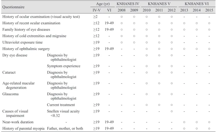
(3) KC Yoon, et al. Overview of KNHANES Ophthalmologic Methodologies. Ophthalmologic interview Correction of visual acuity Visual acuity <0.8. Visual acuity Auto-refraction. Testing for strabismus & ptosis For 19 years or older participants. For 5 years or older participants For 3 years or older participants. Slit lamp examination (cataract, pterygium, depth of anterior chamber) IOP measurement ① IOP ≥22 mmHg or ② Suspected glaucoma on Fundus photograph without pupil dilatation fundus examination. Visual field examination. ① Participants with diabetes mellitus or random blood sugar ≥200 mg or ② Suspected DR on fundus examination or ③ Difficulty in checking the fundus photograph due to media opacity. Fundus photograph after pupil dilatation. Fig. 1. Representative progression chart for the Korea National Health and Nutrition Examination Survey including ophthalmologic interview, visual acuity testing, slit lamp examination, IOP measurement, fundus photograph, and visual field examination. IOP = intraocular pressure; DR = diabetic retinopathy. From Yoon KC, et al. Korean J Ophthalmol 2011;25:421-33, according to the Creative Commons license [1].. Questionnaire and Ophthalmic Disease Examination Methods Ophthalmic questionnaire The ophthalmic questionnaires included history of ocular examinations; recent ocular examinations; family history of eye diseases; history of cold extremities and migraines; UV exposure time; ophthalmic surgery history; history of diagnosis by ophthalmologist including DED, cataract, ARMD, and glaucoma; symptoms of DED; current treatment for glaucoma; causes of visual impairment in cases of Snellen VA less than 0.32; near work duration; and parental history of myopia. The time points and age groups subjected to the questionnaire survey varied by year and are described in Table 1. Briefly, in KNHANES IV-V (2008-2012), ophthalmologic investigators queried participants about their history regarding ocular examination, cold extremities, migraines, and UV exposure time. The possible responses for UV exposure time included: “<5 hours” or “≥5 hours” in the KNHANES IV (2008-2009) and “<2 hours,” “two-five hours,” or “≥5 hours” in the KNHANES V (2010-2012). History of recent ocular examination and family history of eye disease were evaluated in KNHANES IV to VI (2008-2015), and history of ophthalmic surgery was investigated during KNHANES V-VI (2010-2015). In the KNHANES V (2010-2012), subjects were asked about a history of diagnosis of DED, cataract, ARMD, and glaucoma by ophthalmologists. To make data. collection more accurate, subjects were also asked about symptoms they had experienced related to DED such as dryness, foreign body sensation, or burning and about the current status of medical treatment for glaucoma in the KNHANES V (2010-2012). In the cases of Snellen VA less than 0.32, interviews were conducted about the causes of visual impairment in the last year of KNHANES V (2012). Questions about near-work duration and parental history of myopia were added to the questionnaire in the KNHANES VI (2013-2015). Ophthalmic disease examination methods A list of all examination procedures according to age group and test period is shown in Table 2. Uncorrected VA and/or best-corrected distance VA were measured at a distance of 4 meters using an international standard vision chart based on the Snellen scale (Jin’s vision chart, Seoul, Korea) [52]. Participant VA was measured in each eye, right side followed by left side. Each participant was asked to read numbers in the 0.2 line of the VA chart and to proceed to the next line if he or she correctly read at least three of the five letters. Participant VA was defined as the line with the smallest numbers in which the participant accurately read more than three characters. For those participants who presented with VA score lower than 0.8, corrected VA was measured using autorefraction. Automated refraction was performed using an autorefractive keratometer (KR8800; Topcon, Tokyo, Japan), followed by VA retesting using a pinhole in patients with Snellen VA lower than 0.8. Spherical equivalent refractive error was calculated as sphere +1 / 2 cylinder. These examinations were carried out throughout the KNHANES IV-VI (2008-2015). Strabismus testing conducted from 2008 to 2011 included the cover-uncover test, prism and alternative cover test, and/or Krimsky test. Strabismus was defined as a manifest or latent ocular deviation at distance or near fixation with or without spectacle correction, esodeviation of 10 or more prism diopters, exodeviation of 15 or more prism diopters, or any vertical deviation. Blepharoptosis was determined by measuring the marginal reflex distance 1 in the KNHANES IV and V (20102015). Participants were positioned at physician eye level and instructed to look straight ahead and relax while focusing on a distant target. After shining a penlight into the participant’s eye, the distance from the corneal light reflex. 361.
(4) Korean J Ophthalmol Vol.29, No.6, 2015. Table 1. List of ophthalmologic questionnaires in KNHANES according to age group and test period Age (yr). Questionnaire. IV-V. KNHANES IV. KNHANES V. KNHANES VI. VI. 2008. 2009. 2010. 2011. 2012. 2013. 2014. 2015. History of ocular examination (visual acuity test). ≥2. -. ○. ○. ○. ○. ○. -. -. -. History of recent ocular examination. ≥12. 19-49. ○. ○. ○. ○. ○. ○. ○. ○. Family history of eye diseases. ≥12. 19-49. ○. ○. ○. ○. ○. ○. ○. ○. History of cold extremities and migraine. ≥12. -. ○. ○. ○. ○. ○. -. -. -. Ultraviolet exposure time. ≥19. -. ○. ○. ○. ○. ○. -. -. -. History of ophthalmic surgery. ≥19. 19-49. -. -. ○. ○. ○. ○. ○. ○. Dry eye disease. Diagnosis by ophthalmologist. ≥19. -. -. -. ○. ○. ○. -. -. -. Symptom experience. ≥19. -. -. -. ○. ○. ○. -. -. -. Cataract. Diagnosis by ophthalmologist. ≥19. -. -. -. ○. ○. ○. -. -. -. Age-related macular degeneration. Diagnosis by ophthalmologist. ≥19. -. -. -. ○. ○. ○. -. -. -. Glaucoma. Diagnosis by ophthalmologist. ≥19. -. -. -. ○. ○. ○. -. -. -. Current treatment. ≥19. -. -. -. ○. ○. ○. -. -. -. Snellen visual acuity <0.32. ≥19. -. -. -. -. -. ○. -. -. -. ≥19. 19-49. -. -. -. -. -. ○. ○. ○. ≥19. 19-49. -. -. -. -. -. ○. ○. ○. Causes of visual impairment Near-work duration. -. History of parental myopia Father, mother, or both. KNHANES = the Korea National Health and Nutrition Examination Survey.. Table 2. List of examination methods and output indexes of prevalence of ophthalmic diseases in KNHANES according to age group and test period Examination method. Prevalence. Visual acuity test. Age (yr). KNHANES IV. KNHANES V. KNHANES VI. IV-V. VI. 2008. 2009. 2010. 2011. 2012. 2013. 2014. 2015. Visual impairment. ≥5. 19-49. ○. ○. ○. ○. ○. ○. ○. ○. Autorefractometer. Refractive errors. ≥5. 19-49. ○. ○. ○. ○. ○. ○. ○. ○. Strabismus test. Strabismus. ≥3. -. ○. ○. ○. ○. -. -. -. -. Blepharoptosis test. Blepharoptosis. ≥3. -. ○. ○. ○. ○. ○. -. -. -. Slit lamp biomicroscopy. Cataract. ≥19. -. ○. ○. ○. ○. ○. -. -. -. Pterygium. ≥19. -. ○. ○. ○. ○. -. -. -. -. *. Fundus photography. Diabetic retinopathy. ≥19. -. ○. ○. ○. ○. ○. -. -. -. Age-related macular degeneration. ≥19. -. ○. ○. ○. ○. ○. -. -. -. ≥19. -. ○. ○. ○. ○. ○†. -. -. -. -. 19-49. -. -. -. -. -. ○. ○. ○. Intraocular pressure, fundus Glaucoma examination, visual field test Hardy-Rand-Rittler test. Color vision deficiency. KNHANES = the Korea National Health and Nutrition Examination Survey. Without pharmacological pupil dilation in every diabetes mellitus participants; †All participants aged 40 years or more.. *. to the upper eyelid margin was measured in millimeters. Differential diagnosis of blepharoptosis was made with. 362. particular attention to pseudoptosis associated with eyebrow ptosis and dermatochalasis. Blepharoptosis was de-.
(5) KC Yoon, et al. Overview of KNHANES Ophthalmologic Methodologies. fined as a marginal reflex distance 1 of 2 mm or less. Investigators also conducted structured slit-lamp examinations (Haag-Streit model BQ-900; Haag-Streit AG, Koeniz, Switzerland) to test for diseases in the anterior segment of the eye, such as pterygium (2008-2011) and cataract (2008-2012), and to measure the IOP (2008-2012) and anterior chamber depth using the Van Herick method (2008-2012) [53]. Standardized Lens Opacities Classification System (LOCS) III photographic images were used to assess cataracts. Cataract was defined as nuclear (LOCS III score ≥4 for nuclear opalescence or nuclear color), cortical (LOCS III score ≥2 for cortical cataracts), posterior subcapsular (LOCS III score ≥2 for posterior subcapsular), or mixed (more than one type per eye) based on comparison with these standard photographs [54]. Pseudophakic and aphakic eyes were also documented. Pterygium was defined as a radially-oriented fibrovascular lesion crossing the nasal or temporal limbus. Grading was based on the visibility of the underlying episcleral blood vessels [55]. IOP was measured once in each eye from the right side to left side by a trained ophthalmologist with a Goldmann applanation tonometer (Haag-Streit AG). A digital nonmydriatic fundus camera (TRC-NW6S; Topcon) and Nikon D-80 digital camera (Nikon, Tokyo, Japan) were used to obtain digital fundus images throughout the KNHANES IV and V (2008-2012). Digital images were captured under physiological mydriasis in all participants 19 years of age or older. For each participant, one 45° nonmydriatic digital retinal image centered on the fovea was taken per eye (two images per person). Optic nerve configuration and any retinal pathologic findings were recorded. Patients were considered to have early ARMD if they demonstrated presence of soft indistinct drusen or reticular drusen or presence of hard or soft distinct drusen with pigmentary abnormalities (increased pigmentation or hypopigmentation of the retinal pigment epithelium) in the absence of signs of late ARMD. Late ARMD included the signs of wet ARMD or geographic atrophy. Wet ARMD was defined as retinal pigment epithelial detachment or serous detachment of the sensory retina, subretinal or sub-retinal pigment epithelium hemorrhages, and subretinal fibrous scars. Geographic atrophy was defined as a circular discrete area (175 microns in diameter or greater) of retinal depigmentation with visible choroidal vessels, in the absence of signs of wet ARMD [56]. In participants with a history of DM or random blood glucose level of 200. mg/dL or higher and/or suspicion of DR in nonmydriatic fundus photography findings, seven standard field photographs were obtained from each eye after pharmacological pupil dilatation, as per the Early Treatment for Diabetic Retinopathy Study protocol, throughout years 2008 to 2011 [57]. On the other hand, in the last year of the KNHANES V (2012), fundus photography was performed for every participant with DM without pharmacological pupil dilatation. DR was identified in the presence of any characteristic lesion determined by the Early Treatment for Diabetic Retinopathy Study severity scale: microaneurysms, hemorrhages, hard exudates, cotton wool spots, intraretinal microvascular abnormalities, venous beading, and retinal new vessels [58,59]. The prevalence of DR among individuals with DM was estimated. Each fundus image was graded twice. First, preliminary grading was conducted onsite by ophthalmologists or ophthalmologic residents trained by the Korean Ophthalmologic Society. Retinal specialists with expertise in DR grading and ARMD diagnosis then performed detailed final grading using protocols from the Early Treatment for Diabetic Retinopathy Study and International Age-related Maculopathy Epidemiological Study Group. In the KNHANES IV-V (2008-2012), automated visual field testing (Humphrey Matrix frequency-doubling perimeter; Carl Zeiss Meditec Inc., Dublin, CA, USA) with the N-30-1 screening program was performed on participants with any of the following five conditions: (1) elevated IOP (≥22 mmHg), (2) horizontal or vertical cup-to-disc ratio ≥0.5, (3) presence of optic disc hemorrhage, (4) presence of retinal nerve fiber layer defect, or (5) violation of the inferior-superior-nasal-temporal rule. Frequency doubling technology was repeated once if the initial results were deemed unreliable. Patients were considered to have primary open angle glaucoma if they met any one of the category I or II diagnostic criteria previously described [60]. In 2012, however, frequency doubling technology perimetry was conducted for all participants aged 40 years or older, regardless of glaucoma suspicion. The Hardy-Rand-Rittler pseudoisochromatic color vision test was performed throughout the KNHANES VI (20132015) [61]. There were six screening plates, four for protan-deutan deficiencies and two for tritan deficiencies. If all six boxes showed correct responses, the patient had normal color vision, and no more testing was conducted. If the fifth or sixth plates were not noted, the patient was. 363.
(6) Korean J Ophthalmol Vol.29, No.6, 2015. considered to have defective blue-yellow vision, and the examiner proceeded to show plates 21 to 24. If any of the boxes corresponding to plates 7 to 10 were not noted, the patient had defective red-green vision, and the examiner proceeded to show plates 11 to 20. After testing was complete, the total numbers in each column were summed in the spaces under the responses for plates 20 and 24. Participants were diagnosed as protan or deutan if the total numbers of marks in the protan or deutan columns, respectively, was greater than that in the opposite column. In cases of blue-yellow deficiency, participants were diagnosed as tritan or tetartan if the number of errors in the tritan or tetartan columns, respectively, was greater than that in the other column. Participants were diagnosed as having unclassified red-green or blue-yellow defects if the number of marks were the same in the protan and deutan or tritan and tetartan columns or if errors were made only in the screening plates. Color vision deficiency was graded as mild, medium, or strong, depending on whether the participants saw the symbols on the more saturated plates. There were 10 grading plates for protan/deutan defects: patients who made one or more errors in the two plates with the most saturated colors were graded as severe, and those who made an error in the next three most saturated plates were graded as medium. Those who made errors only in the five least-saturated plates were graded as mild. Similar interpretation was performed in the case of blue-yellow deficiency (Fig. 2).. Conclusion The KNHANES survey provides objective, standardized data on the prevalence of a wide range of diseases including major ophthalmic disorders, comorbidity, and risk factors in the noninstitutionalized population in Korea. The KNHANES data can be used to establish, develop, monitor, and evaluate national health programs and policies for eye diseases. However, because the survey components and methods varied partly by year, data users should be aware of changes in the detailed survey methods and questionnaires of concerned variables. The present report highlights the ophthalmologic methodology and the detailed changes in questionnaires and examination procedures according to survey periods of KNHANES (2008-2015). Therefore, this article can be used as a useful reference in. 364. Protan. Deutan. 11 B-Y 5 O,X defect 6 O,▼ If 5 or 6 are not checked, go to 21-24. 7 X,▶ R-G 8 O,▶ defect 9O 10 X If any of 7-10 are not checked, go to 11-20. 12 Mild R-G 13 defect 14. Total number of checks 1. Protan > Deutan 2. Protan < Deutan 3. Protan = Deutan -unclassified. 15 Medium 16 R-G 17 defect 18 Strong 19 R-G defect 20 Total Tritan. Extent of defect Last group of error occurs Tetartan. Medium 21 B-Y defect 22 Strong 23 B-Y defect 24 Total. Total number of checks 1. Tritan > Tetartan 2. Tritan < Tetartan 3. Tritan = Tetartan -unclassified. Fig. 2. Diagnostic flow diagram showing use of Hardy-Rand-Rittler pseudoisochromatic plates to detect, classify, and estimate the degree of defective color vision.. various types of research using KNHANES data in order to assess the prevalence and risk factors of ophthalmologic disorders.. Conflict of Interest No potential conflict of interest relevant to this article was reported.. Acknowledgements The authors thank the Korea Centers for Disease Control and Prevention, which performed the KNHANES.. References 1. Yoon KC, Mun GH, Kim SD, et al. Prevalence of eye diseases in South Korea: data from the Korea National Health and Nutrition Examination Survey 2008-2009. Korean J Ophthalmol 2011;25:421-33. 2. Park SH, Lee JS, Heo H, et al. A nationwide population-based study of low vision and blindness in South Korea. Invest Ophthalmol Vis Sci 2014;56:484-93. 3. Rim TH, Lee CS, Lee SC, et al. Influence of visual acuity on suicidal ideation, suicide attempts and depression in South Korea. Br J Ophthalmol 2015;99:1112-9. 4. Cho GE, Lim DH, Baek M, et al. Visual impairment of Ko-.
(7) KC Yoon, et al. Overview of KNHANES Ophthalmologic Methodologies. rean population: prevalence and impact on mental health. Invest Ophthalmol Vis Sci 2015;56:4375-81.. 17. Kim TN, Lee JE, Lee EJ, et al. Prevalence of and factors associated with lens opacities in a Korean adult population. 5. Park Y, Shin JA, Yang SW, et al. The relationship between. with and without diabetes: the 2008-2009 Korea National. visual impairment and health-related quality of life in Ko-. Health and Nutrition Examination Survey. PLoS One 2014;. rean adults: the Korea National Health and Nutrition Ex-. 9:e94189.. amination Survey (2008-2012). PLoS One 2015;10:e0132779.. 18. Rim TH, Kim MH, Kim WC, et al. Cataract subtype risk. 6. Rim TH, Nam JS, Choi M, et al. Prevalence and risk fac-. factors identified from the Korea National Health and Nu-. tors of visual impairment and blindness in Korea: the. trition Examination survey 2008-2010. BMC Ophthalmol. Fourth Korea National Health and Nutrition Examination Survey in 2008-2010. Acta Ophthalmol 2014;92:e317-25.. 2014;14:4. 19. Rim TH, Nam J, Kim EK, Kim TI. Risk factors associated. 7. Chon B, Qiu M, Lin SC. Myopia and glaucoma in the. with pterygium and its subtypes in Korea: the Korean Na-. South Korean population. Invest Ophthalmol Vis Sci 2013;. tional Health and Nutrition Examination Survey 2008-. 54:6570-7.. 2010. Cornea 2013;32:962-70.. 8. Choi JA, Han K, Park YM, Park CK. Age-related associa-. 20. Lee WJ, Sobrin L, Lee MJ, et al. The relationship between. tion of refractive error with intraocular pressure in the Ko-. diabetic retinopathy and diabetic nephropathy in a popula-. rea National Health and Nutrition Examination Survey.. tion-based study in Korea (KNHANES V-2, 3). Invest. PLoS One 2014;9:e111879. 9. Jee D, Morgan IG, Kim EC. Inverse relationship between. Ophthalmol Vis Sci 2014;55:6547-53. 21. Jee D, Han Kd, Kim EC. Inverse association between high. sleep duration and myopia. Acta Ophthalmol 2015 Jun 1.. blood 25-hydroxyvitamin D levels and diabetic retinopathy. http://dx.doi.org/10.1111/aos.12776.. in a representative Korean population. PLoS One 2014;. 10. Choi JA, Han K, Park YM, La TY. Low serum 25-hydroxyvitamin D is associated with myopia in Korean adolescents. Invest Ophthalmol Vis Sci 2014;55:2041-7. 11. Kim EC, Morgan IG, Kakizaki H, et al. Prevalence and risk factors for refractive errors: Korean National Health and Nutrition Examination Survey 2008-2011. PLoS One 2013;8:e80361.. 9:e115199. 22. Park YH, Shin JA, Han JH, et al. The association between chronic kidney disease and diabetic retinopathy: the Korea National Health and Nutrition Examination Survey 20082010. PLoS One 2015;10:e0125338. 23. Rim TH, Byun IH, Kim HS, et al. Factors associated with diabetic retinopathy and nephropathy screening in Korea:. 12. Lyu IJ, Kim MH, Baek SY, et al. The association between. the Third and Fourth Korea National Health and Nutrition. menarche and myopia: findings from the Korean National. Examination Survey (KNHANES III and IV). J Korean. Health and Nutrition Examination, 2008-2012. Invest Oph-. Med Sci 2013;28:814-20.. thalmol Vis Sci 2015;56:4712-8. 13. Paik JS, Jung SK, Han KD, et al. Obesity as a potential risk factor for blepharoptosis: the Korea National Health and Nutrition Examination Survey 2008-2010. PLoS One 2015; 10:e0131427. 14. Lee DS, Han K, Kim HA, et al. The gender-dependent association between obesity and age-related cataracts in middle-aged Korean adults. PLoS One 2015;10:e0124262.. 24. Byun SH, Ma SH, Jun JK, et al. Screening for diabetic retinopathy and nephropathy in patients with diabetes: a nationwide survey in Korea. PLoS One 2013;8:e62991. 25. Park YM, Ko SH, Lee JM, et al. Glycaemic and haemoglobin A1c thresholds for detecting diabetic retinopathy: the fifth Korea National Health and Nutrition Examination Survey (2011). Diabetes Res Clin Pract 2014;104:435-42. 26. Yang JY, Kim NK, Lee YJ, et al. Prevalence and factors. 15. Na KS, Park YG, Han K, et al. Prevalence of and risk fac-. associated with diabetic retinopathy in a Korean adult pop-. tors for age-related and anterior polar cataracts in a Korean. ulation: the 2008-2009 Korea National Health and Nutri-. population. PLoS One 2014;9:e96461. 16. Park YH, Shin JA, Han K, et al. Gender difference in the. tion Examination Survey. Diabetes Res Clin Pract 2013; 102:218-24.. association of metabolic syndrome and its components. 27. Jee D, Lee WK, Kang S. Prevalence and risk factors for di-. with age-related cataract: the Korea National Health and. abetic retinopathy: the Korea National Health and Nutrition. Nutrition Examination Survey 2008-2010. PLoS One 2014;. Examination Survey 2008-2011. Invest Ophthalmol Vis Sci. 9:e85068.. 2013;54:6827-33.. 365.
(8) Korean J Ophthalmol Vol.29, No.6, 2015. 28. Park SJ, Lee JH, Woo SJ, et al. Five heavy metallic elements and age-related macular degeneration: Korean National Health and Nutrition Examination Survey, 20082011. Ophthalmology 2015;122:129-37. 29. Kim EC, Han K, Jee D. Inverse relationship between high blood 25-hydroxyvitamin D and late stage of age-related macular degeneration in a representative Korean population. Invest Ophthalmol Vis Sci 2014;55:4823-31. 30. Kim EC, Cho E, Jee D. Association between blood cadmium level and age-related macular degeneration in a representative Korean population. Invest Ophthalmol Vis Sci 2014;55:5702-10.. ciated with intraocular pressure elevation in a non-obese Korean population. PLoS One 2015;10:e112929. 40. Kim DW, Kim YK, Jeoung JW, et al. Prevalence of optic disc hemorrhage in Korea: the Korea National Health and Nutrition Examination Survey. Invest Ophthalmol Vis Sci 2015;56:3666-72. 41. Oh E, Yoo TK, Hong S. Artificial neural network approach for differentiating open-angle glaucoma from glaucoma suspect without a visual field test. Invest Ophthalmol Vis Sci 2015;56:3957-66. 42. Kim YH, Jung SW, Nam GE, et al. High intraocular pressure is associated with cardiometabolic risk factors in. 31. Park SJ, Lee JH, Woo SJ, et al. Age-related macular degen-. South Korean men: Korean National Health and Nutrition. eration: prevalence and risk factors from Korean National. Examination Survey, 2008-2010. Eye (Lond) 2014;28:672-9.. Health and Nutrition Examination Survey, 2008 through. 43. Kim MJ, Kim MJ, Kim HS, et al. Risk factors for open-an-. 2011. Ophthalmology 2014;121:1756-65.. gle glaucoma with normal baseline intraocular pressure in. 32. Cho BJ, Heo JW, Shin JP, et al. Epidemiological association. a young population: the Korea National Health and Nutri-. between systemic diseases and age-related macular degen-. tion Examination Survey. Clin Experiment Ophthalmol. eration: the Korea National Health and Nutrition Examination Survey 2008-2011. Invest Ophthalmol Vis Sci 2014; 55:4430-7. 33. Cho BJ, Heo JW, Shin JP, et al. Association between repro-. 2014;42:825-32. 44. Kim MJ, Park KH, Kim CY, et al. The distribution of intraocular pressure and associated systemic factors in a Korean population: the Korea National Health and Nutrition. ductive factors and age-related macular degeneration in. Examination Survey. Acta Ophthalmol 2014;92:e507-13.. postmenopausal women: the Korea National Health and. 45. Yoo TK, Oh E, Hong S. Is vitamin D status associated with. Nutrition Examination Survey 2010-2012. PLoS One 2014;. open-angle glaucoma? A cross-sectional study from South. 9:e102816.. Korea. Public Health Nutr 2014;17:833-43.. 34. La TY, Cho E, Kim EC, et al. Prevalence and risk factors. 46. Kim NR, Park HJ, Suh YJ, et al. Heritabilities of intraocu-. for age-related macular degeneration: Korean National. lar pressure in the population of Korea: the Korean Nation-. Health and Nutrition Examination Survey 2008-2011. Curr. al Health and Nutrition Examination Survey 2008-2009.. Eye Res 2014;39:1232-9.. JAMA Ophthalmol 2014;132:278-85.. 35. Cho BJ, Heo JW, Kim TW, et al. Prevalence and risk fac-. 47. Lin SC, Singh K, Lin SC. Association between body levels. tors of age-related macular degeneration in Korea: the Ko-. of trace metals and glaucoma prevalence. JAMA Ophthal-. rea National Health and Nutrition Examination Survey 2010-2011. Invest Ophthalmol Vis Sci 2014;55:1101-8. 36. Hwang HS, Lee SB, Jee D. Association between blood lead levels and age-related macular degeneration. PLoS One 2015;10:e0134338. 37. Seo S, Lee CE, Kim DW, et al. Prevalence and risk factors of superior segmental optic hypoplasia in a Korean population: the Korea National Health and Nutrition Examination Survey. BMC Ophthalmol 2014;14:157. 38. Jang HD, Kim DH, Han K, et al. Relationship between intraocular pressure and parameters of obesity in Korean adults: the 2008-2010 Korea National Health and Nutrition Examination Survey. Curr Eye Res 2015;40:1008-17. 39. Chun YH, Han K, Park SH, et al. Insulin resistance is asso-. 366. mol 2015;133:1144-50. 48. Um SB, Kim NH, Lee HK, et al. Spatial epidemiology of dry eye disease: findings from South Korea. Int J Health Geogr 2014;13:31. 49. Na KS, Han K, Park YG, et al. Depression, stress, quality of life, and dry eye disease in Korean women: a population-based study. Cornea 2015;34:733-8. 50. Ahn JM, Lee SH, Rim TH, et al. Prevalence of and risk factors associated with dry eye: the Korea National Health and Nutrition Examination Survey 2010-2011. Am J Ophthalmol 2014;158:1205-1214.e7. 51. Kweon S, Kim Y, Jang MJ, et al. Data resource profile: the Korea National Health and Nutrition Examination Survey (KNHANES). Int J Epidemiol 2014;43:69-77..
(9) KC Yoon, et al. Overview of KNHANES Ophthalmologic Methodologies. 52. Jin YH. A new logMAR vision chart: Jin’s vision chart. J Korean Ophthalmol Soc 1997;38:2036-44. 53. Van Herick W, Shaffer RN, Schwartz A. Estimation of width of angle of anterior chamber: incidence and significance of the narrow angle. Am J Ophthalmol 1969;68:6269. 54. Chylack LT Jr, Leske MC, McCarthy D, et al. Lens opacities classification system II (LOCS II). Arch Ophthalmol 1989;107:991-7. 55. Tan DT, Chee SP, Dear KB, Lim AS. Effect of pterygium. Group. Grading diabetic retinopathy from stereoscopic color fundus photographs: an extension of the modified Airlie House classification. ETDRS report number 10. Ophthalmology 1991;98(5 Suppl):786-806. 58. Klein R, Klein BE, Moss SE, Cruickshanks KJ. The Wisconsin Epidemiologic Study of diabetic retinopathy: XIV. Ten-year incidence and progression of diabetic retinopathy. Arch Ophthalmol 1994;112:1217-28. 59. Klein R, Klein BE, Moss SE, Cruickshanks KJ. The Wisconsin Epidemiologic Study of Diabetic Retinopathy:. morphology on pterygium recurrence in a controlled trial. XVII. The 14-year incidence and progression of diabetic. comparing conjunctival autografting with bare sclera exci-. retinopathy and associated risk factors in type 1 diabetes.. sion. Arch Ophthalmol 1997;115:1235-40.. Ophthalmology 1998;105:1801-15.. 56. Bird AC, Bressler NM, Bressler SB, et al. An international. 60. Foster PJ, Buhrmann R, Quigley HA, Johnson GJ. The. classification and grading system for age-related maculop-. definition and classification of glaucoma in prevalence sur-. athy and age-related macular degeneration: the Internation-. veys. Br J Ophthalmol 2002;86:238-42.. al ARM Epidemiological Study Group. Surv Ophthalmol 1995;39:367-74. 57. Early Treatment Diabetic Retinopathy Study Research. 61. Cole BL, Lian KY, Lakkis C. The new Richmond HRR pseudoisochromatic test for colour vision is better than the Ishihara test. Clin Exp Optom 2006;89:73-80.. 367.
(10)
수치


관련 문서
Department of Naval Architecture and Ocean Engineering, Seoul National University of College
Impact of age at first childbirth on glucose tolerance status in postmenopausal women: the 2008-2011 Korean National Health and Nutrition Examination
Department of Naval Architecture and Ocean Engineering, Seoul National University of College
School of Computer Science & Engineering Seoul
*1st Author, Department of International Trade and Business, Kangwon National University, South Korea. ** Coauthor, Department of International Trade and Business,
School of Mechanical and Aerospace Engineering Seoul National University..
School of Mechanical and Aerospace Engineering Seoul National University..
Department of Naval Architecture and Ocean Engineering, Seoul National University of College
