CASE REPORT
Copyright © 2018 The Korean Retina Society
This is an Open Access article distributed under the terms of the Creative Commons Attribution Non-Commercial License (http://creativecommons.org/licenses/by-nc/3.0/) which permits unrestricted non-commercial use, distribution, and reproduction in any medium, provided the original work is properly cited.
pISSN 2508-1926 eISSN 2508-3589
시신경유두주위 포도종에 병발된 당뇨 시신경유두병증
Diabetic Papillopathy on Peripapillary Staphyloma in a 50-year-old Korean Patient
강혜민 Hae Min Kang
가톨릭관동대학교 국제성모병원 안과
Department of Ophthalmology, Catholic Kwandong University International St. Mary’s Hospital, Incheon, Korea
Purpose: We report a case of diabetic papillopathy in a patient with peripapillary staphyoma that was successfully treated via intravitreal dexamethasone implant injection.
Case summary: A 50-year-old male patient with peripapillary staphyloma exhibited diabetic papillopathy associated with subretinal fluid in the right eye. Intravitreal dexamethasone implant injection resolved both the diabetic papillopathy and subretinal fluid within 1 month, and yielded visual improvement.
Conclusions: Diabetic papillopathy can occur in a patient with peripapillary staphyloma. Intravitreal dexamethasone implants are viable treatment options for diabetic papillopathy.
Keywords: Dexamethasone implant; Diabetic papillopathy; Ozurdex; Peripapillary staphyloma
Address reprint requests to Hae Min Kang, MD, PhD
Department of Ophthalmology, Catholic Kwandong University International St. Mary’s Hospital, #25 Simgok-ro 100beon-gil, Seo-gu, Inchoen 22711, Korea
Tel: 82-32-290-3888, Fax: 82-32-290-3879 E-mail: [email protected]
Received: 2018. 3. 14 Revised: 2018. 6. 1 Accepted: 2018. 6. 25
Introduction
Diabetic papillopathy (DP) is a transient, self-limiting optic disc edema associated with mild to moderate visual loss in patients with diabetes mellitus (DM) [1,2]. DP normally resolves spontaneously within 3 to 4 months [1,2], but may be followed by optic nerve ischemia such as non-arteritic an- terior ischemic optic neuropathy (NAION) and subsequent severe visual loss [3,4]. Recently, we experienced a 50-year- old patient with DP combined with peripapillary staphyloma who was successfully treated by a single intravitreal dexa-
methasone implant (Ozurdex, Allergan Inc., Irvine, CA, USA) injection.
Case Report
A 50-year-old male patient visited the ophthalmology clinic due to sudden visual deterioration of the right eye. He had a past history of subtotal gastrectomy due to gastric cancer and DM for 5 years. He also reported that the visual acuity of the right was not so good as that of the left eye, and had been Journal of Retina 2018;3(2):88-91
https://doi.org/10.21561/jor.2018.3.2.88
89
Kang HM.Diabetic papillopathy on peripapillary staphyloma
https://doi.org/10.21561/jor.2018.3.2.88
diagnosed with a disc anomaly in the right eye several years before. He had stopped taking medication for DM several months at the time of visit. The best-corrected visual acuity (BCVA) of the right eye was 20/63 by the Snellen visual acuity chart, with emmetropia. The anterior segment was not remarkable, and fundus examination showed disc swelling with hemorrhages, peripapillary degenerative change, and serous retinal detachment around the disc in the right eye (Fig. 1A). Spectral domain optical coherence tomography (SD OCT; Spectralis, Heidelberg Engineering, Heidelberg, Germany) showed intraretinal fluid cysts, subretinal fluid in- volving the fovea, and disruption of the photoreceptor ellip- soid zone in the right eye (Fig. 1B). Fluorescein angiography (FA) by the Heidelberg Retina Angiograph system (HRA- 2; Heidelberg Engineering) showed microvascular tortuosity around the disc and blocked fluorescence due to disc hemor- rhages in the early phase (Fig. 1C). At the late phase of FA, dye pooling due to subretinal fluid at the macula and diffuse disc leakage were noted (Fig. 1D). On the same day, the pa- tient visited the endocrinology clinic, and his random blood glucose measurement was 340 mg/dL, his blood pressure was 129 mmHg systolic, and 79 mmHg diastolic. The results of the laboratory work-up were as follows: hemoglobin A1c 8.7%, blood urea nitrogen 17.8 mg/dL, creatinine 0.6 mg/dL, and microalbuminuria 1,0551.3 μg/L. Based on the patient’s poor glycemic control and our ophthalmologic evaluation,
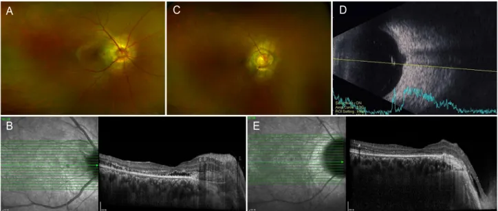
we diagnosed him with DP associated with subretinal fluid. We informed the patient that the DP could improve spontaneously over time, but the patient chose active treat- ment rather than observation. We decided to treat the DP to prevent further visual deterioration of the right eye, and he preferred long-term agents for intravitreal injection. We performed an intravitreal Ozurdex injection in the right eye to achieve faster resolution of DP, especially regarding the subretinal fluid. Three days after injection, the patient’s disc swelling, hemorrhage, and macular edema were significantly improved (Fig. 2A). SD OCT showed that the subretinal fluid had nearly resolved (Fig. 2B). Three weeks later, the disc swelling and hemorrhages were completely resolved and the underlying disc anomaly was clearly identifiable (Fig. 2C). A normal-appearing optic nerve head was surrounded by deep excavation (Fig. 2C), and B-ultrasonography showed obvious excavation, suggesting underlying peripapillary staphyloma (Fig. 2D). Macular edema was completely resolved (Fig. 2E).
DP did not recur for 1 year after initial treatment, and BCVA was maintained up to 20/50 by the Snellen visual acuity chart in the right eye.
A
B
C D
Figure 1. (A) Dilated fundus examination of a 50-year-old male patient with visual deterioration in the right eye showed disc swelling with some hemorrhages as well as macular thickening in the right eye. The best-corrected visual acuity (BCVA) of the right eye was 20/63 by Snellen visual acuity chart. (B) Spectral domain optical coherence tomography (Spectralis, Heidelberg Engineering, Heidelberg, Germany) showed profuse macular thickening with intraretinal and subretinal fluid in the right eye. (C) Early phase fluorescein angiography (FA) revealed microvascular tor- tuosity around the disc and blocked fluorescence due to disc hemorrhage. (D) At the late phase of FA, dye pooling within the serous fluid around macula and diffuse leakage from disc were noted.
90
JOURNAL OF RETINA
https://doi.org/10.21561/jor.2018.3.2.88
Discussion
Peripapillary staphyloma is a rare nonhereditary congenital optic disc anomaly consisting of a deep excavation of the area of the fundus surrounding the optic disc [5,6]. Peripap- illary staphyloma is usually unilateral, and the optic nerve head is situated within an excavated defect of the sclera lined by retina and choroid [5,6]. Vision is usually poor in the af- fected eye [5,6].
Transient disruption in the circulation of the optic nerve head seems to lead to hypoperfusion and ischemia in NAION [7-9]. A small cup-to-disc ratio, known as a ‘crowd- ed optic disc’ is considered a ‘structural disc at risk’ for NAION. When localized swelling occurs in a fixed space anterior to the rigid lamina cribrosa, the capillaries become more easily compressed and secondary ischemia may result.
Although the exact pathogenesis is still under investigation, DP is considered a form of NAION, and similar to NAION, a crowded disc with a smaller cup-disc ratio is suggested as a risk factor for DP [1,2]. In our patient, DP developed in an eye that was also affected by peripapillary staphyloma.
Although DP has not previously been reported in a case of
peripapillary staphyloma, structural alterations in such a disc anomaly may also predispose an individual to transient optic disc hypoperfusion. Our case suggests that disc anomaly may be a risk factor for DP, and further investigations using a larger number of cases are warranted.
In our patient, prompt treatment by intravitreal dexameth- asone implant injection resulted in rapid improvement of DP and associated macular edema, preventing further visual de- terioration. DP usually resolves spontaneously within an av- erage 5 to 7 months after diagnosis [2,10]. Although DP can improve spontaneously, early interventions have been report- ed using intravitreal anti-vascular endothelial growth factor and triamcinolone acetonide [11,12]. Case reports in which clinicians used intravitreal injections [11,12] and periocular triamcinolone acetonide injections [13] showed rapid resolu- tion of DP and visual recovery within 1 month, suggesting that such treatments are effective in these patients. In our case, intravitreal dexamethasone implant injection led to rapid resolution of DP and associated subretinal fluid, within 3 days after injection, and DP completely resolved 3 weeks after intravitreal injection. Our results show that dexameth- asone implants can be also considered as treatment options
C D
A
B E
Figure 2. (A) The patient’s disc swelling and macular edema significantly improved 3 days after intravitreal dexamethasone implant injection in a 50-year-old patient with diabetic papillopathy. (B) Subretinal fluid was also nearly resolved, although the photoreceptor layer was disrupted by spectral domain optical coherence tomography (SD OCT). (C) Three weeks after intravitreal dexamethasone implant injection, both disc swelling and associated hemorrhages resolved and the underlying disc anomaly was clearly noticeable, showing peripapillary excavation. (D) B-ultraso- nography showed excavation at the optic nerve head. (E) Macular edema was also completely resolved, although severe photoreceptor disrup- tion remained on SD OCT.
91
Kang HM.Diabetic papillopathy on peripapillary staphyloma
https://doi.org/10.21561/jor.2018.3.2.88
for DM to promote faster recovery and prevent further visual deterioration. In our case, the patient feared further visual deterioration in the eye with DP, because he already suffered from poor vision due to the underlying disc anomaly. How- ever, DP is known to resolve spontaneously, and therefore we do not suggest that every DP patient requires treatment.
Based on our case and previous case reports [11-13], we sug- gest that if a patient with DP needs or desires faster recovery, active treatment can be considered.
This is the first case report of DP in a patient with under- lying peripapillary staphyloma. Intravitreal dexamethasone implant injection yielded good treatment results, with rapid resolution of DP.
Conflicts of Interest
The author declares no conflicts of interest relevant to this article.
References
1. Regillo CD, Brown GC, Savino PJ, et al. Diabetic papillopathy.
Patient characteristics and fundus findings. Arch Ophthalmol 1995;113:889-95.
2. Bayraktar Z, Alacali N, Bayraktar S. Diabetic papillopathy in type II diabetic patients. Retina 2002;22:752-8.
3. Hayreh SS. Diabetic papillopathy and nonarteritic anterior isch-
emic optic neuropathy. Surv Ophthalmol 2002;47:600-2; author reply 602.
4. Slagle WS, Musick AN, Eckermann DR. Diabetic papillopathy and its relation to optic nerve ischemia. Optom Vis Sci 2009;86:e395- 403.
5. Dutton GN. Congenital disorders of the optic nerve: excavations and hypoplasia. Eye (Lond) 2004;18:1038-48.
6. Kim SH, Choi MY, Yu YS, Huh JW. Peripapillary staphyloma: clin- ical features and visual outcome in 19 cases. Arch Ophthalmol 2005;123:1371-6.
7. Miller NR, Arnold AC. Current concepts in the diagnosis, patho- genesis and management of nonarteritic anterior ischaemic optic neuropathy. Eye (Lond) 2015;29:65-79.
8. Barros AE, Amram AL, Derham AM, et al. Management of isch- emic optic neuropathies. Expert Rev Ophthalmol 2017;12:99-109.
9. Berry S, Lin WV, Sadaka A, Lee AG. Nonarteritic anterior ischemic optic neuropathy: cause, effect, and management. Eye Brain 2017;9:23-8.
10. Ostri C, Lund-Andersen H, Sander B, et al. Bilateral diabetic papil- lopathy and metabolic control. Ophthalmology 2010;117;2214-7.
11. Chin EK, Almeida DR, Sohn EH. Sustained and expedited reso- lution of diabetic papillopathy with combined PRP and bevaci- zumab. Can J Ophthalmol 2015;50:e88-91.
12. Al-Haddad CE, Jurdi FA, Bashshur ZF. Intravitreal triamcinolone acetonide for the management of diabetic papillopathy. Am J Ophthalmol 2004;137:1151-3.
13. Mansour AM, El-Dairi MA, Shehab MA, et al. Periocular cortico- steroids in diabetic papillopathy. Eye (Lond) 2005;19:45-51.