Prognostic Factors for Endotracheal Silicone Stenting in the Management of Inoperable Post-Intubation Tracheal Stenosis
So Yeon Lim, Hojoong Kim, Kyeongman Jeon, Sang-Won Um, Won-Jung Koh, Gee Young Suh, Man Pyo Chung, and O Jung Kwon
Division of Pulmonary and Critical Care Medicine, Department of Medicine, Samsung Medical Center, Sungkyunkwan University School of Medicine, Seoul, Korea.
Received: June 27, 2011 Revised: July 27, 2011 Accepted: August 26, 2011
Corresponding author: Dr. Hojoong Kim, Division of Pulmonary and Critical Care Medicine, Department of Medicine, Samsung Medical Center, Sungkyunkwan University School of Medicine,
81 Irwon-ro, Gangnam-gu, Seoul 135-710, Korea.
Tel: 82-2-3410-3425, Fax: 82-2-3410-3849 E-mail: [email protected]
∙ The authors have no financial conflicts of interest.
© Copyright:
Yonsei University College of Medicine 2012 This is an Open Access article distributed under the terms of the Creative Commons Attribution Non- Commercial License (http://creativecommons.org/
licenses/by-nc/3.0) which permits unrestricted non- commercial use, distribution, and reproduction in any medium, provided the original work is properly cited.
Purpose: Stenting has been developed to deal with airway stenosis and is applicable in patients with post-intubation tracheal stenosis (PITS) in whom surgery would not be indicated. The purpose of this study was to investigate the prognostic factors in inoperable patients in whom a silicone stent was inserted due to PITS. Materials and Methods: We retrospectively evaluated 55 PITS patients undergoing silicone stenting between January 2001 and December 2009. Results: Silicone stent was in- serted to narrowed trachea after the combination of pre-dilatation including laser cauterization, mechanical bougienation and ballooning. Following airway stabiliza- tion, the stent could be removed successfully in 40% (22/55) of the patients after median 12 months of stenting. However, in 60% (33/55) of patients, the stent could not be removed successfully and surgical management was needed after initial stabi- lization. Multivariate analysis revealed that the stent could be successfully removed more frequently in those who do not have cardiovascular disease [odds ratio (OR)=12.195; p=0.036] and the intervention was performed within 6 months after intubation (OR=13.029; p=0.031). Conclusion: Among those patients undergoing silicone stenting due to PITS, the stent could be successfully removed when patients do not have cardiovascular disease and stented within 6 months after intubation.
Key Words: Intervention, post-intubation tracheal stenosis, prognosis, rigid bron- choscopy, silicone stent
INTRODUCTION
Tracheal stenosis (narrowing of the trachea) is a life-threatening, emergent disease with an increasing frequency.1,2 One of the most common etiologies of benign tra- cheal stenosis is post-procedural tracheal stenosis, such as that following long- term tracheal intubation or following tracheostomy. Although the use of low pres- sure cuffs has reduced the incidence of post-intubation tracheal stenosis (PITS) by 10-fold, the occurrence of PITS has increased, due to early application of tracheos- tomy in the intensive care unit.2,3
The management of PITS is a complex problem that requires a multi-disci- plined approach. Generally, the preferred management is open resection and re-
posed for subglottic stenosis, although it has been used with other airway site assessment. It was defined as follows:
Grade I: ≤50% lumen stenosis; Grade II: 51-70% lumen stenosis; Grade III: 71-99% lumen stenosis; Grade IV: no lumen.4 “Successful group” was defined as the group of the stent being removed successfully (usually after 6-12 months) without re-insertion or tracheostomy during the follow-up. “Unsuccessful group” was defined when the stent removal was intolerable and following re-stenting or surgical intervention was needed.
Stents
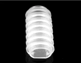
A Natural (M1S Co., Seoul, Korea) stent of 12-14 mm out- er diameter was used for tracheal stenosis (Fig. 1). A new silicone stent, named the Natural stent, was developed by the TNO Company in 2001. This stent is composed of molded silicone and is straight in shape.3,5 It features regu- larly placed ‘C’ circular ribs on its outer surface.3,5 These stent designs increase stent-to-wall contact due to ‘C’ shaped studs, and have a theoretical advantage to reduce the stent migration and granulation tissue overgrowth.3,5 An adequate size and type of stent were selected and used according to the interventionist’s decision.
Airway intervention techniques and follow-up
Airway intervention was performed following standard tech- niques, as described by Dumon.6,7 Briefly, under general an- esthesia, patients were intubated with a rigid bronchoscope tube (Hopkins, Karl-Storz, Germany), and a flexible bron- choscope (EVIS BF 1T240, Olympus, Tokyo, Japan) was introduced through this rigid bronchoscope tube and airway narrowing was examined. The length of stenotic lesion was measured by a scale mark of flexible bronchoscope and a stent of appropriate size (1 cm longer than the stenotic length) was selected by the interventionist. Patients under- went mechanical dilatation prior to stenting, such as dilata- tion with rigid tubes, ballooning (Boston Scientific, Boston, MA, USA), and laser cauterization (LaserSonics, Mipiltas, CA, USA). A stent of an appropriate size was folded longi- tudinally, introduced into a stent pusher (BryanCorp., Wo- burn, MA, USA), and re-positioned using alligator forceps.
Patients were discharged from hospital one to three days following the procedure. We assessed the symptomatic re- lief as interviewing the patients in the next morning of the bronchoscopic intervention. Patients were followed at 1, 3, 6, 9 and 12 months after intervention with chest radiography and spirometry, and three-dimensional CT and flexible anastomosis.2 However, stenting is recommended in pa-
tients with PITS in whom surgery is not indicated due to poor general condition or long involvement of the trachea.2,3 There is an increasing need to investigate the factors that fa- vor stenting in the management of patients with PITS, be- cause the stent can successfully be removed in only 30-40%
of stented patients. Therefore, we investigated such prognos- tic factors in initially inoperable patients in whom stenting was performed.
MATERIALS AND METHODS
Patients
Among 59 patients who underwent bronchoscopic silicone stenting for the treatment of initially inoperable PITS at the Samsung Medical Center, Seoul, Korea between January 2001 and December 2009, 4 patients lost follow-up within 3 months, and 55 patients who had complex type stenotic lesion were included in this study. Twenty-two patients were had already been studied in the prior study by Park, et al.3 Bronchoscopic intervention in patients with PITS was indicated when all of the following conditions were met: 1) The degree of dyspnea was greater than American Thoracic Society grade 3; 2) obstruction in tracheal lumen exceeded 25%; 3) general condition was tolerable for intervention, and 4) distal airways were patent.
The Institutional Review Board of the Samsung Medical Center approved this study. Written informed consent was obtained from each patient.
Definitions
The Myer-Cotton stenosis grading system was initially pro-
Fig. 1. A natural silicone stent.
intervention was conducted in 48 patients, and 7 patients underwent stenting at second intervention after first me- chanical dilatation. Their median age was 60 years (range, 16-84), and 22 patients (40%) were males (Table 1). The intubation-to-intervention time was median 4 months (range, 0.5-480 months). The causes of the tracheal stenosis were post-intubation (72.7%) and post-tracheostomy (27.3%). Baseline spirometry data were available for 20 pa- tients (36.4%); the forced expiratory volume in 1 s (FEV1), forced vital capacity (FVC), and FEV1/FVC were 61% (18- 113%), 68% (22-123%), and 64% (23-89%), respectively.
Bronchoscopic findings and interventions
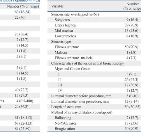
Bronchoscopic findings and interventions are summarized in Table 2. The luminal narrowing was classified by the Myer and Cotton grading system.4 Grade I was observed in 5 patients (9.1%), grade II was evident in 26 patients (47.3%), grade III in 17 patients (30.9%), and grade IV in 7 patients (12.7%).
Outcomes and complications
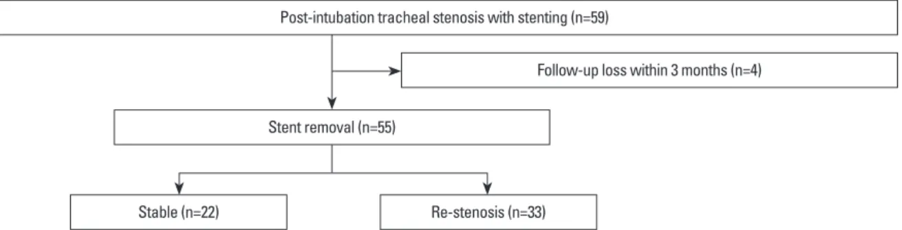
The overall clinical outcomes are shown in Fig. 2. Success- bronchoscopy were performed before stent removal. Stent
removal was planned 12 months after the intervention when the patients were stable and airway related problem did not develop at least for 6 months.
Statistical analysis
For statistical analysis, PAWS 17.0 (SPSS Inc., Chicago, IL, USA) was used. Group comparisons of categorical variables were made using the Pearson chi-square test or Fisher’s ex- act test. To assess the relationship between continuous vari- ables, the Mann-Whitney U test was used. Multivariate lo- gistic regression analysis was used to determine indepen- dent predictors of failure to attain a stent-free airway. Among the variables used in this model, predictive factors with a p- value less than 0.15 were selected for multivariate logistic regression analysis. A two-tailed p-value <0.05 was consid- ered statistically significant.
RESULTS
Clinical characteristics of patients
Total 55 patients were included. The stent insertion at first Table 1. Baseline Characteristics of the Study Population (n=55)
Variable Number (% or range)
Age (yrs) 60 (16-84)
Gender (male) 22 (40)
Cause of intubation
Medical
Respiratory failure 20 (36.4)
Cardiac failure 7 (12.7)
Neurological problem 8 (14.5)
Burn 1 (1.8)
Drug intoxication 5 (9.1)
Surgical
Operation 5 (9.1)
Trauma 8 (14.5)
Suicide 1 (1.8)
Cause of tracheal stenosis
Post-intubation 40 (72.7)
Post-tracheostomy 15 (27.3)
Intubation-to-intervention time, months 4 (0.5-480) Tracheostomized state at the first visit 20 (36.3) Baseline spirometer data (n=20)
FEV1 (% predicted) 61 (18-113)
FVC (% predicted) 68 (22-123)
FEV1/FVC (% predicted) 64 (23-89) FEV1, forced expiratory volume in 1 second; FVC, forced vital capacity.
Data are presented as n (%) or median (range).
Table 2. Bronchoscopic Findings and Parameters of Inter- vention (n=55)
Variable Number
(% or range) Stenosis site, overlapped (n=67)
Subglottis 9 (16.4)
Upper trachea 39 (70.9)
Mid-trachea 13 (23.6)
Lower trachea 6 (10.9)
Stenosis type
Fibrous stricture 50 (90.9)
Malacia 1 (1.8)
Fibrous stricture+malacia 4 (7.3) Characteristics of the lesion at first bronchoscopy Myer and Cotton Grade
I 5 (9.1)
II 26 (47.3)
III 17 (30.9)
IV 7 (12.7)
Luminal diameter before procedure, mm 5 (0-10) Luminal diameter after procedure, mm 12 (9-14)
Length of stent, mm 50 (30-85)
Method of airway dilatation (overlapped)
Ballooning 7 (12.7)
Nd-YAG laser 13 (23.6)
Bougienation 50 (90.9)
Nd-YAG, neodymium-yttrium aluminum garnet.
Data are presented as n (%) or median (range).
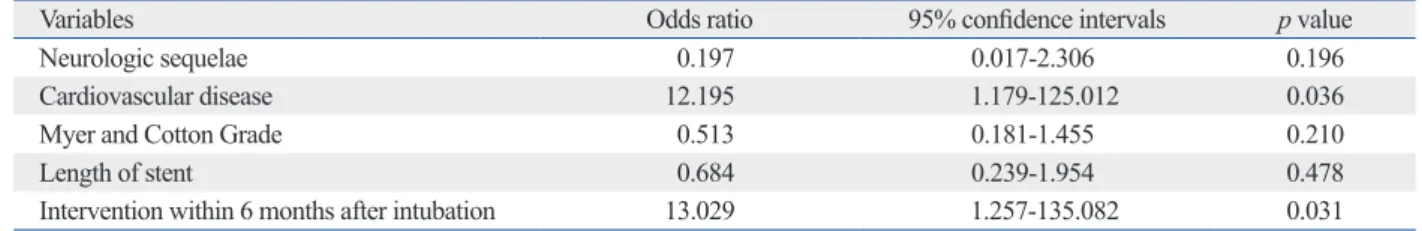
the variables used in the model, no cardiovascular disease (OR=12.195; p=0.036; 95% CI=1.179-125.012) and initia- tion of treatment within 6 months (13.029; 0.031; 1.257- 135.082) were independently associated with successful stent removal (Table 4).
DISCUSSION
The treatment of initially inoperable PITS requires a multi- disciplinary approach, including initial conservative treat- ment, interventional bronchoscopy, and surgical manage- ment such as tracheal resection and end to end anastomosis, slide tracheoplasty, and open expansion tracheoplasty with stent fixation, especially for long segment tracheal steno- sis.8 Surgical resection and re-anastomosis have been the first choice of treatment when the patients’ condition is tol- erable, and interventional bronchoscopy has been applied if the general condition is impossible for an operation.9 How- ever, even with developments in the management of criti- cally ill patients, surgical treatment is still not indicated in PITS patients with poor neurological, cardiovascular, or re- spiratory condition.10 In these patients, interventional bron- choscopy is a good alternative and has led to satisfactory ful stent removal occurred in 22 patients (40%), and dura-
tion of stent placement was median 12 months (Fig. 3).
However, re-stenosis was done in 33 patients (60%).
Among them, persistent stent placement occurred in 23 pa- tients (41.8%). Surgical management, such as tracheal re- section with end-to-end anastomosis, occurred in 10 pa- tients (18.2%).
Pneumothorax occurred in one patient. Late complications including stent migration (36.4%), mucostasis (21.8%), and granulation tissues formation at the end of the stent (49.1%) were observed, and repeated bronchoscopic interventions were required to treat these complications.
Comparison of the “successful group” and
“unsuccessful group”
Patients were grouped according to whether the stent could be successfully removed (successful group) or whether the stent remained or received surgical intervention (unsuccess- ful group) (Table 3). The patients of unsuccessful group had cardiovascular disease (p=0.008), neurological sequelae (p=0.018), high Myer and Cotton grade (p=0.075), and the delay of treatment more than 6 months (p=0.005). Multivari- ate logistic regression model was used to determine indepen- dent predictors of the successful removal of the stent. Among
Fig. 2. Outcomes of bronchoscopic interventions in 59 post-intubation tracheal stenosis patients.
Fig. 3. A representative case of temporary stenting for post-intubation tracheal stenosis. (A) Marked narrowing of the trachea was noted in a 59-year-old male patient after neurosurgical operation. (B) After silicone stent was inserted, the trachea was widened. (C) The stent was removed 1 year after the inter- vention. The widened diameter of trachea was maintained successfully.
A B C
Post-intubation tracheal stenosis with stenting (n=59)
Follow-up loss within 3 months (n=4)
Stable (n=22) Re-stenosis (n=33)
Stent removal (n=55)
tion tracheal stenosis and was performed in one of the largest centers for interventional bronchoscopy in Asia. We demon- strated that the factors contributing to successful stent remov- al included no history of cardiovascular disease and initiation of treatment within 6 months after intubation. To our best results in selected patients with benign airway stenosis. A
positive outcome of interventional bronchoscopy was re- ported in 32 PITS patients by Park, et al.3
The current study was conducted to reveal prognostic fac- tors for tracheal stenting in initially inoperable post-intuba-
Table 3. Subgroup Analysis between “Successful group” (Stable after Removal) and “Unsuccessful group” (Restented or Operated Patients)
Variables Successful group (% or range)
(n=22) Unsuccessful group (% or range)
(n=33) p value
Age, yrs (range) 60 (24-84) 61 (16-80) 0.286
Gender, male 9 (40.9) 13 (39.4) 1.000
Cardiovascular disease 1 (4.5) 12 (36.4) 0.008
Neurologic sequelae 1 (4.5) 11 (33.3) 0.018
Presence of tracheostomy 5 (22.7) 11 (33.3) 0.547
FEV1, predicted %
Before stenting (n=17) 67 (18-84) 47 (20-113) 0.753
After stenting (n=39) 92.5 (40-126) 89 (22-124) 0.364
After removal of stent (n=14) 92.5 (38-127) - -
Change after removal of stent 26 (1-44) - -
Stenotic site 1.000
Subglottis 2 (9) 7 (21.2)
Upper trachea 15 (68.2) 24 (72.7)
Mid trachea 5 (22.7) 9 (27.3)
Lower trachea 2 (9) 4 (12.1)
Characteristics of the lesion at first bronchoscopy
Myer and Cotton Grade 0.075
I 3 (13.6) 2 (6.1)
II 14 (63.6) 12 (36.4)
III 3 (9.1) 14 (42.4)
IV 2 (9.1) 5 (15.2)
Luminal diameter before procedure, mm 5 (0-6) 4 (0-10) 0.609
Luminal diameter after procedure, mm 12 (9-14) 12 (9-14) 0.536
Stenosis type
Fibrous stricture 21 (95.5) 30 (90.9) 1.000
Fibrous stricture and malacia 1 (4.5) 3 (9.1) 0.522
Length of the stent 4.5 (3.5-6) 5 (3-8.5) 0.145
Stenosis-to-intervention time 3 (1-10) 5 (1-60) 0.110
Intervention within 6 months 20 (90.9) 20 (60.6) 0.005
Visit of emergency room 13 (59) 21 (63.6) 0.570
Emergent bronchoscopy 10 (45.5) 16 (48.5) 0.782
Duration of follow-up 13 (6-156) 20 (7-97) 0.918
FEV1, forced expiratory volume in 1 second.
Data are presented as n (%) or median (range).
Table 4. Multivariate Logistic Regression Analysis for Determining the Factors of Successful Stent Removal
Variables Odds ratio 95% confidence intervals p value
Neurologic sequelae 0.197 0.017-2.306 0.196
Cardiovascular disease 12.195 1.179-125.012 0.036
Myer and Cotton Grade 0.513 0.181-1.455 0.210
Length of stent 0.684 0.239-1.954 0.478
Intervention within 6 months after intubation 13.029 1.257-135.082 0.031
other undisclosed factors, which should be revealed by future studies. Second, in majority of patients (35 patients, 64%), spirometer data were missing due to patients’ condition. Oth- er objective measurements should be sought in future study.
In conclusion, among patients undergoing silicone stent- ing due to initially inoperable PITS, the stent could be suc- cessfully removed when the patients did not have cardiovas- cular disease and stented less than 6 months after intubation.
REFERENCES
1. Gamsu G, Webb WR. Computed tomography of the trachea and mainstem bronchi. Semin Roentgenol 1983;18:51-60.
2. Pereszlenyi A, Igaz M, Majer I, Harustiak S. Role of endotracheal stenting in tracheal reconstruction surgery-retrospective analysis.
Eur J Cardiothorac Surg 2004;25:1059-64.
3. Park HY, Kim H, Koh WJ, Suh GY, Chung MP, Kwon OJ. Natu- ral stent in the management of post-intubation tracheal stenosis.
Respirology 2009;14:583-8.
4. Myer CM 3rd, O’Connor DM, Cotton RT. Proposed grading sys- tem for subglottic stenosis based on endotracheal tube sizes. Ann Otol Rhinol Laryngol 1994;103(4 Pt 1):319-23.
5. Ryu YJ, Kim H, Yu CM, Choi JC, Kwon YS, Kwon OJ. Use of silicone stents for the management of post-tuberculosis tracheo- bronchial stenosis. Eur Respir J 2006;28:1029-35.
6. Kim H. Stenting therapy for stenosing airway disease. Respirolo- gy 1998;3:221-8.
7. Colt HG, Dumon JF. Airway stents. Present and future. Clin Chest Med 1995;16:465-78.
8. Lang FJ, Hurni M, Monnier P. Long-segment congenital tracheal stenosis: treatment by slide-tracheoplasty. J Pediatr Surg 1999;34:
1216-22.
9. Marel M, Pekarek Z, Spasova I, Pafko P, Schutzner J, Betka J, et al. Management of benign stenoses of the large airways in the uni- versity hospital in Prague, Czech Republic, in 1998-2003. Respi- ration 2005;72:622-8.
10. Brichet A, Verkindre C, Dupont J, Carlier ML, Darras J, Wurtz A, et al. Multidisciplinary approach to management of postintubation tracheal stenoses. Eur Respir J 1999;13:888-93.
11. de Mello-Filho FV, Antonio SM, Carrau RL. Endoscopically placed expandable metal tracheal stents for the management of complicated tracheal stenosis. Am J Otolaryngol 2003;24:34-40.
12. Benjamin B. Prolonged intubation injuries of the larynx: endo- scopic diagnosis, classification, and treatment. Ann Otol Rhinol Laryngol Suppl 1993;160:1-15.
13. Marshak G, Doyle WJ, Bluestone CD. Canine model of subglottic stenosis secondary to prolonged endotracheal intubation. Laryn- goscope 1982;92(7 Pt 1):805-9.
14. Supance JS, Reilly JS, Doyle WJ, Bluestone CD, Hubbard J. Ac- quired subglottic stenosis following prolonged endotracheal intu- bation. A canine model. Arch Otolaryngol 1982;108:727-31.
15. Nouraei SA, Ghufoor K, Patel A, Ferguson T, Howard DJ, Sandhu GS. Outcome of endoscopic treatment of adult postintubation tra- cheal stenosis. Laryngoscope 2007;117:1073-9.
knowledge, this is the first reported study on the prognosis of PITS, managed by bronchoscopic intervention with silicone stenting. In this study, the silicone stent could be successfully removed in 40% of patients. In 60% of patients, the stent could not be removed and they received surgical manage- ment, demonstrating that the stent can be removed success- fully only in a limited number of patients. The poor progno- sis reflects not only the need for advances in bronchoscopic intervention, but also the shortage in the pathophysiological understanding and early diagnosis of PITS.
In this study, cardiovascular disease had a relevance to un- favorable prognosis. For good prognosis, stent should pro- mote healing of de-epithelialization of the stenotic lesions by allowing mucosal to grow and should not impede airway mucocilliary clearance, resisting bacterial contamination and avoiding excessive pressure that would impede capillary cir- culation.11 However, cardiovascular disease may cause de- fect of good blood supply to the mucosa of trachea. Also, symptomatic dyspnea might be increased when the patients had underlying cardiovascular disease. In addition, many co-morbidities and sequelae result in a poor general condi- tion. These problems may lead to difficulties in both cough- ing and stent-related complications such as mucostasis.
Another important prognostic factor of successful stent removal in this study was the initiation of intervention with- in 6 months after intubation. In the early phase, PITS is ini- tiated by mucosal ulceration and perichondritis, followed by granulation tissue formation.12-14 In the later phase, carti- laginous tracheal rings are damaged and resorbed, leading to the circumferential loss of mechanical support coupled with scar contracture, resulting in the collapse of the whole tracheal segment.15 Because stents provide resistance to scar contracture and provide support in areas of structural weakness because of cartilage loss, this severe stenosis is often indicated to surgical intervention.11 Thus, a favorable outcome would be predicted when the patients were re- ferred prior to damage in the airway cartilages. Consistent with this, we found in the present study that it was an unfa- vorable factor for the successful stent removal, when the initiation of treatment exceeds 6 months.
There are clear limitations in this study. It is a retrospective review of small sample size, which needs a large scaled pro- spective study to avoid the lack of statistical significance in some potentially important confounders. Therefore, the deci- sion to remove a stent will be made by considering the prog- nostic factors being discussed in this study (no cardiovascu- lar disease and initiation of treatment within 6 months) and