Dentoskeletal features in individuals with ectopic eruption of the permanent maxillary first molar
Objective: The aim of the study was to analyze the prevalence and distribution of ectopic eruption of the permanent maxillary first molar (EEM) in individuals scheduled for orthodontic treatment and to investigate the association of EEM with dental characteristics, maxillary skeletal features, crowding, and other dental anomalies. Methods: A total of 1,317 individuals were included and randomly divided into two groups. The first 265 subjects were included as controls, while the remaining 1,052 subjects included the sample from which the final experimental EEM group was derived. The mesiodistal (M-D) crown width of the deciduous maxillary second molar and permanent maxillary first molar, maxillary arch length (A-PML), maxillomandibular transverse skeletal relationships (anterior and posterior transverse interarch discrepancies, ATID and PTID), maxillary and mandibular tooth crowding, and the presence of dental anomalies were recorded for each subject, and the statistical significance of differences in these parameters between the EEM and control groups was determined using independent sample t-tests. Chi-square tests were used to compare the prevalence of other dental anomalies between the two groups.
Results: The prevalence of maxillary EEM was 2.5%. The M-D crown widths, ATID and PTID, and tooth crowding were significantly greater, while A-PML was significantly smaller, in the EEM group than in the control group. Only two subjects showed an association between EEM and maxillary lateral incisor anomalies, which included agenesis in one and microdontia in the other.
Conclusions: EEM may be a risk factor for maxillary arch constriction and severe tooth crowding.
[Korean J Orthod 2015;45(4):190-197]
Key words: Ectopic eruption, Tooth size, Crowding, Transverse maxillary deficiency Manuela Mucedero
aMatteo Rozzi
aGiulia Cardoni
bMaria Rosaria Ricchiuti
aPaola Cozza
aa
Department of Clinical Sciences and Traslation Medicine, University of Rome “Tor Vergata”, Rome, Italy
b
Private Practice, Rome, Italy
Received November 30, 2014; Revised January 14, 2015; Accepted January 23, 2015.
Corresponding author: Manuela Mucedero.
Clinical Fellow, Department of Clinical Sciences and Traslation Medicine, University of Rome “Tor Ver gata”, Fondazione Policlinico Tor Vergata PTV, Department of Orthodontics, Viale Oxford 81, 00133 Rome, Italy.
Tel +39-3478427270 e-mail [email protected]
© 2015 The Korean Association of Orthodontists.
The authors report no commercial, proprietary, or financial interest in the products or companies described in this article.
This is an Open Access article distributed under the terms of the Creative Commons Attribution Non-Commercial License (http://creativecommons.org/licenses/by-nc/4.0) which permits unrestricted non-commercial use, distribution, and reproduction in any medium, provided the original work is properly cited.
pISSN 2234-7518 • eISSN 2005-372X
http://dx.doi.org/10.4041/kjod.2015.45.4.190
INTRODUCTION
Ectopic eruption of the permanent maxillary first molar (EEM) is a local eruption disturbance characterized by eruption that is mesial to the normal path.
1,2Data reported in the literature shows an EEM prevalence rate of 0.75 − 6%.
3-7The permanent molar is initially blocked from complete eruption by the deciduous second mo- lar because of the close contact between the two teeth.
This condition causes atypical resorption on the dis tal surface of the deciduous second molar,
1with a signi- ficant effect on malalignment of teeth, particularly the per manent teeth.
8,9Several etiological theories have been reported in the literature.
1,4,10-16Pulver
4found that EEM depended on a combination of factors, including macrodontia of the permanent maxillary teeth and first molars, maxi- llary hypoplasia, posterior position of the maxilla in relation to the cranial base, abnormal eruption angle of the permanent maxillary first molar, and delayed calci- fication of some affected permanent molars. Bjerklin and Kurol
1suggested the tendency toward a shorter maxilla and macrodontia of the permanent molars
with a more pronounced mesial angle of eruption in child ren with irreversible ectopic eruption compared with those in children with normal eruption, while no signi ficant differences were observed between children with reversible ectopic eruption and the controls. Stu- dies by Yuen et al.
10and Canut and Raga
11repor ted an association of ectopic eruption with a short and posteriorly positioned maxilla, while other studies defined ectopic eruption as a multifactorial pro cess.
12-14More recently, Bjerklin et al.
15analyzed the association between EEM and three other dental anomalies, defining the four conditions as different manifestations of a single syndrome with incomplete penetrance. Baccetti
16suggested the importance of local factors such as tooth size − arch length discrepancy in the etiology of this eruption anomaly.
Subsequently, Becktor et al.
17proposed that irreversible EEM can be an early indicator of lateral canine eruption, which leads to root resorption. Salbach et al.
5reported a significant association between EEM and other forms of malocclusion, such as crowding, lateral malocclusion, and mandibular prognathism.
Despite extensive analysis evaluating the association
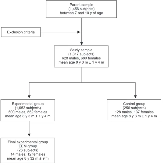
Parent sample (1,456 subjects) between 7 and 10 y of age
Exclusion criteria
Study sample (1,317 subjects) 628 males, 689 females mean age 8 y 3 m + 1 y 4 m
Experimental group (1,052 subjects) 500 males, 552 females mean age 8 y 3 m + 1 y 4 m
Control group (256 subjects) 128 males, 137 females mean age 8 y 3 m + 1 y 4 m
Final experimental group EEM group (26 subjects) 14 males, 12 females mean age 8 y 32 m + 9 m
Figure 1. Study flow chart.
EEM, Ectopic eruption of the
permanent maxillary first
molar; y, years; m, months.
between EEM and maxillary arch length, no studies con- sidered the association of EEM with maxillary transverse deficiency and maxillary and mandibular tooth crow- ding.
Therefore, this study was conducted to analyze the prevalence and distribution of EEM in a large cohort of individuals scheduled for orthodontic treatment and to investigate the association of EEM with dental characteristics, maxillary skeletal features, and crowding using a control group for comparison. The prevalence of other dental anomalies in the EEM group was also assessed.
MATERIALS AND METHODS
The parent sample for this study comprised 1,456 in- dividuals in the early mixed dentition stage recruited from the Department of Orthodontics at the University of Rome “Tor Vergata.” All subjects were observed prior to orthodontic treatment and at the prepuberal stage of skeletal growth using the cervical vertebral maturation method (CS1 − CS2).
18In addition, dental casts and panoramic radiographs were examined for each subject.
Subjects with craniofacial anomalies, cleft lip and/or palate, sequelae of traumatic injuries to the permanent teeth, odontomas, and/or cysts, were not included, as were subjects with Class II dental restorations, extensive caries, or premature loss of the deciduous maxillary second molars. Eventually, 1,317 subjects (628 boys and 689 girls) aged 7 − 10 years were included.
According to the methodology of previous studies,
16,19the study sample was randomly divided into two groups.
The first 265 subjects, including 128 boys and 137 girls, were used as controls; the reference values for all exa- mined parameters were calculated for this group. The remaining 1,052 subjects, including 500 boys and 552 girls with a mean age of 8 years and 3 months ± 1 year and 4 months, comprised the sample from which the final experimental group was derived; this group
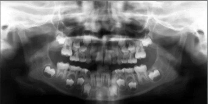
was investigated for the presence of EEM. In total, 26 subjects, including 14 boys and 12 girls with a mean age of 8 years and 2 months ± 9 months, were diagnosed with EEM in the experimental group and were identified as the EEM group (Figure 1). EEM was identified when the permanent first molar was initially blocked from complete eruption by the adjacent primary molar, which showed premature resorption on its distal surface (Figure 2). Two possible evolutions of EEM may follow:
a reversible type, wherein the permanent molar frees itself and erupts to normal occlusion, and an irreversible type.
2,19,20These two forms were not distinguished in the present study for the early mean age of the sample. The unilateral or bilateral intraosseous ectopic position of the permanent first molar was evaluated on panoramic radiographs.
The EEM and control groups were matched in terms of origin, age, and gender distributions (Table 1). All subjects were Caucasian, and there was a possibility of the presence of EEM in the control group.
Initial dental casts were available for each subject.
These casts were used to analyze the maxillary arch length, maxillary and mandibular arch diameters, tooth crowding, and crown widths using a tridimensional scanner (D800; 3Shape A/S, Copenhagen, Denmark;
scan time, 25 s; resolution, two cameras, 5.0 megapixels;
ultrahigh point accuracy, <15 microns). The virtual three-dimensional models were measured and analyzed using specific software (3Shape OrthoAnalyzer
TM2010;
3Shape A/S).
The following parameters were analyzed (Figure 3):
1) Mesiodistal (M-D) crown width of the deciduous se cond molar, as calculated on digital models by recor ding the distance between the central point of the mesial marginal crest and the central point of the distal marginal crest
212) M-D crown width of the permanent first molar, as calculated on digital models by recording the distance between the central point of the mesial marginal crest and the central point of the distal
Figure 2. Panoramic radiograph showing bilateral ectopic eruption of the permanent maxillary first molars in an 8-year-old subject.
Table 1. Dermographic data for the EEM and control groups
EEM group Control group
Subjects 26 265
Male 14 128
Female 12 137
Age 8 y 2 mo ± 9 mo 8 y 4 mo ± 1 y 2 mo Values are presented as number or mean ± standard deviation.
EEM, Ectopic eruption of the permanent maxillary first
molar; y, years; mo, months.
marginal crest.
21In the EEM group, measurements were obtained after extraction of the deciduous second molars in subjects with permanent first molars that were locked distal to the deciduous se- cond molars
13) Maxillary and mandibular tooth crowding, as evalu - ated on digital models using the space analysis me - thod of Tweed
21; the necessary space in subjects with several unerupted permanent teeth was cal - culated using the prediction tables of Moyers
224) Maxillary arch length (A-PML), as calculated on digital models from the central point of the incisive papilla to the tangent of the most distal point of the deciduous maxillary second molars on the right
and left sides, using the median palatal raphe as the midsagittal arch plane
105) Maxillomandibular transverse skeletal relationships, as calculated on digital models by recording the in - tercanine and intermolar distances for the deciduous teeth
The maxillary intercanine width was measured as the distance between the most mesial points on the pala- tal surfaces of the maxillary deciduous canines. The maxillary intermolar width was evaluated as the dis- tance between the central fossae of the deciduous ma- xillary right and left second molars. The mandibular in tercanine width was measured as the distance between the cusp tips of the deciduous mandibular canines. The mandibular intermolar width was evaluated as the dis- tance between the tips of the distobuccal cusps of the deciduous mandibular right and left second molars.
23The anterior transverse interarch discrepancy (ATID) was calculated as the difference between the maxillary and mandibular intercanine widths. In subjects with normal occlusion, the cusp tips of the deciduous mandibular canines occlude with the most mesial points on the palatal surface of the deciduous maxillary canines; con- sequently, the maxillary and mandibular intercanine widths are equal in these subjects. The posterior transverse interarch discrepancy (PTID) was calculated as the difference between the maxillary and mandibular intermolar widths. In subjects with normal occlusion, the distobuccal cusp of the deciduous mandibular second molar occludes with the central fossa of the deciduous maxillary second molar; consequently, the maxillary and mandibular intermolar widths are equal in these subjects. A smaller maxillary width compared with the mandibular width indicates a transverse discrepancy between the dental arches.
All measurements were performed with the investigator
Figure 3. Linear measurements on digital models.
M-D crown widths, Mesiodistal crown widths; A-PML, anteroposterior maxillary length; Max-intercanine width, ma xillary intercanine width; Max-intermolar width, ma- xillary intermolar width; Mand-intercanine width, man- dibular intercanine width; Mand-intermolar width, man- dibular intermolar width.
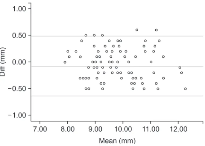
Figure 4. Results of Bland-Altman difference plot analy- ses. Example for M-D crown widths. Diff, difference.
Diff(mm)
7.00 8.00 9.00 10.00 11.00 12.00 Mean (mm)
0.50
0.00
0.50
1.00
1.00
(MM) blinded to the group investigated.
Furthermore, the presence of other associated den tal anomalies was evaluated in both groups using pano- ramic radiographs.
Statistical analysis
The reproducibility of EEM diagnosis was found to be 100% when the records of 100 subjects were re- examined 5 months after the first examination. Re- producibility of the measurements on radiographs and digital models was also estimated at this time by repeating all measurements and assessments for the abovementioned 100 subjects. Statistical analysis was completed using the Statistical Package for Social Sciences (version 13.0; SPSS Inc., Chicago, IL, USA). The two sets of coord inates were compared using paired t-tests, evaluated using Bland-Altman plots,
24and confirmed by Pearson and linear regression analyses (Figure 4). No significant systematic error was found between the measurement sessions (p > 0.05), and the method error was 0.3 mm for the digital model measurements. The Shapiro-Wilk test was used as a normality test; all the measured values followed a normal distribution. The statistical significance of differences between the EEM and control groups in the M-D crown widths, A-PML, ATID and PTID, and
maxillary and mandibular tooth crowding was tested using independent sample t-tests (p < 0.01). Considering the large variance and small sample size, the Kruskal- Wallis test was used to confirm the results of the t-tests.
Chi-square tests with Yates’ correction were performed to compare the prevalence of other dental anomalies between the two groups.
RESULTS
The prevalence of maxillary EEM was 2.5% (26 of 1,052 subjects), with six and 20 subjects showing unilateral and bilateral EEM, respectively (1:5). Twenty boys and six girls showed EEM, indicating an approximate M:F ratio of 5:1.
The results of descriptive statistics for all measurements in both groups are shown in Table 2.
Dental characteristics
The permanent maxillary first molars were significantly larger in the EEM group (12.1 and 11.9 mm for the right and left molars, respectively) than in the control group (10.3 and 10.2 mm for the right and left molars, respectively; p < 0.01). The mean size of the deciduous second molar was greater in the EEM group (9.6 mm and 9.5 mm for the right and left molars, respectively)
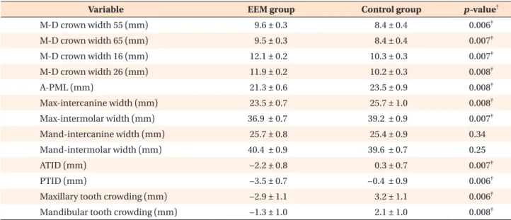
Table 2. Statistical analysis of the EEM and control groups *
Variable EEM group Control group p-value
†M-D crown width 55 (mm) 9.6 ± 0.3 8.4 ± 0.4 0.006
‡M-D crown width 65 (mm) 9.5 ± 0.3 8.4 ± 0.4 0.007
‡M-D crown width 16 (mm) 12.1 ± 0.2 10.3 ± 0.3 0.007
‡M-D crown width 26 (mm) 11.9 ± 0.2 10.2 ± 0.3 0.008
‡A-PML (mm) 21.3 ± 0.6 23.5 ± 0.9 0.008
‡Max-intercanine width (mm) 23.5 ± 0.7 25.7 ± 1.0 0.008
‡Max-intermolar width (mm) 36.9 ± 0.7 39.2 ± 0.9 0.007
‡Mand-intercanine width (mm) 25.7 ± 0.8 25.4 ± 0.9 0.34
Mand-intermolar width (mm) 40.4 ± 0.9 39.6 ± 0.7 0.25
ATID (mm) −2.2 ± 0.8 0.3 ± 0.7 0.007
‡PTID (mm) −3.5 ± 0.7 −0.4 ± 0.9 0.006
‡Maxillary tooth crowding (mm) −2.9 ± 1.1 3.2 ± 1.1 0.006
‡Mandibular tooth crowding (mm) −1.3 ± 1.0 2.1 ± 1.0 0.008
‡*Descriptive statistics and statistical comparisons of dental characteristics, maxillomandibular transverse skeletal relation- ships, and tooth crowding between the EEM and control groups.
Values are presented as mean ± standard deviation.
EEM, Ectopic eruption of the permanent maxillary first molar; M-D crown widths, mesiodistal crown widths; A-PML, anteroposterior maxillary length; Max-intercanine width, maxillary intercanine width; Max-intermolar width, maxillary intermolar width; Mand-intercanine width, mandibular intercanine width; Mand-intermolar width, mandibular intermolar width; ATID, anterior transverse interarch discrepancy; PTID, posterior transverse interarch discrepancy.
†