Predictive Factors of Ectopic Eruption of the Maxillary First Permanent Molar
Jimin Sun, Okhyung Nam, Misun Kim, Hyoseol Lee, Sungchul Choi
Department of Pediatric Dentistry, School of Dentistry, Kyung-Hee University
In order to provide a diagnostic basis for predicting the possibility of the self-correction of ectopic first perma- nent molars, differences among normal eruption, reversible and irreversible ectopic eruption of maxillary first permanent molars were retrospectively analyzed. The angles of the long axes and the occlusal lines between the maxillary first permanent molar and the adjacent tooth were measured by panoramic radiographs.
The occlusal relationship of second primary molars was also investigated. There is a statistically significant difference between the ectopic eruption group and normal group (p < 0.05), but not between the reversible and irreversible ectopic eruption groups (p > 0.05). The angles between the second primary molar and the first per- manent molar, the second primary molar and the second permanent molar in ectopic groups showed a smaller degree than those of the control group. Mesial step was found more frequently in the ectopic eruption group than the normal group.
In conclusion, the angulation of the first permanent molar and tooth germ of the maxillary second permanent molar showed close relation with ectopic eruption of the maxillary first permanent molar and ectopic first perma- nent molar is likely to occur in class Ⅲ patients with maxillary deficiency.
Key words :First permanent molar, Irreversible ectopic eruption, Reversible ectopic eruption
Abstract
Ⅰ. Introduction
Ectopic eruption of the maxillary first permanent mo- lar (FPM) usually presents as an interruption of the eruption path due to the mesial tooth eruption against its normal path1). The ectopic FPM usually contacts the distal prominence of the crown of the adjacent primary second molar and may cause atypical resorption and re- sult in premature loss of the adjacent primary molar2). The prevalence of ectopic maxillary FPM varies from 2%
to 6%, and is predominant in males3). And the frequency is higher in children with cleft lip and palate than in
normal children4).
Ectopic eruption of the maxillary FPM has a multifac- torial etiology5). The following possible etiologic factors were suggested by Pulver6) : larger than normal maxil- lary primary and permanent molars, larger affected FPMs and primary second molars, smaller maxillae, posterior position of the maxillae in relation to the cra- nial base, abnormal angulation of eruption of the maxil- lary FPM, and delayed calcification of some affected FPMs. Bjerklin and Kurol7) showed that mesial angula- tion and large mesiodistal width of the FPM were the two most significant factors.
Corresponding author : Sungchul Choi
Department of Pediatric Dentistry, School of Dentistry, Kyung-Hee University, 26, Kyungheedae-ro, Dongdaemun-gu, Seoul, 02447, Republic of Korea Tel: +82-2-958-9371 / Fax: +82-2-965-7247 / E-mail: [email protected]
Received November 17, 2015 / Revised November 26, 2015 / Accepted November 26, 2015
Ectopic maxillary FPM is classified into two types: re- versible and irreversible5). In the reversible type, the FPM spontaneously corrects its abnormal eruption path and erupts in a normal position. In the irreversible type, it remains in the impacted position until treatment is provided or premature exfoliation of the adjacent prima- ry molar occurs1). This can lead to several complications, such as mesial tipping or rotation of the permanent mo- lar, unfavorable occlusion, and inadequate space for the succeeding second premolar8-10).
The severity of complications can differ between the two types of ectopic FPM. There have been many stud- ies to analyze the cause of ectopic eruption, but a few attempts to establish the diagnostic tool for predicting self-correction of ectopic FPMs4-7,11). In this study, the differences between reversible and irreversible ectopic eruption were investigated to provide a diagnostic basis for predicting the possibility of self-correction of ectopic first permanent molars.
Ⅱ. Materials and Methods 1. Sample collection
The study proposal was reviewed and approved by the
Ethics Committee of Kyung Hee Dental Hospital, Kyung Hee University (KHD-IRB-1506-2).
Children aged 5-9 years who attended the Department of Pediatric Dentistry, Kyung Hee University Dental Hospital, Seoul, Korea, between 2010 and 2014 were retrospectively analyzed. Based on the presence of lock- ing between the FPM and the cervix of the primary sec- ond molar on panoramic radiographs, the patients were classified into the following two groups: normal eruption and ectopic eruption. Ectopic eruption was divided into reversible and irreversible types by evaluating consecu- tive panoramic radiographs (Fig. 1).
2. Radiographic analysis
To evaluate the differences among the three groups (normal eruption, reversible ectopic eruption, and irre- versible ectopic eruption), the angles of the long axes and the occlusal lines were measured using PiView STAR DICOM viewer software (version 5.0.6.1; IN- FINITT Healthcare Co., Ltd., Seoul, Korea). Each mea- surement was performed by two operators to minimize the error, and the mean value was used.
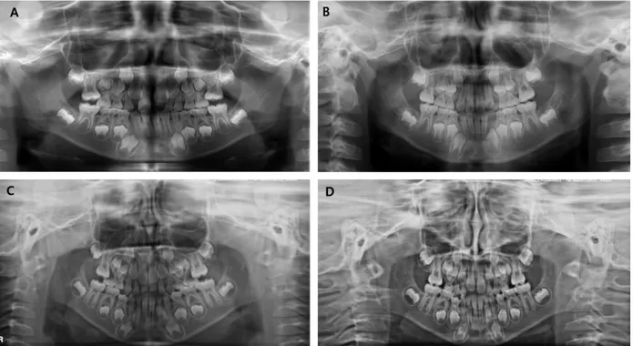
Fig. 1. Panoramic radiographs of reversible and irreversible ectopic eruption. Initial (A) and follow-up (B) panoramic radiograph classified as reversible ectopic eruption. Initial (C) and follow-up (D) panoramic radiograph classified as irreversible ectopic eruption.
1) Angles between the long axes of molars
The angles between the long axes of the maxillary pri- mary second molar and the FPM, the maxillary primary second molar and the tooth germ of the maxillary second permanent molar, and the FPM and the tooth germ of the maxillary second permanent molar were measured.
The long axis was set up with the line extending occlu-
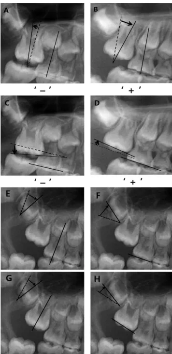
so-cervically parallel at the center of the tooth. In the case of second primary molar with the distal resorption, the center of the tooth was measured by presumed distal outline. The angle was defined as (+) when it showed distal angulation compared to the reference molar, and (-) when their relations were opposite (Fig. 2).
Fig. 2. Measurement methods of the angles on panoramic radiograph. A, B : Measurement method of the positive and negative value of angle between the long axes. C, D : Measurement method of the positive and negative value of angle between occlusal lines. E, F : Measurement method of the angle between second primary molar and the tooth germ of maxillary second permanent molar. G, H : Measurement method of the angle between FPM and the tooth germ of maxillary second permanent molar.
2) Angles between the occlusal lines of molars
The angles between the occlusal lines of the maxillary primary second molar and the maxillary FPM, the max- illary primary second molar and the tooth germ of the maxillary second permanent molar, and the maxillary FPM and the tooth germ of the maxillary second perma- nent molar were measured. The angle was defined as (+) when the distal part of the occlusal line of the molar was positioned higher than an imaginary line parallel to the occlusal line of the reference molar, and (-) when the opposite relationship was evident (Fig. 2).
3. Occlusal relationship in the primary dentition
The occlusal relationship in the primary dentition was evaluated using the relationship between the terminal plane of the maxillary and mandibular primary second molars. It was classified into the mesial step, distal step, and flush terminal plane.
4. Statistical analysis
The differences of angles among groups were analyzed by one-way ANOVA with 5% significance level. Paired t- tests were conducted to compare each group. SPSS ver- sion 15.0 (SPSS Inc., Chicago, IL, USA) was used for all statistical analyses.
Ⅲ. Results
The study population included 127 children (74 male and 53 girls). The normal eruption group consisted of 80 children (47 boys and 33 girls) and the ectopic eruption group included 47 children (27 boys and 20 girls).
Forty-seven children presented with 59 ectopic eruptions of the maxillary FPMs; of these, 28 cases were re- versible type and 31 were irreversible type.
Ectopic FPM occurred more frequently in boys than in girls. (Boy : girl ratio, 1.35 : 1) Ectopic FPM occurred unilaterally in 57.4% and bilaterally in 42.6%. Of uni- lateral ectopic FPMs, 59.3% (n = 16) were on the right side and 40.7% (n = 11) were on the left.
A paired t-test was performed to determine the error of the values measured by two operators. All measure- ments were compared respectively and no significant dif- ferences were found (p > 0.05).
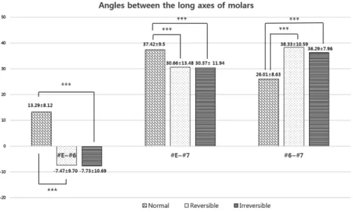
The angles between the long axes of molars are shown in Figure 3. The ectopic eruption group showed a (-) an- gle between the primary second molar and the FPM.
And ectopic eruption group showed smaller angles than the normal eruption group, with the exception of the an- gle between the long axes of the FPM and the tooth germ of the maxillary second permanent molar. These differences were statistically significant between the ec- topic eruption and normal eruption groups (p < 0.001),
Fig. 3. Angles between the long axes of molars. Values are mean ± standard deviation. #E : the second primary molar, #6 : the maxillary first permanent molar, #7 : the tooth germ of the second permanent molar. Paired t-test analysis (*** : p < 0.001, ** : p < 0.01, * : p < 0.05)
but not between the reversible and irreversible ectopic eruption groups (p > 0.05).
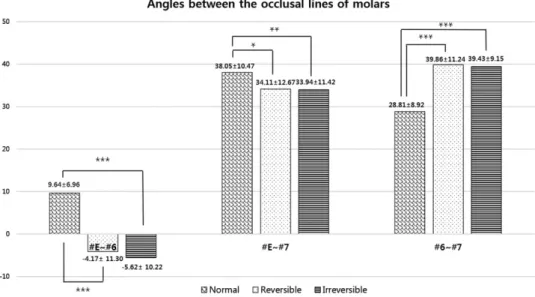
Figure 4 shows the angles between the occlusal lines in the molars. The ectopic eruption group showed a (-) angle between the primary second molar and the FPM.
Ectopic eruption group showed smaller angles than the normal eruption group, with the exception of the angle between the long axes of the FPM and the tooth germ of the maxillary second permanent molar. These differences were statistically significant (p < 0.05) between the ec- topic eruption and normal eruption groups but not be- tween the reversible and irreversible ectopic eruption groups (p > 0.05).
Data on the occlusal relationships of the primary den- tition are given in Table 1. In the normal eruption group, the primary relationship was mesial step (56%), followed by flush terminal plane (40%) and distal step (4%). In the reversible ectopic eruption group, the pri- mary relationship was mesial step (79%), followed by flush terminal plane (21%). And the irreversible ectopic eruption group showed the same trend: mesial step (77%), flush terminal plane (23%). Interestingly, there is no ectopic eruption in distal step.
Ⅳ. Discussion
The distinction between reversible- and irreversible- type ectopic eruption of the FPM is important in terms of early diagnosis and treatment of ectopic eruption of the maxillary FPM13). Although the cone beam computed tomography (CBCT) provides high resolution, accurate, real size images without superimposition. It would be clinically useful to develop a method to predict the possi- bility of self-correction of ectopic eruption using radi- ographs. Because CBCT is expensive, not widely avail- able and high-dose radiation would expose to the young patient compared to conventional techniques14). Therefore, in this study, panoramic radiographs was used for the evaluation, which are commonly used in dental examinations. It would be clinically useful to de- velop a way to predict the possibility of self-correction of ectopic eruption using panoramic radiographs, which are commonly used in dental examinations.
In this study, panoramic radiographs were used for evaluation. The points from which measurements should be taken are easily determined using panoramic radi- ographs. However, there is the possibility of errors in
Fig. 4. Angles between the occlusal lines of molars. Values are mean ± standard deviation. #E : the second primary molar, #6 : the maxillary first perma- nent molar, #7 : the tooth germ of second permanent molar. Paired t-test analysis (*** : p < 0.001, ** : p < 0.01, * : p < 0.05)
measurements because distortion may occur during im- age construction from such radiographs15-18). Samawi and Burke17) reported that the molar teeth expressed the smallest amount of distortion in both arches in panoramic radiographs. Although the method for mea- suring the angle on the panoramic radiograph has the possibility of errors, this is the most common and easy way which the clinician routinely gets.
Ectopic eruption was classified as reversible or irre- versible ectopic eruption by a series of radiograph. In the case of irreversible ectopic eruption, most were treated after diagnosis19). However, when it was difficult to maintain second primary molar due to the pain or mobil- ity, the treatment of space gaining after exfoliation or comprehensive orthodontic treatment was planned.
In accordance with previous reports, male predomi- nance in ectopic FPM was confirmed in our results20,21). Reversible ectopic eruption made up 47% of the total number of cases, in contrast to a previous report that 59% of ectopic FPMs are spontaneously corrected1).
Bjerklin and Kurol7) studied possible etiologic factors and suggested that increase in mesial angle and mesiodistal width of FPM were significant factors of irre- versible ectopic eruption of the FPM. Based on this study, the angle of long axis and occlusal line of maxil- lary FPM and adjacent teeth was measured using panoramic radiograph.
The angles of the long axes and occlusal lines between the primary second molar and FPM differed significantly between the normal and ectopic eruption groups, sug- gesting that the mesial angulation of the maxillary FPM is more pronounced in those with reversible and irre- versible ectopic eruption. Impacted FPM appeared to be hardly self-corrected as the angles between the long axes and between occlusal lines decreased. However, there were no significant differences between the reversible- and irreversible-type ectopic eruption groups.
Regarding the primary second molar and the tooth germ of the maxillary second permanent molar, the measured values differed significantly between the con- trol and ectopic eruption groups, suggesting that the mesial angulation of the tooth germ of the maxillary sec- ond permanent molar may be related to reversible and irreversible ectopic eruption of the FPM. However, there were no significant differences between the reversible- and irreversible-type ectopic eruption groups. The mea- surement method has a limitation in terms of construct- ing the long axis and occlusal line of the tooth germ of
second permanent molar, because the second permanent molar is in a tooth-germ state when the FPM erupts.
The angles of the long axes and occlusal lines between the FPM and the tooth germ of the second permanent molar can be calculated using the angle between the pri- mary second molar and FPM, and that between the pri- mary second molar and the tooth germ of the second permanent molar. However, the measurements show a different value compared to the calculated values. This is thought to be due to errors in measurement. Normal eruption group showed smaller angles than the ectopic eruption group and these differences were statistically significant, suggesting that the degree of mesial angula- tion of the molar is more severe in the FPM than the tooth germ of second permanent molar.
Regarding the occlusal relationship in the primary dentition, mesial step was more frequently observed in the ectopic eruption group compared to the normal erup- tion group. To diagnose malocclusion in the primary dentition, the relationship between the terminal planes of the primary second molars is used. In addition, the terminal plane is commonly used to predict future occlu- sion in the permanent dentition22,23). The likelihood of de- veloping a class I molar relationship in the permanent dentition is greatest when there is a mild mesial step during the primary dentition stage. In contrast, if there is an exaggerated mesial step, a class III permanent mo- lar relationship is likely to develop. The presence of a distal step is highly predictive of the development of a class II permanent molar relationship23). Thus, the ec- topic eruption group had a higher likelihood of a future class III permanent molar relationship, which is consis- tent with the findings of Salbach24).
Ⅴ. Conclusion
Within the limitations of the study, the angulation of the FPM and the tooth germ of the maxillary second permanent molar were found to affect ectopic eruption of the maxillary FPM. The ectopic FPM appeared to be hardly self-corrected as the FPM and the tooth germ of second permanent molar were more mesially angulated.
However, there were no significant differences between reversible- and irreversible-type ectopic eruptions. The high frequency of mesial step in the ectopic eruption group indicates that ectopic FPM is likely to occur in class III patients with maxillary deficiency.
References
1. Kurol J, Bjerklin K : Ectopic eruption of maxillary first permanent molars : a review. ASDC J Dent Child, 53:209-214, 1986.
2. Yuen S, Chan J, Tay F : Ectopic eruption of the maxillary permanent first molar : the effect of increased mesial angulation on arch length. J Am Dent Assoc, 111:447-451, 1985.
3. Young DH : Ectopic eruption of the first permanent molar. J Dent Child, 24:153-162, 1957.
4. Barberia-Leache E, Suarez-Clua MC, Saavedra- Ontiveros D : Ectopic eruption of the maxillary first permanent molar : Characteristics and occurrence in growing children. Angle Orthod, 75:610-615, 2005.
5. Chintakanon K, Boonpinon P : Ectopic eruption of the first permanent molars : prevalence and etiologic factors. Angle Orthod, 68:153-160, 1998.
6. Pulver F : The etiology and prevalence of ectopic eruption of the maxillary first permanent molar.
ASDC J Dent Child, 35:138-146, 1968.
7. Bjerklin K, Kurol J : Ectopic eruption of the maxil- lary first permanent molar : etiologic factors. Am J Orthod, 84:147-155, 1983.
8. Cossman MH : Ectopic eruption : first molar impaction in the mixed dentition. Dent Dig, 76:349- 353, 1970.
9. Kurol J, Bjerklin K : Resorption of maxillary second primary molars caused by ectopic eruption of the maxillary first permanent molar : a longitudinal and histological study. ASDC J Dent Child, 49:273-279, 1982.
10. Cheyne VD, Wessels KE : Impaction of permanent first molar with resorption and space loss in region of deciduous second molar. J Am Dent Assoc, 35:
774-787, 1947.
11. Bjerklin K : Ectopic eruption of the maxillary first permanent molar. An epidemiological, familial, etio- logical and longitudinal clinical study. Swed Dent J Suppl, 100:1-66, 1994.
12. Campbell OA : Ectopic eruption of the first perma- nent molar. J N J Dent Assoc, 62:62-65, 1991.
13. Toutountzakis N, Katsaris N : Ectopic eruption of the maxillary first permanent molar. Orthod Epitheorese, 2:117-128, 1990.
14. Sheikhi M, Dakhil-Alian M, Bahreinian Z : Accuracy and reliability of linear measurements using tangential projection and cone beam computed tomography. Dent Res J, 12:271-277, 2015.
15. Philipp RG, Hurst RV : The cant of the occlusal plane and distortion in the panoramic radiograph.
Angle Orthod, 48:317-323, 1978.
16. Tronje G, Welander U, McDavid WD, Morris CR : Image distortion in rotational panoramic radiogra- phy. III. Inclined objects. Acta Radiol Diagn, 22:
585-592, 1981.
17. Samawi SS, Burke PH : Angular distortion in the orthopantomogram. Br J Orthod, 11:100-107, 1984.
18. Stramotas S, Geenty JP, Petocz P, Darendeliler MA : Accuracy of linear and angular measurements on panoramic radiographs taken at various positions in vitro. Eur J Orthod, 24:43-52, 2002.
19. Oh MH, Lee SE, Park JH, et al. : Treatment of bilateral ectopic eruption of the first permanent molars. J Korean Acad Pediatr Dent, 40:48-52, 2013.
20. Bjerklin K, Kurol J : Prevalence of ectopic eruption of the maxillary first permanent molar. Swed Dent J, 5:29-34, 1981.
21. Kurol J, Bjerklin K : Ectopic eruption of maxillary first permanent molars : familial tendencies. ASDC J Dent Child, 49:35-38, 1982.
22. Baume LJ : Physiological tooth migration and its significance for the development of occlusion ; The biogenesis of accessional dentition. J Dent Res, 29:331-337, 1950.
23. Dean JA, Avery DR, Mcdonald RE : Dentistry for the child and adolescent, 9th ed., Mosby, 518-520, 2011.
24. Salbach A, Schremmer B, Grabowski R, Stahl de Castrillon F : Correlation between the frequency of eruption disorders for first permanent molars and the occurrence of malocclusion in early mixed denti- tion. J Orofac Orthop, 73:298-306, 2012.
주요어: 상악 제1대구치, 가역성 이소맹출, 비가역성 이소맹출
상악 제1대구치 이소 맹출의 예측 인자
선지민∙남옥형∙김미선∙이효설∙최성철 경희대학교 치의학전문대학원 소아치과학교실
본 연구는 이소맹출한 상악 제1대구치의 자율적 수정 가능성을 예측할 수 있는 진단적 기초를 제공하기 위하여 정상군과 가역성과 비가역성 이소맹출의 차이점을 비교 평가하고자 후향적으로 분석하였다. 상악 제1대구치와 인접치아 간의 장축과 교합평면간의 각도를 파노라마상에서 측정하였고, 제2유구치의 교합관계도 조사하였다. 이소맹출군과 정상군은 통계학적으 로 유의한 차이를 보였지만(p < 0.05), 가역성과 비가역성 이소맹출군 간에는 통계학적으로 유의한 차이는 없었다(p >
0.05). 제2유구치와 제1대구치 그리고 제2대구치 치배 간의 각도는 정상군에 비해 이소맹출 군에서 작은 값을 보였으며, 근 심 계단형이 이소맹출군에서 더 높은 빈도로 관찰되었다.
본 연구의 결과를 토대로 상악 제1대구치의 이소맹출은 상악 제1대구치와 제2대구치 치배의 각도와 관련이 있으며, 이소 맹출한 상악 제1대구치는 상악 저성장의 3급 부정교합 환자에서 보다 빈번하게 나타나는 경향이 있는 것을 알 수 있었다.
국문초록