• 검색 결과가 없습니다.

Mannosylerythritol lipids ameliorate ultraviolet A-induced aquaporin-3 downregulation by suppressing c-Jun N-terminal kinase phosphorylation in cultured human keratinocytes

N/A
N/A
Protected

Academic year: 2022

Share "Mannosylerythritol lipids ameliorate ultraviolet A-induced aquaporin-3 downregulation by suppressing c-Jun N-terminal kinase phosphorylation in cultured human keratinocytes"

Copied!
8
0
0

로드 중.... (전체 텍스트 보기)

전체 글

(1)

Author contributions: I.H.B., C.S.L., and D.Y.K. designed the study. I.H.B., S.H.L., S.O., H.W.C., P.A.M., J.W.Y., J.Y.K., and E.S.L. performed the experiments.

I.H.B., T.R.L., C.S.L., and D.Y.K. analyzed the data and contributed to writ- ing the manuscript. All authors read and approved the final version of the manuscript.

This is an Open Access article distributed under the terms of the Creative Commons Attribution Non-Commercial License, which permits unrestricted non-commercial use, distribution, and reproduction in any medium, provided the original work is properly cited.

Copyright © Korean J Physiol Pharmacol, pISSN 1226-4512, eISSN 2093-3827

INTRODUCTION

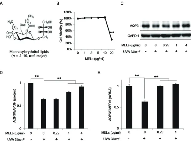
Mannosylerythritol lipids (MELs) are extracellular glycolipids that are produced from different vegetable oil substrates by the yeast strains of genus Pseudozyma [1]. MELs comprise a hydro- philic headgroup containing 4-O--D-mannopyranosyl eryth-

ritol or 1-O--D-mannopyranosyl erythritol and fatty acid-con- taining hydrophobic chains [1]. MELs were originally developed as a highly biodegradable biosurfactant [2], but were subsequently found to have additional biological properties such as anti- inflammation and apoptosis of melanoma cells [3-7]. As the pos- sible applications of MELs expanded, some researchers focused

Original Article

Mannosylerythritol lipids ameliorate ultraviolet A-induced aquaporin-3 downregulation by suppressing c-Jun N-terminal kinase phosphorylation in cultured human keratinocytes

Il-Hong Bae 1,3 , Sung Hoon Lee 1 , Soojung Oh 1 , Hyeongwon Choi 1 , Paulo A. Marinho 1 , Jae Won Yoo 1 , Jae Young Ko 1 , Eun-Soo Lee 1 , Tae Ryong Lee 1 , Chang Seok Lee 2, *, and Dae-Yong Kim 3, *

1

R&D Center, Amorepacific Corporation, Yongin 17074,

2

Department of Beauty and Cosmetic Science, Eulji University, Seongnam 13135,

3

Department of Vet- erinary Pathology, College of Veterinary Medicine, Seoul National University, Seoul 08826, Korea

ARTICLE INFO

Received October 26, 2018 Revised December 17, 2018 Accepted December 24, 2018

*Correspondence Chang Seok Lee

E-mail: [email protected] Dae-Yong Kim

E-mail: [email protected] Key Words

Aquaporin-3

JNK mitogen-activated protein kinases Mannosylerythritol lipid

PPAR gamma Ultraviolet

ABSTRACT Mannosylerythritol lipids (MELs) are glycolipids and have several phar-

macological efficacies. MELs also show skin-moisturizing efficacy through a yet-

unknown underlying mechanism. Aquaporin-3 (AQP3) is a membrane protein that

contributes to the water homeostasis of the epidermis, and decreased AQP3 expres-

sion following ultraviolet (UV)-irradiation of the skin is associated with reduced skin

moisture. No previous study has examined whether the skin-moisturizing effect of

MELs might act through the modulation of AQP3 expression. Here, we report for the

first time that MELs ameliorate the UVA-induced downregulation of AQP3 in cultured

human epidermal keratinocytes (HaCaT keratinocytes). Our results revealed that UVA

irradiation decreases AQP3 expression at the protein and messenger RNA (mRNA)

levels, but that MEL treatment significantly ameliorated these effects. Our mitogen-

activated protein kinase inhibitor analysis revealed that phosphorylation of c-Jun

N-terminal kinase (JNK), but not extracellular signal-regulated kinase or p38, medi-

ates UVA-induced AQP3 downregulation, and that MEL treatment significantly sup-

pressed the UVA-induced phosphorylation of JNK. To explore a possible mechanism,

we tested whether MELs could regulate the expression of peroxidase proliferator-

activated receptor gamma (PPAR-), which acts as a potent transcription factor for

AQP3 expression. Interestingly, UVA irradiation significantly inhibited the mRNA

expression of PPAR- in HaCaT keratinocytes, whereas a JNK inhibitor and MELs

significantly rescued this effect. Taken together, these findings suggest that MELs

ameliorate UVA-induced AQP3 downregulation in HaCaT keratinocytes by suppress-

ing JNK activation to block the decrease of PPAR-. Collectively, our findings suggest

that MELs can be used as a potential ingredient that modulates AQP3 expression to

improve skin moisturization following UVA irradiation-induced damage.

(2)

on using MELs as a cosmetic ingredient; to date, several studies have found that MELs can moisturize the skin. Morita et al. [8,9]

reported that MELs exerted recovery effect on the viability of so- dium dodecyl sulfate (SDS)-damaged human skin cells in a three- dimensional skin equivalent model. In addition, Yamamoto et al.

[10] reported that topical application of MELs to human forearm skin resulted in increased level of water-retention and decreased level of transepidermal water loss for approximately 2 h.

Aquaporin-3 (AQP3) contributes to water homeostasis in the epidermis and is responsible for transporting water and glycerol at the plasma membranes of keratinocytes in the epidermal basal layer [11-13]. Decreased AQP3 expression in the skin is associated with dry skin, reduced elasticity, decreased glycerol levels, and impaired wound healing with defective barrier function [13,14].

AQP3 expression in the skin naturally declines with age [15], but it may also be reduced by harmful external stimuli, such as ultra- violet (UV) irradiation and reactive oxygen species [16]. Numer- ous studies have sought to identify materials that may ameliorate the UV irradiation-induced downregulation of AQP3 expression and to evaluate the possible mechanisms underlying such effects [16-19].

The mitogen-activated protein kinase (MAPK) pathways, namely the extracellular signal-regulated kinase (ERK), c-Jun N- terminal kinase (JNK), and p38 pathways, play important roles in regulating and eliciting various cellular responses, such as proliferation, differentiation, development, transformation, and apoptosis [20]. In epidermal keratinocytes, a diverse range of ex- tracellular stimuli, including UV radiation, strongly induce the phosphorylation of MAPKs [16-18]. AQP3 expression is report- edly regulated by the MAPK pathways [16,21] as well as by the transcription factor, peroxidase proliferator-activated receptor gamma (PPAR-), which may be modulated by MAPKs or UV ir- radiation [22-24].

To our knowledge, no previous study had examined whether MELs are involved in modulating AQP3 expression in UV- irradiated keratinocytes. Here, we evaluated whether MELs can ameliorate UVA-induced AQP3 downregulation in cultured human keratinocytes, and then explored a possible underlying mechanism.

METHODS

Reagents

The MELs used in the present study were produced and provided by DKBIO (Daejeon, Korea). Briefly, MELs were syn- thesized in a yeast strain of Pseudozyma sp., extracted from the culture broth with ethyl acetate, and purified through a fatty acid removal process. Purified MELs were dissolved with dimethyl sulfoxide for use in the experiments. MELs are mixtures consist- ing of MEL-A, MEL-B, MEL-C, and MEL-D. The MELs used in

this study are composed of predominant MEL-B with a purity of about 95% and their molecular weight range is 500 to 850. MAPK inhibitors were purchased from Tocris (#1879; Minneapolis, MN, USA) and used at concentrations selected with reference to previ- ous studies (100 nM SP600125 for JNK, 10 M PD98059 for ERK, and 5 M SB203580 for p38) [16,25].

Cell culture

HaCaT keratinocytes (an immortalized cell line derived from normal human keratinocytes) were purchased from the Ameri- can Type Culture Collection (Manassas, VA, USA) and cultured in Dulbecco’s modified Eagle’s medium (DMEM; 12-604F, Lonza) supplemented with 10% fetal bovine serum (#10082-147;

Thermo Fisher Scientific, Waltham, MA, USA), 100 U/ml potas- sium penicillin, and 100 mg/ml streptomycin sulfate (17-602E;

Lonza, Basel, Switzerland) at 37°C in a humidified incubator con- taining 5% CO

2

. The cells were washed with phosphate-buffered saline (21-031-CVR; Corning, Corning, NY, USA), detached with Accutase (SCR005; EMD Millipore, Burlington, MA, USA), and plated to 60-mm dishes at 0.3 × 10

6

cells/well in fresh culture me- dium. Cells grown to 70%–80% confluence were serum-starved for 4 h and then treated with the indicated concentrations of re- agents.

UVA irradiation

UVA irradiation (365 nm) was performed using a micropro- cessor-controlled, cooled UV irradiation system (Bio-Sun UV- H; Vilber Lourmat, Collégien, France). As previously described [26], cells were covered with serum and phenol red-free DMEM (#21063-029; Thermo Fisher Scientific) during irradiation. After irradiation, cells were returned to fresh DMEM (12-604F; Lonza) for culture.

Cell viability assay

Cell viability was determined using a Cell Counting Kit-8 (CCK-8) (CK04-05; Dojindo, Kumamoto, Japan). HaCaT kerati- nocytes seeded to a 96-well plate at a density of 5 × 10

3

cells/well were incubated with MELs for 24 h. Then, 10 l of CCK-8 solu- tion and 90 l of DMEM (#31053-028; Thermo Fisher Scientific) were added to each well and the plate was incubated at 37°C for 2 h. The absorbance was measured at 450 nm using a microplate reader (Synergy2; BioTek, Winooski, VT, USA).

RNA extraction and quantitative real-time polymerase chain reaction (qRT-PCR)

Total RNA was extracted using the RNeasy Mini Kit (#74101;

Qiagen, Venlo, the Netherlands). One microgram of RNA was

reverse transcribed into complementary DNA (cDNA) using a

(3)

SuperScript III First-Strand Synthesis System according to the manufacturer’s instructions (#18080-051; Invitrogen, Carlsbad, CA, USA). For qRT-PCR, we used an ABI 7500 Fast Real-Time PCR system (Applied Biosystems, Foster City, CA, USA) with the TaqMan Gene Expression Master Mix (#4369016; Thermo Fisher Scientific) and pre-inventoried TaqMan primers for AQP3 (Hs00185020_m1), PPAR- (Hs01115513_m1), and glyceralde- hyde-3-phosphate dehydrogenase (GAPDH, detected as a nor- malization control for cDNA quantity; Hs02786624_g1). The level of relative messenger RNA (mRNA) expression was calculated using the comparative ΔΔCt method [27].

Western blots analysis

Cells were lysed using RIPA Lysis and Extraction Buffer (#89900; Thermo Fisher Scientific) containing a complete mini protease inhibitor (#11836153001; Roche, Basel, Switzerland) and a phosphatase inhibitor cocktail (P5726; Sigma-Aldrich, St. Louis,

MO, USA). The cell lysates were centrifuged for 20 min at 13,000 rpm and 4°C, and the supernatants were collected for analysis.

Protein concentrations were determined using a BCA protein assay kit (#23225; Thermo Fisher Scientific). Equal amounts of protein (30 g/well) were loaded to a 4%–12% gradient Bis- Tris gel (Nu PAGE; Invitrogen), separated by SDS-PAGE, and transferred to a nitrocellulose membrane (LC2000; Invitrogen).

The membrane was blocked with TBST buffer (0.2% Tween 20 in Tris-buffered saline) containing 5% bovine serum albumin at room temperature for 1 h and probed overnight at 4°C with each of the following primary antibodies: AQP3 (ab125129; Abcam, Cambridge, UK), PPAR- (NBP1-61399; Novus, Centennial, CO, USA), GAPDH (#2118; Cell Signaling Technology [CST], Dan- vers, MA, USA), JNK (#9252; CST), p-JNK (#4668; CST), ERK (#4695; CST), p-ERK (#4370; CST), p38 (#8690; CST), and p-p38 (#4511; CST). After three washes with TBST, the membrane was incubated with horseradish peroxidase-conjugated secondary an- tibodies at room temperature for 1 h. Chemiluminescent signals

Fig. 1. AQP3 expression is downregulated by UVA irradiation. (A) HaCaT keratinocytes were irradiated with the indicated doses of UVA or left non-

irradiated (control) and harvested after 24 h for Western blot analysis. (B) The protein level of AQP3 was quantified relative to that of GAPDH using

the Image J software from National Institutes of Health. The results are presented as the mean expression level obtained from three independent

experiments ± SD (**p < 0.01). (C) UVA 3 J/cm

2

-irradiated and non-irradiated control cells were harvested at the indicated time points, and qRT-PCR

analysis of the AQP3 mRNA level was performed. The data were normalized with respect to the expression of GAPDH, and the results are presented as

the mean expression ± SD (n = 3, **p < 0.01). (D) HaCaT keratinocytes were treated with the indicated doses of UVA and cell viability was determined

using a CCK-8 kit (Dojindo, Kumamoto, Japan) 24 h after irradiation of indicated doses of UVA. The viability of cells treated with UVA 3 J/cm

2

decreased

to 90% without significance. AQP3, aquaporin-3; UVA, ultraviolet A; N.S., not significant; GAPDH, glyceraldehyde-3-phosphate dehydrogenase; SD,

standard deviation; qRT-PCR, quantitative real-time polymerase chain reaction; mRNA, messenger RNA.

(4)

were detected with an ECL substrate (RPN2232; GE Healthcare, Hammersmith, UK) and visualized using a chemiluminescent detection system (LAS-3000; Fuji Film, Tokyo, Japan).

Statistical analysis

Data are expressed as means ± standard deviation, and the Stu- dent’s t-test was used for statistical comparisons. A p-value less than 0.05 was considered statistically significant (*, < 0.05; **, <

0.01).

RESULTS

UVA-irradiation downregulates AQP3 expression at the protein and mRNA levels

Prior to define the effect of MELs on UVA-induced AQP3

downregulation, we decided to determine the optimal dose of UVA for significantly downregulating AQP3 expression in our experimental system. First, we irradiated HaCaT keratinocytes with various doses of UVA (0.5, 1, 2, or 3 J/cm

2

) and analyzed the protein level of AQP3 after 24 h (Fig. 1A). The AQP3 protein level was dose-dependently reduced under UVA irradiation, and significantly decreased to 50% of the level seen in the UVA non-irradiation control group under 3 J/cm

2

of UVA irradiation (UVA 3 J/cm

2

) (Fig. 1B). Similarly, qRT-PCR revealed that the mRNA expression of AQP3 was significantly reduced to 54% of the control level at 24 h after irradiation with UVA 3 J/cm

2

. A time-course experiment revealed that a significant decrease in AQP3 mRNA expression could be detected as early as 6 h after UVA irradiation, and this level continued to decrease gradually until 24 h post-irradiation (Fig. 1C). Since cell viability was not significantly different from the control group at 24 h after UVA 3 J/cm

2

irradiation (Fig. 1D), we subsequently used UVA 3 J/cm

2

to significantly reduce AQP3 expression in HaCaT keratinocytes.

Fig. 2. MELs ameliorate the UVA-induced downregulation of AQP3 expression in HaCaT keratinocytes. (A) A structure of MELs. Two fatty acids were attached to –OH groups on mannosylerythritol as an ester bond. The fatty acid contains mainly 6-18 carbon atoms and the major is 8 carbon atoms. (B) HaCaT keratinocytes were treated with the indicated concentrations of MELs for 24 h and cell viability was determined using a CCK-8 kit (Dojindo, Kumamoto, Japan). DMSO was used as the vehicle control. (C) Cells treated with UVA 3 J/cm

2

and the indicated concentrations of MELs for 24 h were subjected to Western blot analysis of AQP3 proteins. (D) The protein levels of AQP3 were quantified relative to that of GAPDH using the Image J software. (E) The mRNA expression of AQP3 was analyzed by qRT-PCR. The results are presented as the mean expression level obtained from three independent experiments ± SD (**p < 0.01). MELs, mannosylerythritol lipids; UVA, ultraviolet A; AQP3, aquaporin-3; DMSO, dimethyl sulfoxide;

GAPDH, glyceraldehyde-3-phosphate dehydrogenase; mRNA, messenger RNA; qRT-PCR, quantitative real-time polymerase chain reaction; SD, stan-

dard deviation.

(5)

MELs ameliorate the UVA-induced downregulation of AQP3

Next, we examined whether MELs (Fig. 2A) could ameliorate the UVA-induced downregulation of AQP3 expression. Non- cytotoxic concentrations of MELs (0.25, 1, and 4 g/ml) were selected based on the results of our cell viability assay (Fig. 2B) and applied to cells for 24 h following UVA 3 J/cm

2

irradiation.

As shown in Fig. 2C, the UVA-induced downregulation of AQP3 protein expression was markedly and dose-dependently amelio- rated by 1 and 4 g/ml of MELs. These levels were increased by 124% and 142%, respectively, compared to the UVA 3 J/cm

2

-irra- diated group (Fig. 2D). AQP3 mRNA expression was increased by 164% when UVA 3 J/cm

2

-irradiated cells were treated with 1 g/

ml MELs compared to those receiving UVA 3 J/cm

2

-irradiation alone (Fig. 2E). This indicates that MEL 1 g/ml is sufficient to alleviate the UVA-induced reduction of AQP3.

Phosphorylation of JNK is involved in the UVA- induced downregulation of AQP3

Since the MAPK pathway is reportedly involved in modulat- ing AQP3 expression [16,21], we investigated whether UVA- induced AQP3 downregulation was associated with changes in the MAPKs, JNK, ERK, and p38 MAPK. Serum-starved HaCaT keratinocytes were harvested at 15 min and 30 min after UVA 3 J/

cm

2

irradiation. Our analyses revealed that the phosphorylation levels of the three MAPKs were significantly increased at both time points compared to those in non-irradiated cells (Fig. 3A).

Next, we treated serum-starved cells with an inhibitor of each MAPK for 1 h followed by UVA 3 J/cm

2

irradiation, and collected cell lysates 24 h later. We found that the UVA-induced down- regulation of AQP3 protein expression was markedly ameliorated by the JNK inhibitor, which brought this level up to 138% that of the UVA 3 J/cm

2

-irradiated group (Fig. 3B). Similarly, the mRNA expression level of AQP3 was 136% higher in the JNK inhibitor-

Fig. 3. Involvement of JNK phosphorylation in the UVA-induced downregulation of AQP3. (A) Cells were irradiated with UVA 3 J/cm

2

and har-

vested at the indicated time points. Protein samples were subjected to Western blotting for phospho-JNK, total JNK, phospho-ERK, total ERK, phos-

pho-p38, total p38, and GAPDH. (B) Cells pretreated with 100 nM SP600125 (a JNK inhibitor) for 1 h were irradiated with UVA 3 J/cm

2

and harvested af-

ter 24 h. The protein level of AQP3 was analyzed by Western blotting and quantified relative to the level of GAPDH using the ImageJ software. (C) The

mRNA expression of AQP3 was examined by qRT-PCR. (D) Cells pretreated with 10 μM PD98059 (an ERK inhibitor) or 5 μM SB203580 (a p38 inhibitor)

for 1 h were irradiated with UVA 3 J/cm

2

and harvested after 24 h. The protein level of AQP3 was analyzed by Western blotting and quantified relative

to the level of GAPDH using the ImageJ software. Values represent the mean expression level ± SD (n = 3, *p < 0.05, **p < 0.01). JNK, c-Jun N-terminal

kinase; UVA, ultraviolet A; ERK, extracellular signal-regulated kinase; GAPDH, glyceraldehyde-3-phosphate dehydrogenase; AQP3, aquaporin-3; mRNA,

messenger RNA; qRT-PCR, quantitative real-time polymerase chain reaction; SD, standard deviation.

(6)

treated group than in the UVA 3 J/cm

2

-irradiated group (Fig.

3C). In contrast, the inhibitors of ERK or p38 failed to rescue the UVA-induced decrease of AQP3 protein expression (Fig. 3D).

MELs ameliorate UVA-induced AQP3 downregulation by suppressing phosphorylation of JNK

Based on the above findings, we assessed the potential involve- ment of the MAPK pathway in the ability of MELs to ameliorate the UVA-induced downregulation of AQP3 expression. Serum- starved HaCaT keratinocytes were pretreated with 0.5 or 1 g/ml MELs for 1 h, subjected to UVA 3 J/cm

2

irradiation, and then har- vested 15 m later. Indeed, we found that the UVA-induced upreg- ulation of phosphorylated JNK was suppressed by MEL pretreat- ment, whereas the levels of phosphorylated ERK and p38 were unchanged (Fig. 4A). Interestingly, a few studies have reported the relationship between JNK activation and PPAR- expression [22-24]. Particularly, JNK activation decreased PPAR- mRNA expression and subsequently led to AQP3 downregulation in he- patic stellate cells. These findings suggest the possibility that the

MELs could up-regulate AQP3 expression through JNK inactiva- tion and subsequent the increased PPAR- expression in UVA- irradiated HaCaT cells. Thus, we decided to focus on PPAR-

expression after MELs treatment in UVA-irraidated HaCaT cells.

In line with a previous study, we found that the mRNA expres- sion of PPAR- was downregulated by UVA 3 J/cm

2

irradiation in HaCaT keratinocytes; however, we observed for the first time that this phenomenon was significantly mitigated by MELs or the JNK inhibitor (Fig. 4B). Taken together, MELs ameliorated UVA- induced AQP3 downregulation through JNK inactivation and PPAR- up-regulation in cultured human keratinocytes (Fig. 4C).

DISCUSSION

AQP3 is well-known to be involved in skin hydration [28].

Thus, it seems logical that restoring a reduced AQP3 expression level (i.e., that triggered by environmental factors, such as UV irradiation) to its normal level could help improve skin hydra- tion. Numerous studies have sought to identify materials that can

Fig. 4. The effects of MELs on JNK phosphorylation and PPAR-γ expression in UVA-irradiated HaCaT keratinocytes. (A) Cells pretreated with the

indicated concentrations of MELs for 1 h were irradiated with UVA 3 J/cm

2

and harvested after 15 min. The protein levels of phospho-JNK, total JNK,

phospho-ERK, total ERK, phospho-p38, total p38, and GAPDH were evaluated by Western blot analysis. (B) Cells treated with UVA 3 J/cm

2

and the in-

dicated concentrations of MELs or a JNK inhibitor for 16 h were prepared for qRT-PCR analysis of PPAR-γ mRNA expression. Values represent the mean

expression level obtained from three independent experiments ± SD (*p < 0.05, **p < 0.01). (C) Proposed model showing how MELs ameliorate the

UVA-induced downregulation of AQP3. MELs, mannosylerythritol lipids; JNK, c-Jun N-terminal kinase; PPAR-γ, peroxidase proliferator-activated recep-

tor gamma; UVA, ultraviolet A; ERK, extracellular signal-regulated kinase; GAPDH, glyceraldehyde-3-phosphate dehydrogenase; qRT-PCR, quantitative

real-time polymerase chain reaction; SD, standard deviation.

(7)

effectively ameliorate UVB-induced AQP3 downregulation in HaCaT keratinocytes [16-19]. However, UVA irradiation can also induce photo-aging of skin [29] and was reported to reduce AQP3 expression in cultured human keratinocytes [26]. Hwang and Shim [26] showed that AQP3 mRNA expression was significantly downregulated in UVA 5 J/cm

2

-irradiated normal human epider- mal keratinocytes, but the authors did not examine any potential underlying mechanism. In the present study, we confirm that UVA irradiation markedly reduces AQP3 expression at both the protein and mRNA levels in HaCaT keratinocytes. Furthermore, we demonstrate that, of the tested MAPK inhibitors, only the JNK inhibitor ameliorates the UVA-induced downregulation of AQP3 in our system. This suggests that the UVA-activated JNK pathway may be an underlying mechanism through which this irradia- tion downregulates AQP3. The UVB-induced downregulation of AQP3 expression has been linked with activation of the ERK or p38 pathways [16-18]. These reports found that a JNK inhibitor did little to mitigate the UVB-induced downregulation of AQP3.

Although further systematic investigations are warranted, the previous and present findings suggest that the MAPK involved in reducing AQP3 expression in UV-irradiated keratinocytes may differ by the type of UV irradiation.

Various biological effects have been reported for MELs, such as induction of differentiation in mouse melanoma cells and rat pheochromocytoma cells [4,5], enhancement of plasmid DNA transfection efficiency [7], and anti-inflammatory activity [3].

Thus, MELs are thought to be versatile candidates for use in vari- ous research and industrial fields. In addition, MELs may have potential for cosmetic use in the areas of skin care, damaged hair care, anti-oxidative effects, and facial color make-up [8-10,30,31].

Therefore, it is important to explore their yet-unidentified func- tions. The present study demonstrates a new biological function of MELs, in that they can alleviate the UVA-induced decrease of AQP3 expression in keratinocytes by suppressing JNK activation.

We believe that our findings strengthen the value of MELs as a skin-moisturizing ingredient when combined with its previously reported efficacies in restoring damaged cells and augmenting water retention in the stratum corneum to hydrate dry skin [9,10].

Given that AQP3 expression was found to be downregulated by severe UVA irradiation of normal human skin fibroblasts [21,32], future efforts are warranted to study the effects of MELs against the UVA-induced downregulation of AQP3 in human fibroblasts.

JNK is reportedly involved in regulating PPAR- expression.

For example, Camp et al. [22] found that JNK activation reduced the effect of PPAR- ligands by decreasing PPAR- mRNA ex- pression. Meanwhile, Tardelli et al. [24] found that activation of the JNK pathway suppressed PPAR- to downregulate AQP3 expression in hepatic stellate cells carrying the genetic polymor- phism I148M of the patatin-like phospholipase domain-contain- ing 3 (PNPLA3 I148M). In the abovementioned study, restoration of AQP3 expression was triggered by SP600125-mediated JNK in- hibition and/or PPAR- stimulation [24]. These findings led us to

investigate whether PPAR- expression was altered in our system.

Consistent with a previous study showing that PPAR- mRNA expression was reduced in UVA-irradiated human adipose tissue- derived mesenchymal stem cells [23], we found that PPAR-

mRNA expression was downregulated in UVA-irradiated HaCaT keratinocytes. Moreover, this downregulation of PPAR- mRNA expression was markedly counteracted by treatment with a JNK inhibitor or MELs. Based on our present findings, we propose that MELs suppress the UVA-induced phosphorylation of JNK, which prevents the UVA-induced decrease of PPAR- expression to ameliorate the reduction of AQP3 expression (Fig. 4C).

In summary, we herein show for the first time that MELs can ameliorate UVA-induced AQP3 downregulation in cultured hu- man keratinocytes. We further reveal that UVA-induced AQP3 downregulation can be ameliorated via the ability of JNK to modulate PPAR- expression in cultured human keratinocytes.

Collectively, our findings support the potential development of MELs for use in moisturizing skin that has been damaged by UVA irradiation.

ACKNOWLEDGEMENTS

This research was supported and funded by Amorepacific R&D Center, and the Basic Science Research Program through the National Research Foundation of Korea (NRF) funded by the Ministry of Education (Grant Number: 2018R1D1A1B07049402).

CONFLICTS OF INTEREST

The authors declare no conflicts of interest.

REFERENCES

1. Morita T, Fukuoka T, Imura T, Kitamoto D. Mannosylerythritol lipids: production and applications. J Oleo Sci. 2015;64:133-141.

2. Kitamoto D, Yanagishita H, Shinbo T, Nakane T, Kamisawa C, Na- kahara T. Surface active properties and antimicrobial activities of mannosylerythritol lipids as biosurfactants produced by Candida antarctica. J Biotechnol. 1993;29:91-96.

3. Morita Y, Tadokoro S, Sasai M, Kitamoto D, Hirashima N. Bio- surfactant mannosyl-erythritol lipid inhibits secretion of inflam- matory mediators from RBL-2H3 cells. Biochim Biophys Acta.

2011;1810:1302-1308.

4. Zhao X, Wakamatsu Y, Shibahara M, Nomura N, Geltinger C, Na- kahara T, Murata T, Yokoyama KK. Mannosylerythritol lipid is a potent inducer of apoptosis and differentiation of mouse melanoma cells in culture. Cancer Res. 1999;59:482-486.

5. Wakamatsu Y, Zhao X, Jin C, Day N, Shibahara M, Nomura N, Na-

kahara T, Murata T, Yokoyama KK. Mannosylerythritol lipid induc-

es characteristics of neuronal differentiation in PC12 cells through

(8)

an ERK-related signal cascade. Eur J Biochem. 2001;268:374-383.

6. Im JH, Yanagishita H, Ikegami T, Takeyama Y, Idemoto Y, Koura N, Kitamoto D. Mannosylerythritol lipids, yeast glycolipid biosur- factants, are potential affinity ligand materials for human immuno- globulin G. J Biomed Mater Res A. 2003;65:379-385.

7. Ueno Y, Inoh Y, Furuno T, Hirashima N, Kitamoto D, Nakanishi M.

NBD-conjugated biosurfactant (MEL-A) shows a new pathway for transfection. J Control Release. 2007;123:247-253.

8. Morita T, Fukuoka T, Imura T, Kitamoto D. Production of manno- sylerythritol lipids and their application in cosmetics. Appl Micro- biol Biotechnol. 2013;97:4691-4700.

9. Morita T, Kitagawa M, Suzuki M, Yamamoto S, Sogabe A, Yanagi- dani S, Imura T, Fukuoka T, Kitamoto D. A yeast glycolipid biosur- factant, mannosylerythritol lipid, shows potential moisturizing ac- tivity toward cultured human skin cells: the recovery effect of MEL- A on the SDS-damaged human skin cells. J Oleo Sci. 2009;58:639- 642.

10. Yamamoto S, Morita T, Fukuoka T, Imura T, Yanagidani S, Sogabe A, Kitamoto D, Kitagawa M. The moisturizing effects of glycolipid biosurfactants, mannosylerythritol lipids, on human skin. J Oleo Sci. 2012;61:407-412.

11. Sugiyama Y, Ota Y, Hara M, Inoue S. Osmotic stress up-regulates aquaporin-3 gene expression in cultured human keratinocytes. Bio- chim Biophys Acta. 2001;1522:82-88.

12. Hara-Chikuma M, Verkman AS. Aquaporin-3 functions as a glyc- erol transporter in mammalian skin. Biol Cell. 2005;97:479-486.

13. Ma T, Hara M, Sougrat R, Verbavatz JM, Verkman AS. Impaired stratum corneum hydration in mice lacking epidermal water chan- nel aquaporin-3. J Biol Chem. 2002;277:17147-17153.

14. Hara M, Ma T, Verkman AS. Selectively reduced glycerol in skin of aquaporin-3-deficient mice may account for impaired skin hydra- tion, elasticity, and barrier recovery. J Biol Chem. 2002;277:46616- 46621.

15. Li J, Tang H, Hu X, Chen M, Xie H. Aquaporin-3 gene and protein expression in sun-protected human skin decreases with skin ageing.

Australas J Dermatol. 2010;51:106-112.

16. Cao C, Wan S, Jiang Q, Amaral A, Lu S, Hu G, Bi Z, Kouttab N, Chu W, Wan Y. All-trans retinoic acid attenuates ultraviolet radiation- induced down-regulation of aquaporin-3 and water permeability in human keratinocytes. J Cell Physiol. 2008;215:506-516.

17. Jeon BK, Kang MK, Lee GT, Lee KK, Lee HS, Woo WH, Mun YJ.

EPA attenuates ultraviolet radiation-induced downregulation of aquaporin-3 in human keratinocytes. Arch Pharm Res. 2015;

38:1552-1560.

18. Ji C, Yang Y, Yang B, Xia J, Sun W, Su Z, Yu L, Shan S, He S, Cheng L, Wan Y, Bi Z. Trans-Zeatin attenuates ultraviolet induced down-reg-

ulation of aquaporin-3 in cultured human skin keratinocytes. Int J Mol Med. 2010;26:257-263.

19. Shan SJ, Xiao T, Chen J, Geng SL, Li CP, Xu X, Hong Y, Ji C, Guo Y, Wei H, Liu W, Li D, Chen HD. Kanglaite attenuates UVB-induced down-regulation of aquaporin-3 in cultured human skin keratino- cytes. Int J Mol Med. 2012;29:625-629.

20. Zhang W, Liu HT. MAPK signal pathways in the regulation of cell proliferation in mammalian cells. Cell Res. 2002;12:9-18.

21. Cao C, Sun Y, Healey S, Bi Z, Hu G, Wan S, Kouttab N, Chu W, Wan Y. EGFR-mediated expression of aquaporin-3 is involved in human skin fibroblast migration. Biochem J. 2006;400:225-234.

22. Camp HS, Tafuri SR, Leff T. c-Jun N-terminal kinase phosphory- lates peroxisome proliferator-activated receptor-gamma1 and nega- tively regulates its transcriptional activity. Endocrinology. 1999;

140:392-397.

23. Lee J, Lee J, Jung E, Kim YS, Roh K, Jung KH, Park D. Ultraviolet A regulates adipogenic differentiation of human adipose tissue- derived mesenchymal stem cells via up-regulation of Kruppel-like factor 2. J Biol Chem. 2010;285:32647-32656.

24. Tardelli M, Bruschi FV, Claudel T, Moreno-Viedma V, Halilbasic E, Marra F, Herac M, Stulnig TM, Trauner M. AQP3 is regulated by PPAR and JNK in hepatic stellate cells carrying PNPLA3 I148M.

Sci Rep. 2017;7:14661.

25. Silvers AL, Bachelor MA, Bowden GT. The role of JNK and p38 MAPK activities in UVA-induced signaling pathways leading to AP-1 activation and c-Fos expression. Neoplasia. 2003;5:319-329.

26. Hwang HS, Shim JH. Brazilin and Caesalpinia sappan L. extract protect epidermal keratinocytes from oxidative stress by inducing the expression of GPX7. Chin J Nat Med. 2018;16:203-209.

27. Livak KJ, Schmittgen TD. Analysis of relative gene expression data using real-time quantitative PCR and the 2

−ΔΔCT

method. Methods.

2001;25:402-408.

28. Hara-Chikuma M, Verkman AS. Roles of aquaporin-3 in the epi- dermis. J Invest Dermatol. 2008;128:2145-2151.

29. Kligman LH. The hairless mouse and photoaging. Photochem Pho- tobiol. 1991;54:1109-1118.

30. Morita T, Kitagawa M, Yamamoto S, Sogabe A, Imura T, Fukuoka T, Kitamoto D. Glycolipid biosurfactants, mannosylerythritol lipids, repair the damaged hair. J Oleo Sci. 2010;59:267-272.

31. Takahashi M, Morita T, Fukuoka T, Imura T, Kitamoto D. Glycolip- id biosurfactants, mannosylerythritol lipids, show antioxidant and protective effects against H

2

O

2

-induced oxidative stress in cultured human skin fibroblasts. J Oleo Sci. 2012;61:457-464.

32. Xie H, Liu F, Liu L, Dan J, Luo Y, Yi Y, Chen X, Li J. Protective role of AQP3 in UVA-induced NHSFs apoptosis via Bcl2 up-regulation.

Arch Dermatol Res. 2013;305:397-406.

수치

Fig. 1. AQP3 expression is downregulated by UVA irradiation.  (A) HaCaT keratinocytes were irradiated with the indicated doses of UVA or left non- non-irradiated (control) and harvested after 24 h for Western blot analysis
Fig. 2. MELs ameliorate the UVA-induced downregulation of AQP3 expression in HaCaT keratinocytes
Fig. 3. Involvement of JNK phosphorylation in the UVA-induced downregulation of AQP3.  (A) Cells were irradiated with UVA 3 J/cm 2  and har- har-vested at the indicated time points
Fig. 4. The effects of MELs on JNK phosphorylation and PPAR-γ expression in UVA-irradiated HaCaT keratinocytes

참조

관련 문서

Fluorescent microscope image to evaluate wound healing in vitro in the scratch assay using a confluent monolayer of HaCaT keratinocytes... NSAIDs

흥사단

Study of Thermal Effects and Structural Deformation Induced by Pulsed Laser Absorption in Human Skin.. By Jae-Young Kim Advisor

In this study, the expression profiles of miRNAs were compared and analyzed for establishment of miRNAs related cancer cell growth inhibition in normal human oral

In a third chapter, I analyse Korean and Chinese 'Human' related neologism in six ways, 'neologism by incorporation', 'neologism by derivation', 'neologism by borrow',

P01B24 Preliminary Drop Time Analysis of a Control Rod Using CFD Code Myoung Hwan Choi, Jin Seok Park, and Won Jae Lee(KAERI)..

PI3K inhibition decreased antioxidants/GD-induced apoptosis in A549 cells, and PI3K inhibitor LY294002 had inhibitory effect on antioxidants/GD-induced caspase-3

V notched failure induced by high in situ stress, ~400 m deep, Winnipeg, V notched failure induced by high in situ stress, 400 m deep, Winnipeg, Canada