on Retinal Photic Injury after Systemic Hyperthermia
Jin Hyoung Kim, PhD
2, Jeong Hun Kim, MD
1, Young Suk Yu, MD
1, Seon Mi Jeong, BS
1, Kyu-Won Kim, PhD
2Department of Ophthalmology, Seoul National University College of Medicine,
Seoul Artificial Eye Center & Clinical Research Institute, Seoul National University Hospital1, Seoul, Korea;
Research Institute of Pharmaceutical Sciences, College of Pharmacy, Seoul National University2, Seoul, Korea
Purpose: This study aimed to determine the relationship between the heat shock protein 70 from hsps70.1 and 70.3 on retinal photic injury after systemic hyperthermia.
Methods: Eight-week-old female C57BL/6 mice were kept at a constant temperature of 41~42℃ for 25~30 minutes. After dark-adaptation for 8 hours, intense light of 11000 lux was maintained for 6 hours. Histology and immunohistochemistry for the inducible heat shock protein 70 (hsp70), the constitutive heat shock protein 70 (hsc70), and western blot analysis, reverse transcriptase-polymerase chain reaction for hsp70.1 and
hsp70.3 were performed just before photic injury and after 1, 4, 7, and 14 days.Results: Light-induced retinal degeneration was prevented by thermotolerance. After hyperthermia, hsp70 was densely expressed in the inner segment of the photoreceptor layer on the photic injury. Hsp70 expression increased for 4 days after photic injury and slowly decreased thereafter. mRNA from hsp70.3 was induced earlier than that of hsp70.1.
Conclusions: Retinal photic injury was prevented by hyperthermia-induced hsp70. Hsp70 from hsp70.3 may be a rapid and short-lived responder, and that from hsp70.1 is a slower and more sustained responder.
Hsp70 from hsp70.3 may be an initial retinal chaperone while hsp70 from hsp70.1 may be a sustained chaperone. Korean Journal of Ophthalmology 19(2):116-121, 2005
Key Words: Hsp70.1, Hsp70.3, Hyperthermia, Retinal photic injury
Received: April 29, 2005 Accepted: May 31, 2005
Reprint requests to Young Suk Yu, MD. Department of Ophthal- mology College of Medicine, Seoul National University Hospital, #28 Yeongeon-dong, Jongno-gu, Seoul 110-744, Korea. Tel: 82-2-2072- 2438, Fax: 82-2-741-3187, E-mail: [email protected]
*This work was supported by grant No. R01-2004-000-10212-0 from the Basic Research Program of the Korea Science & Engineering Foundation.
Heat shock proteins (hsps) play a major role in protecting stressed cells.
1,2They have a wide range of functions, which include protecting against external stress and injury and helping to regulate metabolism during normal development, differentiation, and growth.
3-7In addition, cells acquire thermotolerance after heat stress through transcriptional activation of hsp genes.
8-10Usually, hsp accumulation and the development of thermotolerance culminate 5~10 hours after the priming thermal stress.
11-13On the basis of molecular weight, hsps are classified into families of hsp90 (90-kilodalton, kd), hsp70 (70-kd), hsp60 (60-kd), and small hsps (25- to 30-kd). The hsp70 family consists of several proteins including inducible 70-kd hsps (hsp70) and constitutive 70-kd hsps (hsc70), which have a
distinct distribution in the retina. These proteins are thought to be molecular chaperons that fold or unfold newly synthesized proteins during translation and transport through organelle membranes.
14,15Although our previous results show that hsp70 plays an important role in retinal development and photic injury,
15-17functions of hsp70 in ocular tissues are still unclear.
13-17Hsp70 in mice is known to originate from hsp70.1, 70.2, or 70.3.
18-21In mice, the end products of each gene, hsp70, are highly homologous. This motivates the question of why different forms exist. Our previous results suggested that hsp70s from hsp70.1 and hsp70.3 play different roles.
16,17The main difference may be the timing of action. The data suggested that hsp70 from hsp70.1 may be a delayed responder to retinal light stress.
16,17This study aimed to determine the relationship between the
hsp70s from hsp70.1 and 70.3 on retinal light stress. To
maximize hsp70 production, retinal light stress was
maintained for 8 hours after the onset of hyperthermia, when
thermotolerance culminates. The expression pattern of hsp70
from hsp70.1 or hsp70.3 was evaluated with the time
sequence.
Materials and Methods 1. Hyperthermia
The animals were handled in accordance with the ARVO statement for the Use of Animals in Ophthalmic and Vision Research. Eight-week-old female C57BL/6 mice were kept in a 12-hour cycle of 40 lux light and darkness between 22℃
and 24℃, for two weeks before the experiment. Each mouse was then kept at a constant temperature of 41-42℃. Body temperature was monitored intermittently with a rectal thermistor probe and reached 41-42℃ for 25-30 minutes.
After drying, the mice were allowed to recover in another darkened chamber at 30℃ for 2 hours to prevent reactive hypothermia. Subsequently, the mice were kept at the same condition as before the experiment.
2. Photic injury
After hyperthermia, the mice were dark-adapted for 8 hours. Intense diffuse, cool, white fluorescent light was then applied with an equidirectional intensity of 11000 lux for 6 hours while maintaining the temperature between 22℃ and 24℃ by constant air flow. Just before photic injury and 1, 4, 7, and 14 days after light stress, six mice were sacrificed at each time.
3. Tissue preparation for histology and immuno- histochemistry
Both eyes from each animal were removed and fixed by immersion in Carnoy's solution for 2 hours at room temperature. Each eye was then dehydrated in a graded ethanol series and embedded in paraffin using standard techniques. The paraffin-embedded eyes were sectioned at 4 µm and mounted on slides coated with bovine serum albumin for hematoxylin and eosin staining, or 0.5% Elmer's glue for the immunohistochemistry. Only the sagittal sections parallel to the superior-inferior axis of the eye, including the optic nerve, were collected.
4. Histology & immunohistochemistry
The sections were deparaffinized, rehydrated and stained with hematoxylin and eosin according to normal histologic procedures. For immunohistochemistry, slide-mounted sections were deparaffinized, rehydrated, and treated with 0.3% H
2O
2to eliminate any endogenous peroxidase activity. The sections were treated with 10% normal rabbit serum to block the nonspecific antigenic sites and then incubated overnight at 4℃ with anti-hsp70 (5 µg/ml) and anti-hsc70 antibodies (0.25 µg/ml). The next day, the sections were incubated with biotinylated anti-mouse IgG antibodies (1 µg/ml), followed by a reaction with peroxidase-conjugated streptavidin (1 µg/ml). The sections were rinsed, dehydrated, cleared in
xylene, and then mounted in Permount. The bound antibodies were visualized using the DAB-nickel detection system (Vector Laboratories).
5. Western blot analysis
The dissected retinal tissue was homogenized in a sodium dodecyl sulfate-polyacrylamide gel electrophoresis (SDS- PAGE) buffer (2% SDS, 20% glycerol, 5% β-mercapto- ethanol, 0.01% bromophenol blue, 10 mM Tris-HCL [pH 8.0], 1 mM phenylmethylsulfonyl fluoride and 1 mM EDTA) and incubated for 3 minutes at 95℃. Aliquots of 2 µg were then separated on an 8% SDS-polyacrylamide Minigel (Bio-Rad). Samples of recombinant human hsp70 (>90%
hsp70; SPP755; StressGen Biotechnologies) were included as controls. The proteins in the gel were transferred electrophoretically to nitrocellulose (Hoefer Mighty Small Transphor System) and the filter was split into sections. After blocking with 5% blotto (5% nonfat milk in 10mM phosphate-buffered saline [pH 7.4], 0.1% Tween-20 [PBS-T]) for 1 hour, the membranes were incubated for another hour in 5% blotto containing either of the hsp70 (1 µg/ml;
SPA-815; StressGen) primary antibodies, followed by incubation for 45 minutes in 5% blotto containing either the peroxidase-conjugated goat anti-rat (1 µg/ml; Kirkegaard &
Perry Laboratories, KPL) or goat anti-mouse (1 µg/ml; KPL) secondary antibodies, where appropriate. The immune complexes were visualized using an ECL detection system (Amersham).
6. Reverse transcriptase-polymerase chain reaction (RT-PCR)
The hsp70.1 and hsp70.3 mRNA was detected in the RNA
extracts from the retinal tissues of two eyeballs from the
same mouse using TRIzol reagent and Rnase-free Dnase I
(Life Technologies, Gaithersburg, MD, USA). Duplicate
cDNAs were reverse transcribed from two sets of these
RNAs using random hexamer primers from an RNA PCR kit
(Perkin-Elmer Corp., Norwalk, CT, USA). The negative-
control cDNAs were produced from the extracted RNAs in
the absence of reverse transcriptase. The PCR products were
generated using primers corresponding to the 3'-untranslated
regions of hsp70.1 (upper primer 5'-TGCTTGGGCACCGAT
TACTGTCAAGG-3' and lower primer 5'-GGCAGCTAGA-
CTATATGTCTTCCCAGGCTACTG-3') and hsp70.3 (upper
primer 5'-AGATATGTGG-CCTTGAGGACTGTCATTATTT
C-3' and lower primer 5'-CTGGGGCAGTGCTGAA-TTGAA
GAATATA-3') identical to a previous report.
21Each PCR
utilized one-eighth of the volume of any one batch of cDNA,
amplified for 40 cycles (94℃, 1 min; 53℃, 2 min; 72℃, 30
sec) at pH 8.3 and with 1.5 mM MgCl
2. The hsp70.1
amplicon is 285 bp and the hsp70.3 amplicon is 220 bp. The
control RT-PCRs were run using primers for hsc70 mRNA
(positive control) and hsc70t mRNA (negative control).
Fig. 2. Immunohistochemistry for hsp70 and hsc70 in the retina. Hsp70 expression was most prominent in the inner segment of photoreceptors (black arrows) for 4 days after photic injury following hyperthermia. Hsc70 expression was detected in most retinal layers (black arrows with dotted line), and nearly constant without temporal or spatial difference. Hematoxylin & Eosin staining, magnification × 400 (A, A’: before photic injury; B, B’: 1 day after photic injury; C, C’: 4 days; D, D’: 7 days; E, E’: 14 days).
Results 1. Histology
Hyperthermia-induced thermotolerance prevented retinal degeneration by photic injury. When photic injury occurred without hyperthermia, diffuse disarrangement and deterio- ration in the outer nuclear layer (ONL) was observed after 14 days. However, retinal photic injury after systemic hyper- thermia caused no definite disarrangement in the retina (Fig.
1).
2. Immunohistochemistry
Hsp70 immunoreactivity was detected in some retinal layers (ONL, outer limiting membrane, and photoreceptors) with differing intensities. Hsp70 expression was most prominent in the inner segment of the photoreceptors. Hsp70 immunoreactivity in the inner segment increased for 4 days after photic injury and slightly decreased thereafter (Fig. 2).
Hsc70 immunoreactivity was detected in all retinal layers.
Hsc70 immunoreactivity was almost constant exhibiting little temporal and spatial variation (Fig. 2).
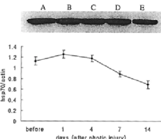
3. Western blot analysis
Hsp70 level was enhanced even before light stress, which originated from hsp-induction through systemic hyperthermia.
The production of hsp70 was detected at each time sequence.
Although the change with time was subtle, hsp70 increased for 4 days after photic injury, and then slowly decreased (Fig. 3).
Fig. 1. Histology of the retina after photic injury with or without hyperthermia. On photic injury without previous hyperthermia, diffuse disarrangements in the ONL and photoreceptor cell layer was observed (white arrows in B). However, photic injury after hyperthermia made no definite change in retina structure (white arrows with dotted line in B’). Hematoxylin & Eosin staining, magnification ×400 (A, A’: before photic injury; B, B’: 14 days after photic injury, L; light stress only, L+H; light stress after systemic hyperthermia)
Fig. 4. Semiquantitative analysis of mRNA expression, RT-PCR, for hsp70.1 and hsp70.3. mRNA expression of hsp70.1 reached a peak 4 days after light stress and slowly decreased until day 14. The mRNA expression in hsp70.3 was most prominent at day 1 and slowly decreased thereafter. (A, A’: before photic injury; B, B’: 1 day after photic injury; C, C’: 4 days; D, D’: 7 days; E, E’: 14 days) Fig. 3. Western blot analysis for hsp70. In A, enhanced hsp70
was due to hsp induction by systemic hyperthermia. The expression of hsp70 was sustained for 7 days. Although changes were subtle, the quantitative measurement showed that production of hsp70 increased for 4 days after photic injury and slowly decreased thereafter. (A: before photic injury; B, C, D and E: 1, 4, 7 and 14 days after photic injury, respectively.)
4. RT-PCR
Semiquantitative analysis of mRNA expression, RT-PCR, showed some interesting results. mRNAs of hsp70.1 and hsp70.3 were expressed more after light stress than before.
mRNA from hsp70.3 was induced earlier than that of
hsp70.1. mRNA expression of hsp70.3 reached a peak 1 day after photic injury, whereas hsp70.1 expression peaked after 4 days (Fig. 4).
Discussion
Heat shock proteins are important for cellular protection.
Substantial evidence has shown that hsps have a protective
function against various noxious conditions as well as
otherwise lethal heat stress.
1-5Furthermore, hsps must not be
overlooked for their clinical importance. They may act as
immunodominant antigens of infecting organisms, immuno-
reactive agents, and powerful antineoplastic vaccines.
22,23Recently, it was found that it is possible to lengthen the life
span of an organism using hsps.
24-26In ocular tissue,
especially the retina, hsp70 is a major stress protein. Hsp70
in mice is composed of the end products of hsp70.1, 70.2,
and 70.3
18-21and has retinal protective effects against various
stresses.
13-18However, it is not known which gene plays the
major role in retinal photic injury. The main question is
whether or not overexpression of hsp70 can be a therapeutic
method for either apoptotic or necrotic retinal disease
processes. There have been some interesting reports showing
the effect of hsp70 overexpression.
27,28However, no study
has been able to answer this question with certainty. There
may be different results depending on which hsp70, (from
hsp70.1 or 70.3), is overexpressed. Therefore, the first step
to finding the answer is to accurately show the different functions of hsp70s from hsp70.1 and 70.3.
This study showed some interesting results regarding the retinal protective action of hsp70 that might be associated with hsp70 induction by systemic hyperthermia. Upon histologic examination, there was no definite disarrangement observed in retinal photic injury after systemic hyperthermia (Fig. 1). The hsp70 expression pattern in immuno- histochemistry and western blotting supports the possibility of retinal protection by hsp70 against retinal deterioration after light stress (Figs. 2, 3).
The protein expression level cannot be predicted from the mRNA level.
29However, mRNA expression is strong evidence of protein production. Because the hsp70s from hsp70.1 and hsp70.3 are highly homologous,
18-21it is very difficult to distinguish between them at the protein level.
Therefore, the production patterns of hsp70s from hsp70.1 and hsp70.3 were evaluated indirectly from the changes in mRNA expression. mRNA expression of hsp70.1 reached a peak later than that of hsp70.3, 4 days after photic injury, and then slowly decreased. These facts suggest two hypotheses; first, that hsp70 from hsp70.3 may provide initial protection against light stress, as it responds quickly, and second, that hsp70 from hsp70.1 may have a delayed response.
Hsp70.1 and 70.3 both play major roles in protection against retinal photic injury. Specifically, hsp70 from hsp70.1 may be a major protector, although it is a delayed responder.
Hsp70.3 may compensate for the delayed action of hsp70.1.
Therefore, the expression level of the mRNA from hsp70.3 increases immediately after light stress, and is expressed for a relatively short period of time.
There are some questions remaining: (1) Why does the mRNA from hsp70.1 respond so much later? (2) If the hsp70s from hsp70.1 and hsp70.3 interact as complements, why is the mRNA from hsp70.3 sustained for 14 days after light stress, without a compensational decrease? and (3) What is the role of hsp70 from hsp70.1, knowing that its inducement is delayed? Answers to these questions will be the focus of a future study ultimately aimed at treating apoptotic retinal disease.
This study showed that hsp70 has specific functions for retinal protection against various stresses, according to which gene they originate from. It is necessary to determine which gene would be most efficacious as a possible gene therapy for apoptotic or necrotic disease in the retina.
References
1. Lindquist S, Craig EA. The heat-shock proteins. Annu Rev Genetic 1988;22:631-77.
2. Hightower LE. Heat shock, stress protein, chaperone, and proteotoxicity. Cell 1991;66:191-7.
3. Hartl FU. Molecular chaperones in cellular protein folding.
Nature 1996;381:571-9.
4. Welch WJ. Mammalian stress response: cell physiology,
structure/function of stress proteins, and implications for medicine and disease. Physiol Rev 1992;72:1063-81.
5. Morimoto RI, Santoro MG. Stress-inducible responses and heat shock proteins: new pharmacologic targets for cytoprotection. Nat Biotechnol 1998;16:833-8.
6. Rassow J, Von Ahsen O, Boemer U, Pfanner N. Molecular chaperones: towards a characterization of the heat-shock protein 70 family. Trends Cell Biol 1997;7:129-33.
7. Beckmann RP, Mizzen LA, Welch WJ. Interaction of HSP 70 with newly synthesized proteins: implications for protein folding and assembly. Science 1990;248:850-4.
8. Yufu Y, Nishimura J, Ideguchi H, Nawata H. Enhanced synthesis of heat shock proteins and augmented thermotolerance after induction of differentiation in HL-60 human leukemia cells. FEBS Lett 1990;268:173-6.
9. Lee YJ, Hou Z-Z, Curetty L, Corry PM. Expression, synthesis, and phosphorylation of HSP 28 family during development and decay thermotolerance in CHO plateau-phase cells. J Cell Physiol 1992;150:441-6.
10. Yokota S, Kitahara M, Nagata K. Benzylidene lactam compound, KNK437, a novel inhibitor of acquisition of thermotolerance and heat shock protein induction in human colon carcinoma cells. Cancer Res 2000;60:2942-8.
11. Barbe MF, Tytell M, Gower DJ, Welch WJ. Hyperthermia protects against light damage in the rat retina. Science 1988;30:1817-20.
12. Landry J, Bernier D, Chretien P, et al. Synthesis and degradation of heat shock proteins during development and decay of thermotolerance. Cancer Res 1982;42:2457-61.
13. Tytell M, Barbe MF, Brown IR. Induction of heat shock (stress) protein 70 and its m RNA in the normal and light-damaged rat retina after whole body hyperthermia. J Neurosci Res 1994;38:19-31.
14. Dean DO, Kent CR, Tytell M. Constitutive and inducible heat shock protein 70 immunoreactivity in the normal rat eye. Invest Ophthalmol Vis Sci 1999;40:2952-62.
15. Kim JH, Yu YS, Kim JH, et al. Immunoreactivity of constitutive and inducible heat shock protein 70 in human fetal retina. Korean J Ophthalmol 2003;17:14-8.
16. Yu YS, Heo JH, Hwang SW, et al. Effect of the absence of heat shock protein 70.1 (hsp70.1) on retinal photo- receptors in normal and rd mice. Korean J Ophthalmol 2001;15:67-73.
17. Kim JH, Yu YS, Chung H, et al. Effect of the absence of heat shock protein 70.1 (hsp70.1) on retinal photic injury.
Korean J Ophthalmol 2003;17:7-13.
18. Lewden O, Garcher C, Assem M, et al. Changes of the inducible heat shock protein70 mRNA level in rat retina after ischemia and reperfusion. Ophthalmic Res 1998;30:
291-4.
19. Milner CM, Campbell RD. Structure and expression of the three MHC - linked hsp70 genes. Immunogenetics 1990;32:
242-51.
20. Hunt C, Calderwood S. Characterization and sequence of a mouse hsp70 gene and its expression in mouse cell lines.
Gene 1990;87:199-204.
21. Dix DJ, Garges JB, Hong RL. Inhibition of hsp70-1 and hsp70-3 expression disrupts preimplantation embryogenesis and heightens embryo sensitivity to Arsenic. Mol Reprod Dev 1998;51:373-80.
22. Nakagawa T, Okano H, Furuichi T, et al. Novel subtypes of the mouse inositol 1,4,5-trisphosphate receptor are expressed in tissue- and developmentally specific manner.
Proc Natl Acad Sci USA 1991;88:6244-8.
23. Udono H, Srivastava PK. Heat shock protein 70 associated peptides elicit specific cancer immunity. J Exp Med
1993;178:1391-6.
24. Tower J. Trasgenic method for increasing Dorsophilia life span. Mech Ageing Dev 2000;118:1-14.
25. Rea IM, McNerlan S, Pockley AG. Serum heat shock protein and anti-heat shock protein antibody levels in aging.
Exp Gerontol 2001;36:341-52.
26. Minois N, Khazaeli AA, Curtsinger JW. Locomotor activity as a function of age and life span in Drosophilia melano- gaster overexpression hsp70. Exp Gerontol 2001;36:1137- 53.
27. Ding XZ, Tsokos GC, Kiang JG. Overexpression of hsp70 inhibits the phosphrylation of HSF1 by activating protein phosphatase and inhibiting protein kinase C activity. FASEB J 1998;12:451-9.
28. Liossis SNC, Ding XZ, Kiang JG, Tsokos GC. Over- expression of the heat shock protein 70 enhances the TCR/CD3- and Fas/Apo-1/CD95-mediated apoptotic cell death in Jurkat cells. J Immunol 1997;158:5668-75.
29. Futcher B, Latter GI, Monardo P, et al. A sampling of the yeast proteome. Mol Cell Biol 1999;19:7357-68.