CEP290 돌연변이로 인해 발생한 Joubert 증후군 말기 신부전 1례
성균관대학교 의과대학 삼성창원병원 소아청소년과1, 서울대학교 의과대학 진단검사의학과2
서울대학교 의과대학 희귀질환 센터3
김성훈1· 이상택1· 성문우2· 김만진2,3· 이준화1
A Case of End-Stage Renal Disease with Joubert Syndrome due to
CEP290
MutationSung Hoon Kim
1, Sang Taek Lee
1, Moon-Woo Seong
2, Man Jin Kim
2,3, Jun Hwa Lee
1*
Department of Pediatrics1, Samsung Changwon Hospital,Sungkyunkwan University School of Medicine, Changwon, Korea Department of Laboratory Medicine2, Seoul National University Hospital,
Seoul National University College of Medicine, Seoul, Korea Rare Disease Center3, Seoul National University Hospital, Seoul National University College of Medicine, Seoul, Korea
Joubert syndrome (JS) is a rare genetic disorder that is characterized by ataxia, hypotonia, developmental delay, respiratory abnormalities such as apnea-hyperpnea, and abnormal eye movements. The pathog- nomonic diagnostic finding is the “molar tooth sign” (MTS) on brain magnetic resonance imaging (MRI), described as cerebellar vermis hypoplasia or dysplasia, thick and horizontally oriented superior cerebellar peduncles, and an abnormally deep interpeduncular fossa. JS is characterized by genetic heterogeneity:
pathogenic variants in over 30 genes have been identified to date. The CEP290 protein, which is on chromosome 12q21.3, is most frequently mutated in patients with JS, especially with renal involvement.
Here, we report a case of JS in a 14-year-old male patient with end-stage renal disease. To the best of our knowledge, this is the first Korean report of a patient with JS due to CEP290 mutation (c.6012- 12T> A) whose diagnosis was confirmed after repetitive MRI. We suggest consultation with an experi- enced neuro-radiologist and follow-up MRI studies to detect a “hidden” MTS if clinical findings suggest a diagnosis of JS. Furthermore, even in the absence of an MTS, whole exome sequencing should be considered.
Key words: Joubert syndrome, Molar tooth sign, Chronic renal failure, CEP290 mutation
1)
Introduction
Joubert syndrome (JS) is related to agenesis of the cerebellar vermis and was initially described
책임저자: 이준화, 경상남도 창원시 마산회원구 팔용로 158 성균관대학교 의과대학 삼성창원병원 소아청소년과 Tel 055)233-5931, Fax: 055)233-5359 ORCID: 0000-0001-9717-3677
E-mail: [email protected], [email protected]
in 1969 in four siblings of a French-Canadian
family with hyperpnea, abnormal eye movements,
ataxia, and cognitive deficits
1). The prevalence has
been reported to be between 1 per 80,000 and 1
per 100,000 live births, but these numbers may
underestimate the actual prevalence because of
low awareness of this condition due to its rarity,
ongoing identification of additional causative genes
and private mutations, and reports of a broader range of phenotypic findings
2-5). In addition to clinical findings, the so-called molar tooth sign (MTS), caused by unique hypoplasia or agenesis of the cerebellar vermis and brainstem malforma- tion, is recognizable on brain magnetic resonance imaging (MRI) and is a mandatory criterion for diagnosis of JS
2,6-8). JS can be clinically suspected as early as the first few months of life upon ob- servance of hypotonia progressing to cerebellar ataxia, global developmental delay, intellectual dis- ability of variable severity, abnormal ocular move- ments (mainly ocular motor apraxia and nystagmus), and breathing dysregulation (short alternating episodes of apnea and tachypnea or episodic ta- chypnea alone)
2-4). The clinical phenomenology of JS is quite diverse because it can affect multiple organs, most commonly the eye (retinal defects that range in severity from Leber congenital ama- urosis to slowly progressive retinopathies with partially preserved vision), kidney (nephronoph- thisis, cystic dysplastic kidneys), and liver (con- genital liver fibrosis)
2-4).
The genetic bases of JS are extremely complex despite the tremendous acceleration in gene dis- covery enabled by next-generation sequencing techniques
2). At least 30 causative genes have been identified to date
2,3,5,9). These genes univer- sally encode proteins localizing to the primary cilia, the cell organelles that function as environmental sensors and signaling pathways during develop- ment and homeostasis. Thus, JS is considered a ciliopathy
2-4). The gene for the CEP290 protein is on chromosome 12q21.3 and is mutated in about 50% of patients with JS. The CEP290 protein is localized to the base (centrosome) and stalk of primary cilia and is most frequently associated with JS cases with renal involvement of nephro- nophthisis at 12q21.3 and the Meckel-Gruber
syndrome-associated gene TMEM67 at 8q22.1
2-5,9)
.
We report a case of JS caused by CEP290 mu- tation in a male patient with end-stage renal dis- ease whose diagnosis was initially suspected by clinical features and by atypical MTS identified after repetitive MRI.
Case Report
The patient was a male baby and the only child of a healthy 32-year-old mother and a healthy 33-year-old father, neither of whom had a family history of neurological or hereditary disorder. The patient weighed 3,700 g (90th percentile) at birth at a gestational age of 40 weeks by Cesarean sec- tion delivery due to cephalopelvic disproportion.
On the 12th day of life, he was treated for alter- nating episodes of tachypnea and apnea.
He first visited the outpatient clinic of our hos- pital at 8 months of age with developmental delay.
Head control and rolling had not been achieved.
A chromosome study, brain MRI, and metabolic work up were performed and revealed no specific findings. At 15 months of age, he was diagnosed with mitochondrial disease through various tests, including muscle biopsy at other tertiary hospital.
At that time, he could not talk, follow with or turn his eyes toward objects or sound, hold up or control his head, sit upright independently, or hold his bottle.
The patient continued with rehabilitation treat-
ment until he was 7 years 8 months old, when
he returned to the hospital with his first afebrile
seizure due to hyponatremia shortly after hemo-
dialysis. At that time, electroencephalogram (EEG)
test result was normal. He was only able to roll
over at the time of his visit, and he had been in
hemodialysis for two weeks ago due to an un-
Fig. 2. At 9 years 4 months of age, the patient under- went brain computed tomography (CT) that showed intracranial hemorrhage, intraventricular hemorrhage, and midline shifting to the left side due to hypertension (A and B). The molar tooth sign (MTS) was observed on brain CT per- formed at 9 years 4 months of age (C) and on brain magnetic resonance imaging (MRI) at 12 years 2 months of age (D). Axial T2-weighted MRI shows an MTS appearance of the midbrain with thickened superior cerebellar peduncle (arrow) and vermian hypoplagia.
Fig. 1. A photograph of the patient at 14 years in which he sat in a wheelchair due to global developmental delay (A). His facial photograph shows facial dysmorphisms, including prominent forehead, right-dominant bilateral ptosis, high rounded eyebrows, broad and shallow nasal bridge, lower lip eversion with trapezoid-shaped open mouth, and tongue protrusion (B).
Publication of gross photos was permitted by his parents through in- formed consent.
known cause of end stage renal disease. He ex- perienced another seizure, a generalized tonic- clonic (GTC) type, at 7 years 10 months of age immediately after dialysis. On physical examina- tion at that time, he had facial dysmorphisms, in- cluding prominent forehead, right-dominant bila- teral ptosis, high rounded eyebrows, broad and shallow nasal bridge, lower lip eversion with tra- pezoid-shaped open mouth, and tongue protrusion (Fig. 1). He also showed both corneal band kera- topathy, corneal opacity and nystagmus in the ophthalmologic examination. The patient suffered intracranial hemorrhage at 9 years 4 months of age, with a systolic blood pressure of 190 mmHg during hemodialysis. At that time, MTS findings were suspected on brain computed tomography (CT), suggesting the possibility of JS, but genetic testing was not possible due to economic and other problems. Although brain CT or MRI has been performed previously, radiological readings at the age of nine diagnosed hypoplastic cerebellar vermis with MTS characteristics and he was cli- nically suspected as JS at that time based on his clinical features and MTS (Fig. 2).
Finally, at 13 years 10 months of age, he was finally confirmed with JS through whole exome
sequencing (WES) revealed identification of a c.
6012-12T>A, homozygote mutation in CEP290
(Fig. 3). The following method of WES was utilized
for gene analysis. An Agilent SureSelectXT Human
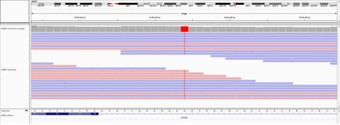
Fig. 3. Identification of a c.6012-12T>A, homozygote mutation in CEP290 (NM_025114.3). The BAM file displayed in The Integrative Genomics Viewer (IGV) shows c.6012-12T>A, homozygote is indisputable in the forward (red in IGV) and reverse (blue in IGV) strands.
all Exon 50 Mb kit (Agilent, Santa Clara, CA, USA) was used to enrich the exon regions of the ge- nome. Paired-end 100-bp sequencing was per- formed using Illumina HiSeq platform (Illumina, San Diego, CA, USA). The produced sequencing data were analysed using NextGENe software v2.4.0.1. (SoftGenetics, State College, PA, USA).
Finally, the detected variants were classified as a pathogenic variant according to the American College of Medical Genetics and Genomics 2015 guidelines
10).
The patient started peritoneal dialysis at age 14 years 2 months, and he still uses a wheelchair for ambulation.
Discussion
Joubert syndrome (JS) is a rare, predominantly autosomal, recessive, ciliopathic condition that is clinically characterized by congenital cerebellar ataxia, hypotonia, global developmental delay, and specific mid-hindbrain malformation (“molar tooth sign” [MTS] on brain MRI because of its resem- blance to the cross-section of a tooth on axial imaging)
2,3,6,11). Its three key diagnostic criteria are (1) radiologic finding of MTS; brain MRI fin- dings demonstrating the hallmark imaging features
of MTS on axial imaging with these three com- ponents: a) midline cerebellar vermis hypoplasia (characterized by incomplete lobulation and en- larged fourth ventricle), b) deepened interpedun- cular fossa (and dysgenesis of the isthmus [part of the brainstem between the pons and inferior colliculus], which is seen as elongation and thin- ning of the ponto-mesencephalic junction), and c) thick, elongated, and straight superior cerebellar peduncles; (2) hypotonia in infancy with later ataxia; and (3) developmental delays/intellectual disability of variable severity. Additionally, one or both of the following are not required but are supportive of diagnosis: a) irregular breathing pattern in infancy (episodic tachypnea and/or apnea) and b) abnormal eye movements (nys- tagmus, jerky eye movements, and oculomotor apraxia or difficulty with smooth visual pursuits)
2,15)
. Although not a pathognomonic finding on
MRI, a sagittal vermis cleft (incomplete fusion of
the halves of the vermis) on axial or coronal MRI
plane is often visualized in patients with JS
3,6).
Because the spectrum of neuroimaging findings
in JS is wide, neuroimaging plays a key role in
diagnosis
8). In the presence of neurological fea-
tures suggestive of JS, the diagnosis is easily
confirmed upon demonstration of the MTS on
brain MRI
2). All patients with JS have some degree of vermis hypoplasia and the MTS, and both are mandatory features for diagnosis
8). However, al- though a high-resolution MRI scan with 3 mm sections is recommended for visualizing the MTS, its shape varies considerably. Therefore, without examination of multiple MRI cuts for subtle MTS findings and vermis hypoplasia, even experienceed neuro-radiologists may miss this diagnostic hall- mark, as in the present case
5). According to the literature, 12% of JS cases were initially diag- nosed with Dandy-Walker malformation; after posterior fossa decompression and re-expansion of the cerebellar hemispheres, these patients were diagnosed with JS
8). This finding emphasizes the importance of follow-up MRI studies in patients with suspected Dandy-Walker malformation or JS to detect a “hidden” MTS
8). The MRI finding of vermis hypoplasia in the absence of other typical clinical features does not lead to diagnosis of JS
3). Neuroimaging does not predict the genetic cause and is considered of limited value in predicting cognitive function in JS, although it may predict the neurodevelopmental outcome
8). A high degree of vermis hypoplasia correlates with a worse neu- rodevelopmental outcome
8).
The three aforementioned cardinal features are necessary for diagnosis of JS, and 29% of patients do not have extra-neurological organ involvement, although the syndrome does manifest several other variable features, as in our case
2,8). The most prominent feature in the newborn period is an abnormal respiratory pattern characterized by episodic hyperpnea, consisting of alternating ta- chypnea and/or apnea
6,12). This abnormal brea- thing pattern of distinctive, short, alternating epi- sodes of apnea and hyperpnea or episodic hyper- pnea alone is sometimes described as ‘panting like a dog’ and may intensify with stress. Some neo-
nates demonstrate worrisome bouts of apnea re- quiring pharmacological intervention, like our pa- tient
6,13). This abnormal respiratory pattern pro- gressively improves with age and usually disap- pears around sixth months of age; however, death occurring before the age of 6 years in JS is most often (35%) due to respiratory failure
13).
Ocular and oculomotor involvement is common in JS, and it manifests variable phenotypes among patients
5,13,14). Abnormal eye movements are typi- cally characterized by oculomotor apraxia, jerky eye movements and head thrusting (resulting from absence or defect in controlled, voluntary, and purposeful eye movements), as well as coloboma, nystagmus, strabismus, and ptosis of the eyelids
5,11,14)
. In addition, the most severe manifestations are congenital blindness in the spectrum of Leber congenital amaurosis
2,5). This retinal degeneration may develop with age and is slowly progressive with partially preserved vision
2,5,14). Overall, retinal involvement is present in about 80% of patients with CEP290 and AHI1 mutations, and severe re- tinal degeneration that is early and aggressive is also seen in these patients
14).
Several studies report renal manifestations in 23-32% of patients with JS, most commonly in those with mutations in CEP290 , TMEM67, and AHI1 . Patients generally demonstrate one of two forms: nephronophthisis (NPHP) or cystic kidneys
15)
. Subjects with renal manifestations show early
onset hypertension, diagnosed shortly after birth
or within the first years of life, before any mea-
surable decrease in estimated glomerular
filtration rate, and renal failure (37.5%) was the
most common cause of death, especially in older
individuals
5,13,15,16). NPHP, the most typical form
of JS, manifests as chronic tubulointerstitial ne-
phropathy and may present in the first or second
decade of life, often progressing to end-stage
renal disease within a decade
5,15,16).
Like other syndromic ciliopathies, JS is charac- terized by extreme genetic heterogeneity. Since the first gene for JS, NPHP1 , was identified in 2004
17), bi-allelic pathogenic variants in over 30 genes encoding proteins of the primary cilium have been identified to date: AHI1 , ARL13B, ARMC9, CC2D2A, CEP104, CEP290, INPP5E, KIF7, NPHP1, OFD1, RPGRIP1L, TCTN1, TCTN2, MKS1, TMEM67 (MKS3), TMEM237, TMEM2167 , etc
3,5,8,9). Although most mutations resulting in JS are inherited in an autosomal recessive manner, as are the majority of ciliopathies, Oral Facial Syndrome 1 (OFD1) de- monstrates X-linked recessive inheritance
5). Of note, because almost all these genes have also been implicated in other ciliopathy disorders, it typically is difficult to identify clear-cut genotype- phenotype correlations for many of these genes
5). However, in a mouse model of JS due to CEP290 mutation, gene therapy has demonstrated reduction of cystic kidney disease burden and rescued re- tinal degeneration
5,18,19).
In conclusion, we describe a male patient with end-stage renal disease, to the best of our know- ledge, who was firstly confirmed to have JS with CEP290 mutation (c.6012-12T>A) by a molecular test after repetitive brain CT and MRI in Korea.
Although the classic symptoms of JS developed in sequence in this patient, it was not diagnosed clinically until the fifth brain CT due to the rela- tively atypical appearance of the MTS. Therefore, because the shape of the MTS varies considerably, MRI with multiple and thin sections should be performed when JS is clinically suspected so as to not miss this diagnostic hallmark. In addition, follow-up MRI studies should be performed to detect a possibly “hidden” MTS. Furthermore, if the possibility of JS is high, genetic molecular test should be considered even if there is no demon-
stration of the MTS.
요 약
쥬버트 증후군(JS, Joubert syndrome)은 대부분 상염색체 열성으로 유전되는 유전성 대사질환으로 임 상증상은 신생아 시기부터 발현된다. 저자들은 신생아 기부터 특징적인 임상 증상이 순차적으로 발현되어 임 상적으로 JS를 의심하였으나 특징적인 뇌 MRI 소견인 molar tooth sign (MTS)이 늦게 나타난 후 전장엑솜 분석(WES, whole exome sequencing)으로 확진 된, 말기 신부전을 동반한 JS 1례를 경험하였기에 보고하 고자 한다. 14세 남자 환자는 출생 직후 반복적인 무 호흡과 과호흡으로 치료받은 병력이 있으며, 생후 8개 월때부터 전반적 발달 지연과 관련되어 처음 병원을 방 문하여 기본적인 발달 지연에 관한 검사를 시행하였으 나 특이 소견 없었고, 이후 15개월 때 근육생검을 포함 한 여러 검사를 통해 사립체(mitochondrial) 질환으로 진단 되었었다. 이후 물리 치료만 하며 관찰 하던 중 안 구진탕과 망막질환이 확인되었다. 생후 7세 8개월에는 처음 발작이 있었으며, 말기 신부전이 있어 8세부터 혈 액투석을 시작한 후, 혈액 투석 직후 수차례 발작이 있 었으나 전해질 불균형으로 인한 발작으로 진단하여 항 뇌전증 약물 치료는 하지 않았다. 9세 4개월 때 고혈압 으로 인한 뇌출혈로 치료 받았으며, 이때 시행한 뇌 CT 상 MTS가 처음 의심되었다. 13세 10개월에 시행한 뇌 MRI 검사상 MTS가 명확히 확인되었고, 전장엑솜 분석으로 JS의 CEP290 mutation (c.6012-12T>A) 이 확인되었다. 환자는 신생아기부터 발현된 특징적인 임상 소견과 말기 신부전 상태, 뇌 CT 또는 MRI소견, 그리고 전장엑솜분석 검사로 JS로 확진하였다. JS는 임상 양상이 다양할 뿐만 아니라 진단에 중요한 MTS 소견이 초기에 보이지 않더라도, 임상적으로 의심된다 면 확진을 위해서 전장엑솜분석을 시행하는 것이 필요 하다.
Acknowledgement
The authors are very grateful to the patient and
his parents for allowing him to use his portrait rights.
References
1) Joubert M, Eisenring JJ, Robb JP, Andermann F. Fa- milial agenesis of the cerebellar vermis. A syndrome of episodic hyperpnea, abnormal eye movements, ataxia, and retardation. Neurology 1969;19:813-25.
2) Romani M, Micalizzi A, Valente EM. Joubert synd- rome: congenital cerebellar ataxia with the molar tooth.
Lancet Neurol 2013;12:894-905.
3) İncecik F, Hergüner MÖ, Altunbaşak Ş, Gleeson JG.
Joubert syndrome: report of 11 cases. Turk J Pediatr 2012;54:605-11.
4) Brancati F, Dallapiccola B, Valente EM. Joubert synd- rome and related disorders. Orphanet J Rare Dis 2010;
5:20.
5) Parisi MA. The molecular genetics of Joubert synd- rome and related ciliopathies: The challenges of genetic and phenotypic heterogeneity. Transl Sci Rare Dis 2019;4:25-49.
6) Maria BL, Hoang KB, Tusa RJ, Mancuso AA, Hamed LM, Quisling RG, et al. "Joubert syndrome" revisited:
key ocular motor signs with magnetic resonance ima- ging correlation. J Child Neurol 1997;12:423-30.
7) Poretti A, Huisman TAGM, Scheer I, Boltshauser E.
Joubert syndrome and related disorders: spectrum of neuroimaging findings in 75 patients. AJNR Am J Neuroradiol 2011;32:1459-63.
8) Poretti A, Snow J, Summers AC, Tekes A, Huisman TAGM, Aygun N, et al. Joubert syndrome: neuroima- ging findings in 110 patients in correlation with cogni- tive function and genetic cause. J Med Gen 2017;54:
521-9.
9) Sang L, Miller JJ, Corbit KC, Giles RH, Brauer MJ, Otto EA, et al. Mapping the NPHP-JBTS-MKS pro- tein network reveals ciliopathy disease genes and path- ways. Cell 2011;145:513-28.
10) Richards S, Aziz N, Bale S, Bick D, Das S, Gastier- Foster J et al. Standards and guidelines for the inter-
pretation of sequence variants: a joint consensus re- commendation of the American College of Medical Genetics and Genomics and the Association for Mole- cular Pathology. Genet Med 2015;17:405-24.
11) Maria BL, Quisling RG, Rosainz LC, Yachnis AT, Gitten J, Dede D, et al. Molar tooth sign in Joubert syndrome: clinical, radiologic, and pathologic signifi- cance. J Child Neurol 1999;14:368-76.
12) Maria BL, Boltshauser E, Palmer SC, Tran TX. Clinical features and revised diagnostic criteria in Joubert synd- rome. J Child Neurol 1999;14:583-90.
13) Dempsey JC, Phelps IG, Bachmann-Gagescu R, Glass IA, Tully HM, Doherty D. Mortality in Joubert synd- rome. Am J Med Genet A 2017;173:1237-42.
14) Brooks BP, Zein WM, Thompson AH, Mokhtarzadeh M, Doherty DA, Parisi M, et al. Joubert Syndrome:
Ophthalmological Findings in Correlation with Geno- type and Hepatorenal Disease in 99 Patients Prospec- tively Evaluated at a Single Center. Ophthalmology 2018;125:1937-52.
15) Fleming LR, Doherty DA, Parisi MA, Glass IA, Bryant J, Fischer R, et al. Prospective Evaluation of Kidney Disease in Joubert Syndrome. Clin J Am Soc Nephrol 2017;12:1962-73.
16) Vilboux T, Doherty DA, Glass IA, Parisi MA, Phelps IG, Cullinanee AR, et al. Molecular genetic findings and clinical correlations in 100 patients with Joubert syndrome and related disorders prospectively evaluated at a single center. Genet Med 2017;19:875-82.
17) Parisi MA, Bennett CL, Eckert ML, Dobyns WB, Glee- son JG, Shaw DW, et al. The NPHP1 gene deletion associated with juvenile nephronophthisis is present in a subset of individuals with Joubert syndrome. Am J Hum Genet 2004;75:82-91.
18) Ramsbottom SA, Molinari E, Srivastava S, Silberman F, Henry C, Alkanderi S, et al. Targeted exon skip- ping of a CEP290 mutation rescues Joubert syndrome phenotypes in vitro and in a murine model. Proc Natl Acad Sci U S A 2018;115:12489-94.
19) Dooley SJ, McDougald DS, Fisher KJ, Bennicelli JL, Mitchell LG, Bennett J. Spliceosome-Mediated Pre- mRNA trans-Splicing Can Repair CEP290 mRNA.
Mol Ther Nucleic Acids 2018;12:294-308.