• 검색 결과가 없습니다.

Torsion of a parasitic myoma that developed after abdominal myomectomy

N/A
N/A
Protected

Academic year: 2022

Share "Torsion of a parasitic myoma that developed after abdominal myomectomy"

Copied!
4
0
0

로드 중.... (전체 텍스트 보기)

전체 글

(1)

Case Report

Obstet Gynecol Sci 2016;59(1):75-78 http://dx.doi.org/10.5468/ogs.2016.59.1.75 pISSN 2287-8572 · eISSN 2287-8580

www.ogscience.org 75

Introduction

Iatrogenic parasitic myomas may develop after seeding by small fibroid masses when morcellation is performed dur- ing uterine surgery. This condition is the most common late complication after laparoscopic myomectomy; the reported incidence is 0.2% to 1.2% [1,2]. Several cases have described parasitic myomas that develop after abdominal myomectomy [3]. The myomas sometimes parasitize the greater omentum.

Such cases are usually found only incidentally during surgery;

some patients exhibit nonspecific symptoms. Acute abdominal pain attributable to torsion of the pedicle of a fibroid that has parasitized the greater omentum is extremely rare as an initial presentation [4]. However, such an event may occasionally cause acute abdominal pain.

Case report

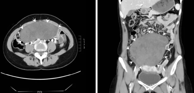
A 38-year-old woman was admitted to our emergency de- partment complaining of a 2-hour history of sudden, severe right lower abdominal pain. She appeared to be acutely ill.

The pain was both severe and progressive. Her medical history was unremarkable. She had undergone a cesarean section 17 years prior, followed by abdominal myomectomy to remove uterine fibroids 7 months prior. During myomectomy, two in- tramural myomas and one subserosal myoma were removed.

Uterine myomas were removed entirely without morcellation;

we minimized myoma tissue disruption. The abdominal cavity contained no visible tissue remnants.

Physical examination revealed lower abdominal rigidity and

Torsion of a parasitic myoma that developed after abdominal myomectomy

In Ae Cho

1

, Jong Chul Baek

1

, Ji Kwon Park

1,2

, Dae Hyun Song

3

, Wan Ju Kim

1

, Yoon Kyoung Lee

1

, Ji Eun Park

1

, Jeong Kyu Shin

1,2

, Won Jun Choi

1,2

, Soon Ae Lee

1,2

, Jong Hak Lee

1,2

, Won Young Paik

1,2

1

Department of Obstetrics and Gynecology,

2

Institute of Health Science,

3

Department of Pathology, Gyeongsang National University School of Medicine, Jinju, Korea

Iatrogenic parasitic myomas are rare. The condition is defined by the presence of multiple smooth-muscle tumorous nodules in the peritoneal cavity. This may be attributable to seeding of myoma particles during uterine surgery. The clinical course is usually indolent. The disease is often asymptomatic and is usually discovered only incidentally. A 38-year-old woman who had undergone abdominal myomectomy 7 months prior presented with acute abdominal pain and a huge pelvic mass. We performed exploratory laparotomy. A parasitic mass 17 cm in diameter with a twisted omental pedicle was identified. En bloc excision of the mass and omentum was performed, followed by total abdominal hysterectomy. Histopathological examination of multiple sections revealed features compatible with an infarcted leiomyoma. Thus, we present a very rare case of an iatrogenic, rapidly growing parasitic myoma complicated by omental torsion (which caused the acute abdominal pain). We also offer a literature review.

Keywords: Abdominal pain; Omental torsion; Parasitic myoma

Articles published in Obstet Gynecol Sci are open-access, distributed under the terms of the Creative Commons Attribution Non-Commercial License (http://creativecommons.

org/licenses/by-nc/3.0/) which permits unrestricted non-commercial use, distribution, and reproduction in any medium, provided the original work is properly cited.

Copyright © 2016 Korean Society of Obstetrics and Gynecology Received: 2015.8.26. Revised: 2015.10.6. Accepted: 2015.10.14.

Corresponding author: Jong Chul Baek

Department of Obstetrics and Gynecology, Gyeongsang National University School of Medicine, 15 Jinju-daero 816beon-gil, Jinju 52727, Korea

Tel: +82-55-750-8146 Fax: +82-55-759-1118 E-mail: [email protected]

http://orcid.org/0000-0003-0919-0800

(2)

www.ogscience.org 76

Vol. 59, No. 1, 2016

guarding, with an associated rebound tenderness suggest- ing that surgery might be required. All vital signs were within normal ranges and the patient’s consciousness was clear. The white blood cell count was mildly elevated, to 11,800/mm

3

, but all remaining laboratory test data were normal. Pelvic

examination revealed a large, tender, non-mobile mass that filled the lower abdomen.

Ultrasonography revealed her uterus (8.6×7.4×6.3 cm in dimension) to contain a 4.4-cm-diameter subserosal fibroid and a 2.7-cm-diameter intramural myoma. A huge mass was

A B

A B

Fig. 1. Preoperative computed tomography imaging of the mass. (A) Axial and (B) coronal computed tomography reveal a well-defined, lobulated heterogeneous mass in the lower abdomen.

Fig. 2. (A) Coronal computed tomography showing a twisted omentum. These findings are consistent with the operative findings (white

arrows). (B) An intra-operative photograph shows the omental mass connected to a twisted vascular pedicle (black arrows).

(3)

www.ogscience.org 77 In Ae Cho, et al. Parasitic myoma presenting as omental torsion

evident in the upper pelvis; this extended into the upper ab- domen to a position 8 cm above the level of the umbilicus.

The mass was slightly heterogeneous in echotexture, and the degree of through-transmission was poor.

A computed tomography (CT) scan of the entire abdomen revealed a well-defined, lobulated heterogeneously enhanced mass 17×15×7 cm in dimension in the abdominal cavity (Fig.

1). Inferiorly, the mass lay separate from the uterus and ova- ries. A small amount of fluid was noted within the pelvic cav- ity. The coronal CT image showed a whirling pattern of fatty streaks within the omentum (Fig. 2A). This is a relatively char- acteristic feature of omental torsion.

We performed an exploratory laparotomy on the basis of a preoperative diagnosis of omental torsion. During this opera- tion, a large encapsulated mass, 17 cm in diameter, was found in the greater omentum. The mass was fed by small vessels extending from the omentum, but was nonetheless easily re- moved. The mass was twisted 1.5 rotations clockwise around the stalk (Fig. 2B). The operative findings were consistent with the CT data. A dark brown parasitic myoma was identified on the twisted pedicle. No other connection was evident be- tween the omental mass and the uterus. En bloc excision of both the mass and the omentum was performed, followed by total abdominal hysterectomy due to the presence of multiple small uterine myomas.

Gross examination revealed a globular soft tissue mass 16×13×6 cm in dimension. The cut surface was fasciculated and dark gray in color. Histopathological examination of mul- tiple sections revealed features compatible with a leiomyoma.

The leiomyomatous tissue exhibited multifocal hemorrhages, congestion, and dilated vessels; all are typical pathological fea- tures of omental torsion. The tumor cells were immunoposi- tive for smooth-muscle actin. These findings are consistent with the presence of an infarcted leiomyoma. Thus, we made a final diagnosis of an iatrogenic parasitic myoma complicated by omental torsion. The patient was discharged from the hospital 6 days after surgery. No abnormality was evident at follow-up 12 months later.

Discussion

Myomas are benign tumors that originate from individual smooth-muscle cells. The tumors can form wherever smooth muscle is present, but they most often develop in the uterus

[5,6]. Parasitic myomas (defined as myomas caused by extra- uterine seeding) were first reported in the early 1900s [7]. Tra- ditionally, it was thought that parasitic myomas were originally pedunculated subserosal myomas that became incidentally separated from the uterus. A new blood supply was attained upon attachment and rooting to other organs [8]. However, the increasing incidence of parasitic myomas after minimally invasive endoscopic procedures may suggest otherwise [9].

Small tissue fragments retained in the abdominal cavity after morcellation are now thought to possibly explain the develop- ment of parasitic fibroids in the peritoneal cavity [9-11]. Such parasitic myomas are thus iatrogenic in nature.

The symptoms do not appear to be specific, but patients ini- tially complain principally of abdominal or pelvic pain, dyspa- reunia, abdominal distension, an increased urinary frequency, and constipation [1,12]. Parasitic myomas develop at various sites and vary in diameter (range, 0.8 to 30 cm). The sites include the port of entry for the operation, intestines, perito- neum, omentum, and abdominal cavity [2,12,13]. The interval between the initial surgery and the repeat surgery during which myomas are discovered ranges from 2 to 108 months [12]. Several cases of parasitic myomas after abdominal hys- terectomy have also been reported, suggesting that factors other than morcellation may be in play [3,14].

The present case is important in that it shows that even an abdominal myomectomy can trigger the development of a parasitic myoma. Multiple myomectomy is likely associated with myoma particle implantation in the abdominal cavity. Our surgical team carefully confirmed that no visible particles re- mained in the abdominal cavity after surgery. However, small particles invisible to the naked eye could have been present.

Seven months later, a single parasitic myoma 17 cm in diam- eter was found in, and derived its blood supply solely from, the omentum. The use of a meticulous technique featuring the complete removal of even very small myoma fragments is essential during both open and laparoscopic procedures.

The omentum is rarely the source of acute abdominal pain.

Torsion of the omentum is an unusual cause of such pain. The typical clinical features of omental torsion include sudden- onset pain of short duration, which is usually constant initially but gradually increases in severity [15]. CT is becoming ever more important in the diagnosis of such torsion, and can dif- ferentiate surgical from nonsurgical conditions.

In February 2015, the Steering Committee on Fibroid Mor-

cellation of the European Society of Gynaecological Endos-

(4)

www.ogscience.org 78

Vol. 59, No. 1, 2016

copy considered how parasitic fibroid development could be prevented after morcellation. Every effort should be made to prevent tissue loss and remove all tissue fragments. The pa- tient should be placed in the reverse Trendelenburg position after morcellation and the abdomen and pelvis should be ex- tensively irrigated after prior irrigation of the peritoneal cavity.

Both the abdomen and pelvis should be meticulously inspect- ed and any residual tissue fragments should be removed [10].

In conclusion, if a patient presents with an abdominal mass or abdominal pain after myomectomy or hysterectomy, the surgeon should be aware that an iatrogenic parasitic myoma may have formed. All necessary intraoperative precautions minimizing the development of such myomas must be taken.

Conflict of interest

No potential conflict of interest relevant to this article was reported.

References

1. Cucinella G, Granese R, Calagna G, Somigliana E, Perino A. Parasitic myomas after laparoscopic surgery: an emerging complication in the use of morcellator? De- scription of four cases. Fertil Steril 2011;96:e90-6.

2. Sinha R, Hegde A, Mahajan C, Dubey N, Sundaram M.

Laparoscopic myomectomy: do size, number, and loca- tion of the myomas form limiting factors for laparoscopic myomectomy? J Minim Invasive Gynecol 2008;15:292- 300.

3. Kho KA, Nezhat C. Parasitic myomas. Obstet Gynecol 2009;114:611-5.

4. Tan CH, Ho BC, Shelat V, Tan CH. Leiomyomatosis peri- tonealis disseminata presenting as omental torsion. Sin- gapore Med J 2012;53:e71-3.

5. Ryan GL, Syrop CH, Van Voorhis BJ. Role, epidemiology, and natural history of benign uterine mass lesions. Clin Obstet Gynecol 2005;48:312-24.

6. Wallach EE, Vlahos NF. Uterine myomas: an overview of development, clinical features, and management. Obstet Gynecol 2004;104:393-406.

7. Kelly HA, Cullen TS. Myomata of the uterus. Philadelphia (PA): Saunders; 1909.

8. Brody S. Parasitic fibroid. Am J Obstet Gynecol 1953;65:1354- 6.

9. Nezhat C, Kho K. Iatrogenic myomas: new class of myo- mas? J Minim Invasive Gynecol 2010;17:544-50.

10. Brolmann H, Tanos V, Grimbizis G, Ind T, Philips K, van den Bosch T, et al. Options on fibroid morcellation: a lit- erature review. Gynecol Surg 2015;12:3-15.

11. Larrain D, Rabischong B, Khoo CK, Botchorishvili R, Ca- nis M, Mage G. “Iatrogenic” parasitic myomas: unusual late complication of laparoscopic morcellation proce- dures. J Minim Invasive Gynecol 2010;17:719-24.

12. Huang PS, Chang WC, Huang SC. Iatrogenic parasitic myoma: a case report and review of the literature. Tai- wan J Obstet Gynecol 2014;53:392-6.

13. Ramesh B, Sharma P, Gunge D. Abdominal wall parasitic myoma following electromechanical morcellation. J Ob- stet Gynaecol India 2014;64(Suppl 1):73-5.

14. Rajab KE, Aradi AN, Datta BN. Postmenopausal leimyo- matosis peritonealis disseminata. Int J Gynaecol Obstet 2000;68:271-2.

15. Park CM, Kim SY. Primary omental torsion diagnosed

during hysterectomy. Obstet Gynecol Sci 2014;57:415-8.

수치

Fig. 1. Preoperative computed tomography imaging of the mass. (A) Axial and (B) coronal computed tomography reveal a well-defined,  lobulated heterogeneous mass in the lower abdomen.

참조

관련 문서

After listening to the opinions of firemen in field activities, we confirmed that the firepower can be secured by reducing the loss of firepower caused

In this thesis, we introduce a Hurwitz polynomial ring and study the irreduciblity of Hurwitz polynomials over  We also investigate the irreduciblilty of

They suggested that vaporization of a thin layer of liquid between the growing bubble and the heating surface caused removal of heat from the surface, thus lowing

(Result) The reports of nausea, vomiting, abdominal discomfort, jaundice and hepatomegaly in acute hepatitis A patients and febrile sensation, chillness,

8.50… thin-walled, closed x-s of arbitrary shape subjected to an applied torque, assumed to be in a state of uniform torsion, axial strain and stress components vanish  n(s)

Just as we achieved good control with a joint-based controller that was based on a linearizing and decoupling model of the arm, we can do the same for the Cartesian case.

We also define period of a Fibonacci sequence modulo an integer, m and derive certain interesting properties related to them.. Afterwards, we derive some new properties of a

If we are forbidden by our culture to think a particular thought or entertain a particular desire, that taboo instantly brings to mind the very thing we