787 Original Article
Korean Circulation J 2005;35:787-789
ISSN 1738-5520
ⓒ 2005, The Korean Society of Circulation CASE REPORT
Drug-Eluting Stent Strut Fracture as a Cause of Restenosis
Jang-Ho Bae, MD, PhD1, Dae-Woo Hyun, MD1, Ki-Young Kim, MD1, Hyun-Ju Yoon, MD1 and Sunao Nakamura, MD2
1Division of Cardiology, Heart Center, College of Medicine, Konyang University, Daejeon, Korea
2New Tokyo Hospital, Chiba, Japan
ABSTRACT
We report a case of in-stent restenosis due to the fracture of a sirolimus-eluting stent, which was confirmed by intravascular ultrasound. It can be suggested that a stent fracture is an important cause of restenosis in this era of drug-eluting stents. (Korean Circulation J 2005;35:787-789)
KEY WORDS:Stent fracture;Coronary restenosis;Drug-eluting stent.
Introduction
There is no doubting the efficacy of drug-eluting stent (DES) in terms of restenosis. However, some degree of restenosis is still evident, although the rate is quite small.1-3) Recently, it has been reported that a stent fracture after post-dilation with a larger balloon was a possible cause of restenosis after the implantation of a sirolimus-eluting stent(SES).4) Here, a case of in-stent restenosis, with a sirolimus-eluting stent, due to a stent strut fracture is reported, which was confirmed by intra- vascular ultrasound, even though nominal pressure(12 atm) was applied during the SES deployment into a not heavily calcified or severe tortuous lesion.
Case
A 62-year-old man, with a history of smoking, pre- sented with resting onset angina in June 2004. He sho- wed no specific abnormalities, with a normal blood pressure on physical examination. The biochemical test results, including cardiac enzyme, were all normal. There was no regional wall motion abnormality, with a 70%
ejection fraction, on routine echocardiogram. Coronary angiography revealed severe luminal stenosis in the mid left anterior(LAD) coronary artery, with borderline
stenosis in the mid left circumflex(LCX) coronary artery(Fig. 1).
The LAD was engaged with a 6 Fr guiding catheter (Vista brite tip L4.0, Cordis Co., Miami, FL) via the femoral approach. A 0.014″ Hi-torque Whisper MS gui- dewire(Guidant, Santa Clara, CA) was able to cross the lesion without difficulty. We predilated the lesion with a 2.0 mm balloon(Silky 2.0×20 mm; Stenttech, Seoul) up to 10 atm, then deployed a 2.5×28 mm Bx Velocity SES(Cypher, Cordis Europe, Roden, The Netherlands) at 12 atm, with a good angiographic result(Fig. 1). He was discharged the next day, and administered life-long aspirin and clopidogrel for at least 6 months. He agreed to undergo follow up angiography at 6 months after the initial intervention.
He remained asymptomatic for 6 months. The follow up coronary angiography, in December 2004, showed a focal stenosis(9 mm long and 71% diameter stenosis) in the middle part of 2.5×28 mm long stent(Fig. 1) on quantitative coronary angiography(QCA). An intra- vascular ultrasound(IVUS; Atlantis SR pro, 40 MHz;
Boston Scientific) examination revealed significant neo- intimal hyperplasia and a fractured segment of the stent strut at the stenotic site(Fig. 2). It also showed a kin- ked and collapsed stent strut just above the distal calcified lesion. The fractured segment of the stent strut was found near the top(lumen side) and also in the middle of the plaque, with neointimal hyperplasia in the area absence of the stent strut. However, there was no inti- mal hyperplasia throughout the rest of the stent length.
Based on the above results, the LAD restenotic lesion was treated with direct 2.5×18 mm Bx Velocity SES (Cypher, Cordis Europe, Roden, The Netherlands) implantation, at 16 atm, with good angiographic and
Received:April 25, 2005 Revision Received:June 13, 2005 Accepted:June 23, 2005
Correspondence:Jang-Ho Bae, MD, PhD,Division of Cardiology, Heart Center, College of Medicine, Konyang University, 685 Gasoowon-dong, Seo-gu, Daejeon 302-718, Korea
Tel: 82-42-600-6400, Fax: 82-42-600-6399 E-mail: [email protected]
788·Korean Circulation J 2005;35:787-789
IVUS results, and the left circumflex coronary artery was treated with a 2.5×23 mm Bx Velocity SES(Cypher, Cordis Europe, Roden, The Netherlands), at 16 atm, with a good angiographic result after pre-dilation with a 2.5 mm balloon(Ryujin 2.5×20 mm; Terumo Europe, Leuven, Belgium)(Fig. 1, 2).
Discussion
It has recently been reported that the fracture of a SES within coronary arteries resulted in very focal in-
stent restenosis, with complete abolition of neointimal hyperplasia over the rest of stent length.4) The two repor- ted cases were both in long(33 mm) stents, post-dilated with a larger balloon at high pressure. Furthermore, the fractures had developed at the point of maximal vessel curvature and the extremities of the overlapping stent, indicating fractures are subject to develop in the area of increased rigidity. However, the stent fracture in this report developed in the middle part of a 28mm long SES, with no severe tortuous or calcified lesion, under nominal pressure(12 atm). We thought the reason for
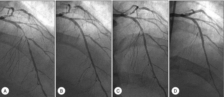
Fig. 1. Angiography of the left anterior descending coronary artery. A: shows 24 mm long, 89% diameter stenosis with 2.62 mm (2.20 mm) proximal (distal) reference diameter by quantitative coronary angiography. B: shows acceptable result without intravascular ultrasound with cypher 2.5’ 28 mm under 12 atm. C: shows a significant focal retenosis in the middle of cypher stent 6 months after the initial procedure. D: shows a nice result after deployment of cypher 2.5’ 18 mm under 16 atm at the instent restenosis lesion.
A B C D
A B
C DE
F
A B C
D E F
Fig. 2. Intravascular ultrasound image of the left anterior descending coronary artery at 6-months follow-up. In A and D, good stent struts apposition to the vessel wall without neointimal hyperplasia in the proximal and distal from the stenosis, respectively. B & C: at the restenotic site, there are significant neointimal hyperplasia in the 12 O’clock, where the fractured stent strut is visible at this cross-section indicating the stent fracture. E &
F: kinked and collapsed stent strut (E) due to the distal calcified lesion (F).
Jang-Ho Bae, et al: Drug-Eluting Stent Fracture and Restenosis·789
the restenosis in this case was stent strut fracture and collapse due to the increased distal resistance caused by the stented distal calcified lesion, causing the stent to kink and fracture.
In-stent restenosis in the era of DES is usually cau- sed by a balloon injury, an unintentional gap between adjacent stents or stent underexpansion, just proximal or distal to the stent.5)6) Based on this, and the previous report, a stent fracture is also an important cause of in-stent restenosis in the era of DES.
With the higher pressure and post-dilation with a larger balloon, a greater chance of a stent fracture would be expected. Also, distal calcium, invisible by angiography, can induce a stent strut fracture or collapse, which is also invisible by angiography. Therefore, we cautiously recommend that higher pressure or post-dilation with a larger balloon is not necessary in the era of DES, as long as the stent apposition has already been confirmed by IVUS examination. IVUS can also be a very impor- tant tool in the era of DES for the early diagnosis of a stent strut fracture.
In conclusion, this, combined with the previous re- port, suggests a stent fracture can be an important cause of in-stent restenosis in the era of DES. However, we
also agree with the need of further studies to show the correlation between a stent fracture, by IVUS, and in- stent restenosis.
REFERENCES
1) Holmes DR Jr, Leon MB, Moses JW, et al. Analysis of 1-year clinical outcomes in the SIRIUS trial: a randomized trial of a sirolimus-eluting stent versus a standard stent in patients at high risk for coronary restenosis. Circulation 2004;109:634-40.
2) Stone GW, Ellis SG, Cox DA, et al. One-year clinical results with the slow-release, polymer-based, paclitaxel-eluting TAXUS stent.
Circulation 2004;109:1942-7.
3) Kim KH, Jung MH, Hong SN, et al. The clinical effects of drug- eluting stents for the treatment of coronary in-stent restenosts.
Korean Circ J 2005;35:443-47.
4) Sianos G, Hofma S, Ligthart JM, et al. Stent fracture and reste- nosis in the drug-eluting stent era. Catheter Cardiovasc Interv 2004;61:111-6.
5) Lemos PA, Saia F, Ligthart JM, et al. Coronary restenosis after sirolimus-eluting stent implantation: morphological description and mechanistic analysis from a consecutive series of cases.
Circulation 2003;108:257-60.
6) Fujii K, Mintz GS, Kobayashi Y, et al. Contribution of stent un- derexpansion to recurrence after sirolimus-eluting stent implan- tation for in-stent restenosis. Circulation 2004;109:1085-8.