Korean Circulation Journal
Introduction
Arterial stiffness is emerging as a marker of cardiovascular disease.
1-3)Over the past decade, many studies have focused on arterial stiffness resulting in clarification of the associated factors. Elevated arterial stiffness is associated with a number of cardiovascular risk factors that include age, hypertension, diabetes mellitus, and end stage renal dise-
Print ISSN 1738-5520 • On-line ISSN 1738-5555
The Brachial Ankle Pulse Wave Velocity is Associated with the Presence of Significant Coronary Artery Disease but Not the Extent
Myung-Joon Chae, MD, In-Hyun Jung, MD, Duck-Hyun Jang, MD, Soo-Yeon Lee, MD,
Joo-Yong Hyun, MD, Jae-Hoon Jung, MD, Dae-Sung Ahn, MD, Dal-Soo Lim, MD, and Sook-Jin Lee, MD
Department of Cardiology, Sejong General Hospital, Bucheon, Korea
Background and Objectives: Arterial stiffness is well known as an important risk factor for cardiovascular disease. At our institution, we assessed the association between arterial stiffness, as determined by brachial ankle pulse wave velocity (baPWV), and the extent of coronary artery disease (CAD), as detected by conventional coronary angiography (CAG) in patients who visited the outpatient clinic for angina without any previous history of heart disease. In addition, we evaluated if the level of baPWV could predict the revascularization as a clinical outcome.
Subjects and Methods: On a retrospective basis, we analyzed the data of 651 consecutive patients who had undergone baPWV and elective CAG for suspected CAD between June 2010 and July 2011, at a single cardiovascular center.
Results: The baPWV was one of the statistically meaningful predictors of significant CAD (diameter of stenosis >50%) in addition to male gender, age, the level of high density lipoprotein-cholesterol, and hemoglobin A
1c in multivariate analysis. However, baPWV was not the significant predictor of revascularization. When the extent of CAD was classified into following 4 groups; no significant CAD, 1-, 2- and 3-vessel disease, there was significant difference of baPWV between the significant and non-significant CAD group, but there was no dif- ference of baPWV among the 3 significant CAD groups, although there was a trend toward the positive correlation.
Conclusion: Although baPWV was an independent predictor of significant CAD, it was neither associated significantly with the extent of CAD nor with the risk of revascularization. Therefore, baPWV has a limited value for portending the severity of CAD in patients with chest pain. (Korean Circ J 2013;43:239-245)
KEY WORDS: Arterial stiffness; Coronary artery disease; Atherosclerosis.
Received: November 22, 2012 Revision Received: January 30, 2013 Accepted: February 20, 2013
Correspondence: In-Hyun Jung, MD, Department of Cardiology, Sejong Ge- neral Hospital, 91-121 Sosabon-dong, Sosa-gu, Bucheon 422-711, Korea Tel: 82-32-340-1447, Fax: 82-32-340-1236
E-mail: [email protected]
• The authors have no financial conflicts of interest.
This is an Open Access article distributed under the terms of the Creative Commons Attribution Non-Commercial License (http://creativecommons.
org/licenses/by-nc/3.0) which permits unrestricted non-commercial use, distribution, and reproduction in any medium, provided the original work is properly cited.
ase
4)5); furthermore, arterial stiffness is increased in patients with atherosclerotic disease.
2)Although arterial stiffness can be evaluat- ed by various parameters, there is no consistent method.
Among these methods, pulse wave velocity (PWV) has been be- coming a widely adopted as an index of arterial stiffness because of its simplicity, reproducibility, and inexpensiveness. A large number of studies have suggested that a relationship exists between incr- eased PWV and coronary artery disease (CAD)
6-8); one of these studies was the Framingham study, which has included over 2000 participants and has demonstrated an association between high aortic PWV and first CVD event in white middle aged and in elderly subjects.
3)Among the several ways evaluating arterial stiffness with PWV,
carotid-femoral PWV (cfPWV), which is a measurement of stiffness
of the thoracic and abdominal aorta has mostly been used in previ-
ous studies. However, the brachial-ankle (ba) PWV has been used in-
creasingly in clinical research and in practice currently, because ease of
use for clinical staff and its comfort to subjects who take the examin-
ation. Thus, this method allows a greater convenience of screening
the general population than other methods of measuring PWV.
9)Although baPWV contains both the components of central and pe- ripheral arterial stiffness when compared to cfPWV, which reflects stiffness of large arteries such as the aorta, it can be thought that th- ere are associations between arterial stiffness measured through baPWV and significant CAD.
6)9-13)On these grounds, we assessed, retrospectively, the association between arterial stiffness, as determined by baPWV, and the pres- ence and extent of CAD, as detected by conventional coronary an- giography (CAG) in patients who visited outpatient clinic for chest pain without any previous history of ischemic heart disease. Fur- thermore, we evaluated how the baPWV is related to the revascu- larization as a clinical outcome.
Subjects and Methods
Study population
From June 2010 to July 2011, 1276 patients visited the outpatient clinic in the Department of Cardiology at Sejong General Hospital for the evaluation of effort chest pain and underwent both CAG and baPWV. Of the 1276 patients, we enrolled adults of 30 years to 80 years old, but excluded patients with a past history of ischemic heart disease, myocardial infarction, percutaneous coronary intervention (PCI), coronary artery bypass surgery (CABG), chronic kidney disease (the estimated glomerular filtration rate <60 mL/min/1.73 m
2or se- rum creatinine >2.0 mg/dL), or heart failure (left ventricular ejection fraction <45%).
Among the patients, those who underwent PCI or CABG urgently or who were diagnosed with unstable angina or myocardial infarc- tion by electrocardiogram (ECG) or elevation of cardiac enzyme (cr- eatine kinase-MB, TnT) were not included. Also, the patients who had a previous history of peripheral artery disease (PAD), symptoms of intermittent claudication, limb ischemic signs, or ankle brachial index (ABI) <0.9 were excluded from this study. Because previous studies have suggested that a decreased ABI is related to an in- creased PWV; severe PAD is related with increased arterial wave re- flection, which may confound our result.
13)Our final study population included 651 subjects and was approved by the institutional review board of Sejong General Hospital.
Other variables of interest including cardiovascular risk factors We obtained basic demographic data from participant’s medical records. Each subject was interviewed closely regarding information on any medical history including hypertension, diabetes mellitus, stroke or myocardial infarction, tobacco use, and current medica- tion. Height and weight were measured for all subjects. Body mass index (BMI) {weight (kg)/height
2(m
2)} was calculated. Mean arteri- al pressure was determined as the diastolic pressure plus one third
pulse pressure. All patients had a thorough physical examination and routine biochemical analysis of the blood and urine. After pa- tients had fasted for at least 12 hours, the following parameters were measured: plasma triglycerides, total cholesterol, low density lipoprotein-cholesterol (LDL-C), high density lipoprotein-cholesterol (HDL-C), glycated hemoglobin A
1c (HbA
1c), fasting blood glucose, uric acid, blood urea nitrogen, and serum creatinine.
Measurements of baPWV
The baPWV measurement was performed using a volume-plethy- smographic device (Omron Healthcare, Kyoto, Japan), which uses a method based on blood pressure cuffs wrapped on the arm near the brachial artery and tibial artery of ankle
14); thus, blood pressure, ABI, ECG, bilateral baPWV, and heart sounds are measured simultane- ously. Subjects were examined in the supine position after at least 5 minutes rest, with ECG electrodes placed on both wrists, and a mi- crophone for detecting heart sounds placed on the left edge of the sternum. Brachial and ankle pulse arterial pressure waveforms were stored for 10 seconds interval sampling times with automatic gain analysis and quality adjustment. The mean of bilateral baPWV value was used for the analysis. The validity and reproducibility of such baPWV measurements have been described elsewhere.
9)The baPWV measurement had been performed within at least 1 week before un- dergoing CAG as a part of the clinical pathway.
Coronary angiography
Coronary angiography was performed in all patients using a stan- dard Judkins technique via the right femoral artery or right radial ar- tery at the physician’s discretion. The coronary angiograms were as- sessed by 3 experienced physicians who were blinded to the inform- ation about an individual patient’s baPWV. We defined significant CAD as a more than 50% narrowing of the lumen by visual estimation. Sub- jects were assigned to the non-significant, 1-, 2-, or 3-vessel disease group based on the presence of a 50% or greater diameter narrow- ing for each of the 3 main coronary arteries (the patients with left main disease were included in the 2-vessel disease group).
Clinical outcomes
We categorized the subjects into three groups in the light of cli-
nical outcomes; non-significant CAD, intermediate-CAD, and revas-
cularization groups. The non-significant CAD group was defined as
follows; normal CAG, <50% of luminal narrowing of main coronary
artery, or small vessel disease. Patients who had >50% luminal nar-
rowing of the main coronary artery, but had a negative stress test
result, were included in the intermediate CAD group. Both of these
groups received medical treatment alone. Subjects who had under-
gone PCI or CABG were categorized into the revascularization group.
Statistical analysis
All data are expressed by mean±SD. Categorical and continuous variables were compared between groups using chi-squared, unpair- ed Student’s t-test, and the analysis of covariance. A multivariate logistic regression analysis was carried out to assess the odds ratios of factors related to significant CAD and of those patients who un- derwent revascularization. A receiver operating characteristic (ROC) curve was used to show a positive correlation between the baPWV and clinical outcomes in patients with angina. Cut-off values were determined as the sum of sensitivity and specificity. Statistical analy- sis was performed using the Statistical Package for the Social Scienc-
es (SPSS) 12.0 statistical package (SPSS Inc., Chicago, IL, USA) and the values of p<0.05 were considered to indicate statistical significance.
Results
Baseline characteristics of the overall study population and of each group of patients classified as significant versus non-significant CAD are summarized in Table 1. The sample was composed of 378 male and 273 female patients. The mean age of participants was 61.5±
9.9 years and the mean BMI was 24.7±3.1 kg/m
2. Three-hundred- seventy-six patients (57.8%) were hypertensive, 182 (28%) were
Table 1. Baseline patient characteristics
Total (n=651) No significant lesion (n=250) Significant lesion (n=401) p
Male (%) 58.1 48.8 63.8 <0.001
Age (year) 61.5±9.9 58.8±10.1 63.2±9.4 <0.001
BMI (kg/m
2) 24.7±3.1 24.7±3.3 24.7±3.0 0.99
Hypertension (%) 57.8 53.6 60.3 0.10
Diabetes (%) 28.0 18.4 33.9 <0.001
Smoking (%) 17.2 16.8 17.5 0.83
Dyslipidemia (%) 55.1 46.0 60.1 <0.001
Medication (%)
ACEI or ARB 24.7 24.0 25.2 0.73
β-blocker 15.2 12.0 17.2 0.07
CCB 23.3 23.2 23.4 0.94
Diuretics 14.6 13.2 15.5 0.43
Statin 22.9 20.0 24.7 0.17
Hemoglobin (g/dL) 14.0±1.5 13.9±1.4 14.0±1.5 0.70
FBS (mg/dL) 111.4±29.1 106.7±24.4 114.2±31.3 0.001
HbA
1c (%) 6.2±1.1 5.9±0.9 6.3±1.2 <0.001
TC (mg/dL) 180.7±41.6 178.6±36.7 182.0±44.3 0.30
HDL-C (mg/dL) 49.2±13.3 52.0±12.8 47.5±13.2 <0.001
LDL-C (mg/dL) 114.0±38.1 110.7±34.6 116.1±40.1 0.07
Triglyceride (mg/dL) 145.5±79.6 133.0±75.0 153.2±81.5 0.002
BUN 16.2±4.3 15.9±3.9 16.4±4.6 0.13
Creatinine 0.9±0.2 0.8±0.2 0.9±0.2 <0.001
Uric acid 5.4±1.5 5.2±1.5 5.5±1.5 0.03
SBP (mm Hg) 134.2±19.0 130.7±18.4 136.4±19.0 <0.001
DBP (mm Hg) 79.2±10.8 78.4±11.2 79.8±10.5 0.12
Heart rate (bpm) 66.4±13.0 66.4±13.3 66.3±12.9 0.92
MAP (mm Hg) 103.3±14.9 101.0 ± 14.5 104.8±14.9 0.002
PP (mm Hg) 54.8±12.9 52.0±12.2 56.6±13.1 <0.001
baPWV (cm/s) 1646.5±367.2 1514.0±287.6 1729.0±387.2 <0.001
ABI 1.1±0.1 1.1±0.1 1.1±0.1 0.15
Data were presented as % or mean±standard deviation. Significant stenosis was defined as >50% luminal narrowing of coronary artery on conventional coronary angiogram. BMI: body mass index, ACEI: angiotensin-converting enzyme inhibitor, ARB: angiotensin II receptor blocker, CCB: calcium channel blocker, FBS: fasting blood sugar, HbA
1c: hemoglobin A
1c, TC: total cholesterol, HDL-C: high density lipoprotein-cholesterol, LDL-C: low density lipoprotein- cholesterol, BUN: blood urea nitrogen, SBP: systolic blood pressure, DBP: diastolic blood pressure, MAP: mean arterial pressure, PP: pulse pressure, baPWV:
brachial-ankle pulse wave velocity, ABI: ankle/brachial pressure index
diabetic, and 112 (17.2%) were current smokers. The Significant CAD group was comprised of 401 (61.6%) subjects, in which baPWV was significantly increased compared to the non-significant CAD group (1729±387.2 cm/s vs. 1514±287.6 cm/s, p<0.001).
Among the significant CAD group, revascularizations were per- formed in 256 patients (63.8%), which included 226 PCI and 30 CABG. In terms of extent of CAD, 207 patients had single vessel dise- ase, 133 patients had 2-vessel disease and 61 patients had 3-vessel disease. Table 2 showed that the more the number of vessel diseas- ed, the more invasive treatment is needed (p<0.001).
We carried out multivariate logistic regression analysis to detect the factors related to significant CAD and revascularization. As sh- own in Table 3, male gender, age, HDL-C, HbA
1C and baPWV were meaningfully related to the existence of significant CAD. When it came to the clinical outcome of revascularization, male gender, age, HDL-C, LDL-C and HbA
1C were significantly associated with the patient who received revascularization (Table 4). However, the level
of baPWV was not independently associated with increased risk of revascularization (odds ratio=1.001, 95% confidence interval: 1.000- 1.001, p=0.191)
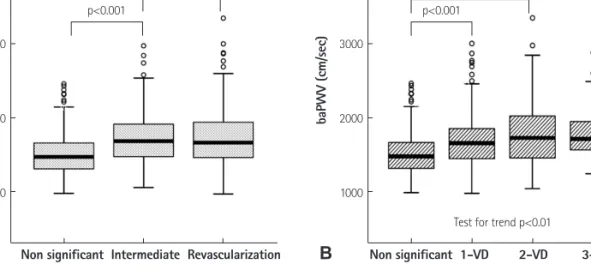
Fig. 1 shows the value of baPWV according to the history of revas- cularization and the presence of CAD. In the analysis of clinical out- comes, the non-significant CAD group’s baPWV was significantly lo- wer than other groups, but the baPWV values were not significantly different between intermediate CAD group and the revasculariza- tion group (Fig. 1A). In the view of extent of CAD, we assorted the subjects into one of 4 groups: non-significant stenosis, 1, 2 and 3- vessel disease. A significant difference of baPWV was not observed between each group except for the subjects without significant st- enosis. However, a trend toward positive association was found be- tween the extent of CAD and baPWV (p<0.01) (Fig. 1B). The ROC curve analysis of the association between baPWV and CAD is shown in Fig. 2. In a ROC curve of baPWV for significant CAD (Fig. 2A), the area under the curve was 0.68 (95% confidence interval=0.64- Table 2. The clinical outcomes in patients with significant coronary disease
Total, n=401 Medical treatment, n=145 PCI, n=226 CABG, n=30
1-vessel disease, n (%) 207 (51.6) 110 (75.9)* 94 (41.6) 3 (10.0)
2-vessel disease, n (%) 133 (33.2) 27 (18.6) 102 (45.1)* 4 (13.3)
3-vessel disease, n (%) 61 (15.2) 8 (5.5) 30 (13.3) 23 (76.7)*
*p<0.001, by chi-square test. PCI: percutaneous coronary intervention, CABG: coronary artery bypass graft Table 3. Multivariate logistic regression analysis of factors related to sig-
nificant coronary artery disease
Odds ratio 95% confidence interval p
Male gender 3.407 2.186-5.309 <0.001
Age 1.039 1.014-1.065 0.002
BMI 0.984 0.924-1.048 0.613
Hypertension 0.949 0.610-1.476 0.815
Diabetes 1.132 0.608-2.111 0.696
Smoking 0.732 0.423-1.267 0.265
Dyslipidemia 1.089 0.712-1.665 0.696
ACE-I or ARB 0.739 0.422-1.296 0.291
Beta-blocker 1.572 0.873-2.831 0.132
CCB 1.047 0.637-1.721 0.856
Diuretics 1.077 0.562-2.062 0.824
HDL-C 0.985 0.970-1.000 0.048
LDL-C 1.004 0.999-1.010 0.132
FBS 0.999 0.989-1.010 0.902
HbA
1c 1.581 1.123-2.225 0.009
SBP 1.006 0.993-1.020 0.373
baPWV 1.001 1.000-1.002 0.019
BMI: body mass index, ACE-I: angiotensin converting enzyme inhibitor, ARB:
angiotensin receptor blocker, CCB: calcium channel blocker, HDL-C: high density lipoprotein-cholesterol, LDL-C: low density lipoprotein-cholesterol, FBS: fasting blood glucose, HbA
1c: hemoglobin A
1c, SBP: systolic blood pres- sure, baPWV: brachial-ankle pulse wave velocity
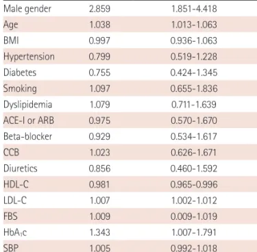
Table 4. Multivariate logistic regression analysis of factors related to the revascularization
Odds ratio 95% confidence interval p
Male gender 2.859 1.851-4.418 <0.001
Age 1.038 1.013-1.063 0.002
BMI 0.997 0.936-1.063 0.937
Hypertension 0.799 0.519-1.228 0.306
Diabetes 0.755 0.424-1.345 0.340
Smoking 1.097 0.655-1.836 0.726
Dyslipidemia 1.079 0.711-1.639 0.720
ACE-I or ARB 0.975 0.570-1.670 0.928
Beta-blocker 0.929 0.534-1.617 0.795
CCB 1.023 0.626-1.671 0.928
Diuretics 0.856 0.460-1.592 0.623
HDL-C 0.981 0.965-0.996 0.015
LDL-C 1.007 1.002-1.012 0.012
FBS 1.009 0.009-1.019 0.067
HbA
1c 1.343 1.007-1.791 0.044
SBP 1.005 0.992-1.018 0.466
baPWV 1.001 1.000-1.001 0.191
BMI: body mass index, ACE-I: angiotensin converting enzyme inhibitor, ARB:
angiotensin receptor blocker, CCB: calcium channel blocker, HDL-C: high
density lipoprotein-cholesterol, LDL-C: low density lipoprotein-cholesterol,
FBS: fasting blood glucose, HbA
1c: hemoglobin A
1c, SBP: systolic blood pres-
sure, baPWV: brachial-ankle pulse wave velocity
0.72). For revascularization (Fig. 2B), the area under the curve was 0.61 (95% confidence interval=0.56-0.65). The cut-off point of 1540 cm/s showed 65% sensitivity and 61% specificity in predicting sig- nificant CAD and the cut-off point of 1570 cm/s showed 60% sensi- tivity and 54% specificity in predicting revascularization.
Discussion
In this study, the baPWV was independently associated with sig- nificant CAD in patients who had no previous history of ischemic he- art disease, heart failure, peripheral arterial occlusive disease or ch-
ronic renal failure. However, it did not show the meaningful differ- ence among the classified groups by the extent of CAD or by the cli- nical outcomes of medical treatment versus revascularization, thou- gh there was a trend toward positive association between the extent of CAD and baPWV.
Increased arterial stiffness hinders the hemodynamic buffering effect of the cardiovascular system, contributing to elevated systolic blood pressure and pulse pressure, coronary arterial disease, and left ventricular hypertrophy.
1)15)Therefore, measuring aortic PWV is sug- gested as the standard method for the evaluation of aortic stiffness in assessing subclinical target organ damage.
2)16)Previous studies Fig. 1. The value of baPWV according to the history of revascularization and the presence of CAD. A: comparison of baPWV between the subjects divided into 3 groups based on clinical outcomes. The baPWV of the patients with normal or minimal CAD was significantly lower than that of the patients who had intermediate CAD or received revascularization, but there was no significant difference in baPWV between the intermediate CAD and revascularization group. B: comparison of baPWV according to the extent of coronary artery disease. There was no significant difference in baPWV between each group ex- cept the subjects without significant stenosis, but there was a linear trend between the number of stenosed vessels and increased baPWV. baPWV: brachi- al-ankle pulse wave velocity, CAD: coronary artery disease, VD: vessel disease, NS: no significant difference.
4000
3000
2000
1000
4000
3000
2000
1000
Test for trend p<0.01
baPWV (cm/sec) baPWV (cm/sec)
Non significant Intermediate Revascularization Non significant 1-VD 2-VD 3-VD p<0.001
p<0.001 p<0.001
p<0.001
NS p<0.001
A B
Fig. 2. Receiver operating characteristic (ROC) curve analysis. A: ROC curve of baPWV for significant coronary artery disease, baPWV cut-off value at 1540 cm/s showed sensitivity 65%, specificity 61% (area under the ROC curve=0.68; 95% confidence interval=0.64-0.72). B: ROC curve of baPWV for revascularization, 1570 cm/s showed sensitivity 60%, specificity 54% (area under the ROC curve=0.61; 95% confidence interval=0.56-0.65). baPWV: brachial-ankle pulse wave velocity, AUC: area under curve.
1.0
0.8
0.6
0.4
0.2
0.0
1.0
0.8
0.6
0.4
0.2
0.0 0.0 0.2 0.4 0.6 0.8 1.0
AUC: 0.68
(95% confidence interval: 0.64-0.72)
AUC: 0.61
(95% confidence interval: 0.56-0.65) 0.0 0.2 0.4 0.6 0.8 1.0
Sensitivity Sensitivity
1-specificity 1-specificity