ASSOCIATION OF GENITAL MYCOPLASMAS INFECTION IN WOMEN WHO HAD PRETERM DELIVERY AND OUTCOMES IN PREMATURE INFANTS
Hyun Kyung Chung, MD
1, So Yun Park, MD
1, Mi Hye Park, MD, PhD
1, Yong Ju Kim, MD, PhD
1, Sun Hee Chun, MD, PhD
1, Su Jin Cho, MD, PhD
2, Eun Ae Park, MD, PhD
2Departments of 1Obstetrics and Gynecology, 2Pediatrics, Ewha Womans University School of Medicine, Seoul, Korea
Objective
We conducted this study to evaluate the relationship between genital Mycoplasmas infection in women who had preterm delivery and the neontal outcomes.
Methods
We studied 116 women with preterm delivery (gestational age, ranged from 24 to 37 weeks) and their 116 premature infants born at Ewha Womans University Mokdong Hospital from August 2009 to December 2010. Culture for genital Mycoplasmas or a polymerase chain reaction was performed in vaginal fl uid obtained from women who had preterm delivery. For perinatal outcomes, chorioamnionitis, funisitis and puerperal infection were analysed. We also evaluated the following neonatal data; birth weight, sex, small for gestational age, Apgar score, cord blood pH, ventilator care, hospital days, incidence of respiratory distress syndrome, bronchopulmonary dysplasia, sepsis, intraventricular hemorrhage and patent ductus arteriosus.
Results
Genital Mycoplasmas were isolated in 36.2% (42 of 116 patients). History of prior preterm birth was signifi cantly higher and the cervical length at admission was signifi cantly shorter in patients with genital Mycoplasmas infection (P < 0.05). Perinatal outcomes according to the Mycoplasmas infection and the neonatal outcomes were not statistically different between study and control groups. The relative risk of respiratory distress syndrome and bronchopulmonary dysplasia were 1.55 (95% confi dence interval [CI], 0.65-3.70), and 1.24 (95% CI, 0.42-3.67), respectively.
Conclusion
There is higher incidence of prior preterm birth and shorter cervical length in patient with genital Mycoplasmas infection. However, genital Mycoplasmas infection does not affect the neonatal outcomes
Keywords:
Genital Mycoplasmas; Mycoplasma hominis; Ureaplasma urealyticum; Preterm birth; Premature infants
Received: 2011.9.21. Revised: 2011.12.5. Accepted: 2011.12.19.
Corresponding author: Sun Hee Chun, MD, PhD Department of Obstetrics and Gynecology, Ewha Womans University School of Medicine, 1071 Anyangcheon-ro, Yangcheon- gu, Seoul 158-710, Korea
Tel: +82-2-2650-2866 Fax: +82-2647-9860 E-mail: [email protected]
Th is is an Open Access article distributed under the terms of the Creative Commons Attribution Non-Commercial License (http://creativecommons.org/licenses/
by-nc/3.0/) which permits unrestricted non-commercial use, distribution, and reproduction in any medium, provided the original work is properly cited.
Copyright © 2012. Korean Society of Obstetrics and Gynecology 조산은 재태연령 37주 미만에 분만되는 것으로 세계적으로 전체 분
만의 10%에서 발생되고 신생아 사망의 70%, 이환의 75%를 차지하고 있는 산과학과 신생아학의 주요 문제이다[1]. 지난 50년 동안 조산 방 지를 위한 여러 가지 치료법 개발에도 불구하고 그 발생빈도는 감소 되지 않고 있어 원인 규명 및 그에 따른 치료 방법은 많은 의학적 관 심이 되고 있다. 조산의 병인론은 다양하며 이 중 생식기 감염은 전 체 조산의 원인 중 30% 이상을 차지한다고 알려져 있다. 이러한 감염 이 조기 진통 및 조기 양막파수를 일으키고, 조산아의 사망과 이환에 영향을 미치는 것으로 알려져 왔으며[2], 감염의 원인균으로 group B streptococcus, Ureaplasma urealyticum과 Mycoplasma hominis 등
pISSN 2233-5188 · eISSN 2233-5196
을 포함하는 Mycoplasmas, Chlamydia trachomatis, Escherichia coli, Klebsiella spp, Hemophylus influenza 등이 알려져 있다. 이 중 가장 흔 한 원인균은 Ureaplasma urealyticum (U. urealyticum)과 Mycoplasma hominis (M. hominis) 같은 Mycoplasmas이다[3].
최근 생식기 감염이 단순히 조기 진통 또는 조산을 일으키기보다는 자궁내 혹은 양수내 감염을 일으켜 이에 대한 반응으로 여러 종류의 염 증성 싸이토카인이 분비되어 결국 태아조직의 손상을 일으키는 태아 염증반응증후군(fetal inflammatory response syndrome)으로 이해되고 있으며 따라서 이미 조기진통을 가지고 있는 임신부의 자궁내 태아는 폐나 뇌의 조직손상이 진행되어 있을 수 있어 출생 후 기관지폐 형성이 상(bronchopulmonary dysplasia, BPD)이나 뇌성마비 등이 발병될 가 능성을 설명하고 있다[4,5]. 이런 관점에서 조기 진통이 발생되기 전인 임신 초기의 생식기감염, 특히 Mycoplasmas와 관련 있는 세균성 질염 (bacterial vaginosis)을 미리 진단하여 조기 항생제 치료를 시행함으로 써 조산 발생을 감소시켰다는 연구보고가 있다[6].
M. hominis와 U. urealyticum은 사춘기 이후의 여성과 남성의 비뇨생 식기계에 집락하며, 임신 여성에서 그 집락률은 40%-90%를 차지한 다[7]. 여성 생식기의 Mycoplasmas 감염은 특히 조기 양막파수 및 조 산, 불임 등에 관여한다고 알려져 있다. 또한 분만 중에는 감염된 산도 를 통해 신생아에게도 여러 가지 질환을 유발할 수 있다고 밝혀졌다 [8]. 최근 Mycoplasmas 감염과 신생아 질환의 관련성에 대한 연구 보 고를 보면, 선천성폐렴, 뇌수막염, 패혈증 등의 원인균으로 관찰되었 다[9,10]. 신생아의 호흡기 미숙으로 기관지폐형성이상과의 연관성과 Mycoplasmas 감염이 연관성이 있다는 연구들이 발표되고 있어 임신 중 Mycoplasmas 감염의 중요성을 다시 한번 인식시켜주었다[11]. M.
hominis와 U. urealyticum에 대한 연구는 국내에서도 활발히 진행되어 조기 진통과 조기 양막파수가 있는 산모에게서의 감염률 등에 대한 보 고는 있었다[12,13]. 그러나 이들 감염이 있는 임신부에서 분만된 조산 아들의 이환이 어느 정도인지에 대한 연구는 아직 미미하다.
이에 저자는 조산아를 분만한 산모의 분만 전 Mycoplasmas의 감염 여부를 파악하고 이들에게서 태어난 신생아의 질환과의 연관성을 분석 하여 조기분만 시 Mycoplasmas 감염이 산과적 합병증 및 신생아의 이 환율에 미치는 영향을 알아보고자 하였다.
연구대상 및 방법
1. 대상
2009년 8월부터 2010년 12월까지 이화여자대학교 이대목동병원 산부인과에 입원하여 분만한 산모는 1,062명이었으며 이 중 임신 37 주 이전에 조산을 한 경우는 190명이었다. 쌍태아를 분만한 26명을 제 외하고 단태아를 분만한 164명에서, Mycoplasmas 검사를 시행하지 못한 43명과 검사 결과 M. hominis 및 U. urealyticum 이외의 균이 발 견된 5명을 제외 후, 최종 116명의 산모 및 이들에게서 태어나 신생아 실이나 신생아집중치료실에 입원한 신생아들을 대상으로 하였다. 조산
은 월경주기가 규칙적인 경우는 마지막 월경 시작일을, 월경주기가 불 규칙한 경우는 초기임신 시 초음파 소견을 기준으로 임신 24주 이후와 37완전 주(37 completed weeks) 이전에 분만한 경우로 정의하였다.
분만 전 산모의 자궁경부 분비물 검체의 Mycoplasmas 배양 또는 중합 요소연쇄반응법(polymerase chain reaction, PCR)을 시행하여 두 검사 중 어느 하나의 검사 결과에서, M. hominis 및 U. urealyticum 둘 중 하 나라도 양성인 경우를 연구군으로, M. hominis와 U. urealyticum에 모 두 음성인 경우를 대조군으로 하였다.
2. 방법
본 연구는 의무기록 분석을 통한 후향적 연구를 시행하였다. 내원 당시 산모의 조기 양막파수는 질경검사에서 질내 양수가 고여있거나 nitrazine test 양성인 경우로 진단하였다. 입원 시 내진 및 경질 초음 파로 자궁경부 길이를 측정하였으며, 초음파 기기는 Prosound SSD- 3500 (Aloka Co. Ltd, Tokyo, Japan)을 사용하였다. 자궁경부 길이는 환자의 방광을 완전히 비운 후 쇄석위로 눕히고 질식 탐촉자를 질 앞 천장에 부드럽게 넣은 후, 영상을 화면의 75% 이상을 차지하게 확대 시켜 자궁경부 내구부터 외구까지 가장 긴 길이를 일직선으로 측정하 였다[14]. 산모의 합병증 중 자간전증은 National High Blood Pressure Education Program Working Group (2000) 기준에 따라 임신 20주 이 후 수축기 혈압 140 mm Hg 이상, 이완기 혈압 90 mm Hg 이상의 혈 압을 보이며 24시간에 300 mg 이상의 단백뇨가 함께 있는 경우로 진 단하였다[15]. 임신성 당뇨는 공복 시와 100 g 포도당 복용 1시간, 2 시간, 3시간 후 정맥 혈장의 혈당치가 각각 105 mg/dL, 190 mg/dL, 165 mg/dL, 145 mg/dL 중 2회 이상에서 기준치를 초과하는 경우[16]
또는 75 g 포도당 복용 2시간 후 정맥 혈장의 혈당치가 140 mg/dL를 초과하는 경우로 진단하였다[17].
검체 채취는 내진 전 질경으로 자궁경부를 확인 후 질벽에 닿지 않 도록 주의하여 자궁경부에 swab (Yuhan Lab Tech Co., Ltd., Seoul, Korea)을 삽입하여 채취하였고 MYCOFAST Evolution 2 (International Microbio, Signes, France)의 transport medium에 접종하여 지침 에 따라 배양검사를 시행하거나 PCR로 검사한 후 M. hominis 및 U.
urealyticum 감염 여부를 판정하였다. PCR의 경우 STI 1 Real-time Test kit (Seegene, Seoul, Korea)를 사용하여 제조사의 지시에 따라 시 행하였으며 GeneAmp PCR system 9700 (Applied biosystems Inc., California, CA, USA)을 사용하였다.
산과적 합병증으로는 임상적 융모양막염(clinical chorioamnionitis), 조직학적 융모양막염(histologic chorioamnionitis) 및 제대염(histologic funisitis), 산욕기 감염 여부를 살펴보았다. 임상적 융모양막염은 산 모의 체온이 37.8oC 이상이면서 자궁압통, 악취나는 질 분비물, 백혈 구증가증(>15,000/μL), 임신부 빈맥(>100회/분), 태아빈맥(>160회/
분) 중 2가지 이상 존재하는 경우로 진단하였다[18]. 조직학적 융모 양막염 및 제대혈관염은 분만 후 태반과 제대를 10% 포르말린에 고 정 후 hematoxylin eosin (H&E) 염색을 실시하여 병리학자에 의해 태 반, 융모양막 및 제대혈관 등에서 다형핵 백혈구(polymorphonuclear
leukocyte)의 침윤이 관찰될 경우로 진단하였다[19]. 산욕기 감염 여부 는 산욕열, 상처감염, 자궁내막염 여부 등을 파악하였다.
신생아의 경우는 출생체중과 부당경량아 여부, 성별, 1분 및 5분 Apgar 점수, 제대동맥혈검사, 인공환기기 사용 여부, 입원 기간 등 을 비교하였다. 부당경량아는 해당 제태 연령체중 백분위가 10백분 위 이하로 정의하였다[20]. 신생아 이환, 즉 신생아호흡곤란증후군 (respiratory distress syndrome, RDS)은 다른 폐 질환의 증거가 없 는 상태에서 임상 소견상 특징적인 호흡곤란, 흉곽함몰, 신음 및 청색 증을 보이고, 산소 요구량의 증가(fraction of inspired oxygen ≥0.4) 를 보이면서 흉부의 방사선학적 검사상 망상과립상, 공기-기관지 음 영 등 특징적 소견이 관찰되는 경우로, 패혈증(sepsis)은 전신 증상이 나 혈액검사상 이상 소견이 동반된 경우로, 뇌실내 출혈(intraventricular hemorrhage, IVH)은 신생아 뇌초음파를 시행하여 Volpe 분류에 따 라 각각 진단하였다[21]. BPD은 2001년 National Institute of Child Health workshop에서 합의된 내용을 바탕으로 방사선학적 소견과는 상관없이 재태기간 32주 미만의 미숙아의 경우 월경 후 주령 36주와 퇴원 시점 중 빠른 시기에 21%가 넘는 산소를 적어도 28일 이상 투여 한 경우로[22], 동맥관개존(patent ductus arteriosus, PDA)은 심장초
음파에 의해 진단되고, 인도메타신이나 수술적 방법을 사용하여 치료 한 경우로 각각 정의하였다.
3. 통계
통계 분석은 SAS ver. 9.1 (Institute Inc., Cary, NC, USA)를 사용하 였고, Mycoplasma 감염 빈도 및 두 군간의 빈도 분석은 카이제곱 검 정을 이용하였으며, 평균은 Student t-검정을 이용하여 산출하였고 P 값이 0.05 미만인 경우에 통계적으로 유의하다고 판단하였다. Proc genmode를 사용하여 상대위험비(relative risk, RR) 값을 산출하였다.
결 과
1. Mycoplasmas 감염 빈도
총 116명의 대상 산모 중 M. hominis와 U. urealyticum에 모두 음성 인 경우가 74명(63.8%), M. hominis 및 U. urealyticum 둘 중 하나라도 양성인 경우가 42명(36.2%)이었다. 이 중 M. hominis 단독감염이 7명 (6.0%), U. urealyticum 단독 감염이 25명(21.6%)이었으며 혼합감염은
Table 1. Mycoplasma hominis and Ureaplasma urealyticum infection
No. of infection Infection rate (%)
M(+) U(+) 10 8.6
M (+) U (-) 7 6.0
M (-) U (+) 25 21.6
M (-) U (-) 74 63.8
M, Mycoplasma hominis; U, Ureaplasma urealyticum.
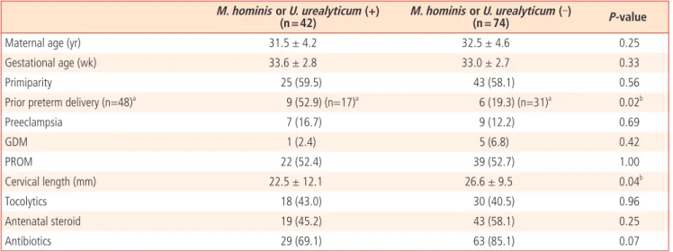
Table 2. Clinical characteristics
M. hominis or U. urealyticum (+)
(n = 42) M. hominis or U. urealyticum (‐)
(n = 74) P‐value
Maternal age (yr) 31.5 ± 4.2 32.5 ± 4.6 0.25
Gestational age (wk) 33.6 ± 2.8 33.0 ± 2.7 0.33
Primiparity 25 (59.5) 43 (58.1) 0.56
Prior preterm delivery (n=48)a 9 (52.9) (n=17)a 6 (19.3) (n=31)a 0.02b
Preeclampsia 7 (16.7) 9 (12.2) 0.69
GDM 1 (2.4) 5 (6.8) 0.42
PROM 22 (52.4) 39 (52.7) 1.00
Cervical length (mm) 22.5 ± 12.1 26.6 ± 9.5 0.04b
Tocolytics 18 (43.0) 30 (40.5) 0.96
Antenatal steroid 19 (45.2) 43 (58.1) 0.25
Antibiotics 29 (69.1) 63 (85.1) 0.07
Values are presented as mean ± standard deviation or number (%).
M, Mycoplasma hominis; U, Ureaplasma urealyticum; GDM, gestational diabetic mellitus; PROM, premature rupture of membrane.
aThis is the number except primiparity.
bP < 0.05
10명(8.6%)을 차지하였다(Table 1).
2. 산모의 임상적 특징
연구군과 대조군 산모의 평균 연령은 31.5 ± 4.2세와 32.5 ± 4.6세, 재태연령은 33.6 ± 2.8주와 33.0 ± 2.7주로 두 군 사이에 유의한 차이 는 없었다(P > 0.05). 이전의 조산 과거력은 연구군에서 22.0%로 대조 군의 8.1%보다 유의하게 높았으며(P = 0.03), 자궁경부 길이도 연구군 이 22.5 ± 12.1 mm로 대조군의 26.6 ± 9.5 mm보다 유의하게 짧았다 (P = 0.04). 조기 양막파수의 경우, 연구군은 16.7%, 대조군은 12.2%
로 두 군 사이에 유의한 차이는 없었다(P > 0.05). 임신성 고혈압이 합 병된 경우는 연구군이 16.7%, 대조군이 12.2%였으며, 임신성 당뇨도 각각 2.4%, 6.8%로 모두 두 군 사이에 유의한 차이는 보이지 않았다
(P > 0.05). 그 밖에 진통억제제, 스테로이드, 항생제 사용여부는 두 군 사이에 통계학적 유의한 차이는 없었다(P > 0.05) (Table 2).
3. 융모양막염과 분만 후 산모의 합병증
임상적 융모양막염은 연구군은 16.7%, 대조군은 12.2%로 두 군 사이 에 발생 빈도의 유의한 차이를 보이지 않았다(P > 0.05). 조직학적 융모양 막염, 제대혈관염의 발생빈도도 또한 두 군 간에 차이가 없었다(P > 0.05).
산욕기 감염 여부에 대해 비교하였을 때도 연구군이 7.5%, 대조군이 2.7%로 통계학적으로 유의한 차이를 보이지 않았다(P > 0.05) (Table 3).
4. 신생아 이환
연구군과 대조군에서 태어난 조산아의 평균 몸무게를 분석한 결
Table 3. Delivery outcomes according to Mycoplasma hominis and Ureaplasma urealyticum infection M. hominis or U. urealyticum (+)
(n = 42) M. hominis or U. urealyticum (‐)
(n = 74) P‐value
Clinical chorioamnionitis 7 (16.7) 9 (12.2) 0.69
Histologic chorioamnionitis 10 (27.0) 31 (43.7) 0.14
Histologic funisitis 5 (13.5) 4 (5.6) 0.14
Puerperal infection 3 (7.5) 2 (2.7) 0.34
Values are presented as number (%).
Table 4. Effect of Mycoplasma hominis and Ureaplasma urealyticum infection on neonatal morbidity M. hominis or
U. urealyticum (+) (n = 42)
M. hominis or U. urealyticum (‐)
(n = 74) P‐value Relative risk 95% CI
Birth weight (g) 2073.8 ± 621.0 2008.0 ± 637.7 0.59
Male sex 28 (66.7) 47 (63.5) 0.89
SGA 3 (7.1) 10 (13.5) 0.15
Apgar score<7 (1 min) 14 (33.3) 28 (37.8) 0.78
Apgar score<7 (5 min) 5 (11.9) 7 (9.5) 0.75
Cord blood pH 7.4 ± 0.2 7.3 ± 0.1 0.12
Ventilator care 7 (16.7) 22 (29.7) 0.18
Hospital daysa (day) 15.0 (8.0‐26) 20 (11‐48) 0.16
≤8
11 (26.2) 13 (17.6) 0.29≤36 23 (54.8) 38 (51.4)
>36 8 (19.1) 23 (31.1)
RDS 8 (19.1) 9 (12.3) 0.48 1.55 0.65‐3.70
Sepsis 3 (7.1) 5 (6.9) 1.00 1.04 0.26‐4.15
IVH 1&2 8 (19.05) 10 (13.70) 0.62 1.39 0.60‐3.25
BPD 5 (11.90) 7 (9.59) 0.76 1.24 0.42‐3.67
PDA 5 (11.90) 14 (19.18) 0.44 0.62 0.24‐1.60
Values are presented as mean ± standard deviation or number (%).
CI, confi dence interval; SGA, small for gestational age; RDS, respiratory distress syndrome; IVH, intraventricular hemorrhage; BPD, bronchopulmonary dysplasia; PDA, patent ductus arteriosus.
aNon‐parametric analysis using Wilcoxon rank‐sum test.
과 각각 2,073.8 ± 621.0 g, 2,008.0 ± 637.7 g으로 통계학적으로 유의 한 차이는 없었다(P > 0.05) 성별 및 부당경량아 비율에서도 두 군 간 의 차이를 보이지 않았다(P > 0.05). 1분 및 5분 Apgar 점수가 7점 이 하인 경우를 살펴본 결과, 연구군은 각각 33.3%와 11.9%였고, 대조 군은 37.8%와 9.5%로 나왔으며, 제대동맥혈 pH를 비교해 본 결과 연 구군은 7.4 ± 0.2, 대조군은 7.3 ± 0.1이었고, 모두 유의한 차이는 없었 다(P > 0.05). 인공환기기 사용 여부, 입원 기간에서도 두 군 간에 차이 를 보이지 않았다(P > 0.05). RDS의 경우 RR가 1.55 (95% confidence interval [CI], 0.65-3.70)였으며, IVH 1, 2단계 및 BPD의 경우 각 각 1.39 (95% CI, 0.60-3.25), 1.24 (95% CI, 0.42-3.67)였다. 그러 나 모두 연구군과 대조군 간에 통계학적으로 유의한 차이는 없었다 (P > 0.05) (Table 4).
고 찰
Mycoplasmas는 세포벽이 없는 가장 작은 세균으로 세포벽에 작 용하는 ß-lactam 항생제에 반응하지 않고 배양도 잘되지 않는다.
Mycoplasma hominis가 1937년 Bartholin 농양으로부터 처음 분리되 어 보고된 이 후, 현재까지 약 12종의 Mycoplasmas가 사람에서 분류 되었는데, 6종의 Mycoplasma와 2종의 Ureaplasma가 비뇨생식기계에 집락을 형성하고 있다[23]. 이 중에서 M. hominis와 U. urealyticum이 산모와 신생아에게 영향을 미치며 다양한 질환을 유발하는 주요 병원 체로 알려져 있다. 이들을 분리하기 위해서는 특수 배지를 이용한 배양 방법을 통해 가능하였으나 최근에는 PCR을 이용하여 진단이 가능해졌 고 이로 인해 검체 분리 시간도 단축할 수 있게 되었다[24].
임신 중 Mycoplasmas 감염의 빈도는 저자에 따라 상당한 차이가 있는데 M. hominis 감염의 경우 McCormack 등[25]은 21.3%의 분 리율을, 그리고 국내 Chang 등[26]은 8%의 분리율을 보고하였고, U.urealyticum의 경우 Mårdh와 Weström [27]은 68.4%, 국내 Chang 등[26]은 51.6%의 분리율을 보고하였다. 본 연구에서는 M. hominis 단 독 감염이 6.0%, U. urealyticum 단독 감염이 21.6%이었으며 혼합감염 은 8.6%로 기존의 연구와 달리 조기분만한 산모를 대상으로 조사한 것 으로 감염율에 차이가 나타난 것으로 보인다.
Mycoplasmas 감염이 조산 및 신생아 질환과 밀접한 관계가 있음을 밝히는 많은 논문들이 보고되었다. Klein 등[28]과 Braun 등[29]은 대 조군에 비해 U. urealyticum과 M. hominis 감염군에서 출산된 태아의 평균 몸무게가 더 적다고 보고하였으나, Embree 등[30]은 Ureaplasma 감염과 저체중아 출산과 관련이 없다고 보고하였다. 2003년 Yoon 등 [31]은 양수내의 U. urealyticum 감염에 따른 산모와 신생아들의 임상 양상을 살펴보았다. 양수내 U. urealyticum은 배양과 PCR을 통하여 검 출하였으며, 그 결과 PCR 양성이면서 배양 검사에서는 음성인 조기 진 통 산모에게서 조산을 하는 비율이 높았으며, 주산기 예후에도 영향을 미치는 것으로 나타났다. 또한 출산된 태아의 몸무게도 더 적었으며 주 요 질병의 이환율도 통계학적으로 의미 있게 높은 것으로 나타났다. 그
러나 본 연구에서는 연구군과 대조군 사이에 출산된 태아의 평균 몸무 게 및 이환율을 비교하였을 때 통계학적으로 의미 있는 차이를 보이지 않았는데 이는 양수내 감염이 아닌 질내 감염 결과로만 비교하였기에 위 결과와 다른 결과가 나온 것으로 생각된다.
이 외에 Ureaplasma 감염과 RDS 및 BPD 발생률과의 관계를 살펴본 연구 발표도 있었다. 여러 연구에서 자궁내 Ureaplasma 감염이 미숙아 에게서 RDS 발생률을 줄이는 것으로 보고 하였으며 이는 태내에서의 하부기도내 감염은 아급성의 폐포내 염증반응을 일으키고 폐성숙과 폐 표면활성제의 생산을 증가시켜 미숙아의 급성 호흡기질환의 발생을 낮 추는 효과를 나타내기 때문이다[32,33]. BPD의 경우에는 발병률을 높 인다는 연구 발표도 있었는데[34-36], U. urealyticum에 의한 BPD의 발생 기전은 기도 상피세포에 집락 형성을 한 후 섬모 운동의 혼란 및 군집을 형성하고 탐식세포를 자극해 IL-6, TNF-alpha 등의 BPD 발생 과 관련된 염증성 인자를 생산하는 것으로 생각되어 진다[37]. 이와 반 대로 재태연령을 비롯한 몇 가지 변수를 보정한 결과 연관성이 관찰되 지 않았다는 발표도 있었다[38]. 본 연구에서는 RDS 및 BPD 발생률의 차이를 비교한 결과 BPD 경우 상대위험도를 계산하였을 때 연구군이 대조군에 비해 1.24배 높았으며 RDS경우 1.55배 높게 나타났으나 통 계학적으로 의미 있는 차이를 보이지 않았다.
신생아 패혈증의 경우 임신부의 산도를 통한 감염으로 인해 일어날 수 있으며, 자궁내 태아에게도 감염된 양수와 태반을 통해 감염을 일으 킬 수 있다[39]. 기존의 연구에서 Ureaplasma 양성 신생아의 혈액배양 검사에서 혈액 중의 Ureaplasma의 동정을 확인하여 보고한 바 있었다 [40]. 그러나 본 연구에서는 산모의 질내 Mycoplasmas 감염이 신생아 패혈증의 발생률을 높이지는 않는 것으로 나타났다.
본 연구는 조산아를 분만한 산모의 분만 전 Mycoplasmas의 감염 여 부를 파악하고 이들에게서 태어난 신생아의 질환과의 연관성을 분석하 여 분만 전 Mycoplasmas 감염이 임신 및 신생아의 예후에 미치는 영 향을 알아보고자 하였다. 대상 산모들의 특징으로는 연구군에서 조산 과거력이 더 많고, 자궁경부 길이가 더 짧은 것으로 확인되었다.
그러나 본 연구는 후향적으로 의무기록을 조사하여 이루어진 연구 로 신생아의 여러 발생 질환이 실제 모체의 Mycoplasmas 감염으로 인 한 것인지 정확히 구분하기 어렵다. 임신 초기의 생식기 감염이 지속적 으로 존재하여 태아 염증반응을 일으켜 조산을 유발할 수 있으며 따라 서 임신 초기의 세균성 질염 진단과 조기 치료가 조산 및 신생아 이환 의 발생을 감소시킬 수 있다는 발표가 있었다[6]. 따라서 세균성 질염 이 있는 산모는 조기 진통이나 조기 양막파수의 위험이 높아질 수 있으 나 본 연구에서는 세균성 질염의 유무를 함께 평가하지 못하였기에 이 로 인한 영향 여부를 분석하지 못한 한계가 있으며 향후 이에 대한 연 구 필요하겠다.
References
1. Challis JR, Lye SJ, Gibb W, Whittle W, Patel F, Alfaidy N. Under-
standing preterm labor. Ann N Y Acad Sci 2001;943:225-34.
2. Romero R, Mazor M. Infection and preterm labor. Clin Obstet Gynecol 1988;31:553-84.
3. Romero R, Shamma F, Avila C, Jimenez C, Callahan R, Nores J, et al. Infection and labor. VI. Prevalence, microbiology, and clinical signifi cance of intraamniotic infection in twin gesta- tions with preterm labor. Am J Obstet Gynecol 1990;163:757- 61.
4. Yoon BH, Romero R, Park JS, Kim M, Oh SY, Kim CJ, et al. The relationship among infl ammatory lesions of the umbilical cord (funisitis), umbilical cord plasma interleukin 6 concentration, amniotic fl uid infection, and neonatal sepsis. Am J Obstet Gy- necol 2000;183:1124-9.
5. Romero R, Gomez R, Ghezzi F, Yoon BH, Mazor M, Edwin SS, et al. A fetal systemic infl ammatory response is followed by the spontaneous onset of preterm parturition. Am J Obstet Gynecol 1998;179:186-93.
6. Lamont RF, Duncan SL, Mandal D, Bassett P. Intravaginal clindamycin to reduce preterm birth in women with abnormal genital tract fl ora. Obstet Gynecol 2003;101:516-22.
7. Anagrius C, Loré B, Jensen JS. Mycoplasma genitalium: preva- lence, clinical signifi cance, and transmission. Sex Transm Infect 2005;81:458-62.
8. Kataoka S, Yamada T, Chou K, Nishida R, Morikawa M, Minami M, et al. Association between preterm birth and vaginal colo- nization by mycoplasmas in early pregnancy. J Clin Microbiol 2006;44:51-5.
9. Cassell GH, Waites KB, Watson HL, Crouse DT, Harasawa R.
Ureaplasma urealyticum intrauterine infection: role in prema- turity and disease in newborns. Clin Microbiol Rev 1993;6:69- 87.
10. Velemínský M, Tosner J. Relationship of vaginal microfl ora to PROM, pPROM and the risk of early-onset neonatal sepsis.
Neuro Endocrinol Lett 2008;29:205-21.
11. Katz B, Patel P, Duffy L, Schelonka RL, Dimmitt RA, Waites KB.
Characterization of ureaplasmas isolated from preterm infants with and without bronchopulmonary dysplasia. J Clin Micro- biol 2005;43:4852-4.
12. Kim MJ, Choi MH, Seong WJ, Koo TB, Park IS. The detection rate of Ureaplasma urealyticum and Mycoplasma hominis in patients with impending preterm birth and mid-trimester cer- vical swab. Korean J Perinatol 2008;19:370-6.
13. Kim SY, Lee YJ, Meen H, Lee SH, Park IY, Ahn HY, et al. Analysis of mycoplasma hominis and ureaplasma urealyticum infection in preterm labor and prom patients. Korean J Obstet Gynecol
2004;47:1469-73.
14. Iams JD, Goldenberg RL, Meis PJ, Mercer BM, Moawad A, Das A, et al. The length of the cervix and the risk of spontaneous premature delivery. National Institute of Child Health and Hu- man Development Maternal Fetal Medicine Unit Network. N Engl J Med 1996;334:567-72.
15. Report of the National High Blood Pressure Education Pro- gram Working Group on High Blood Pressure in Pregnancy.
Am J Obstet Gynecol 2000;183:S1-S22.
16. Classification and diagnosis of diabetes mellitus and other categories of glucose intolerance. National Diabetes Data Group. Diabetes 1979;28:1039-57.
17. WHO Expert Committee on Diabetes Mellitus: second report.
World Health Organ Tech Rep Ser 1980;646:1-80.
18. Gibbs RS, Blanco JD, St Clair PJ, Castaneda YS. Quantitative bacteriology of amniotic fluid from women with clinical in- traamniotic infection at term. J Infect Dis 1982;145:1-8.
19. Hollander D. Diagnosis of chorioamnionitis. Clin Obstet Gyne- col 1986;29:816-25.
20. Lubchenco LO, Hansman C, Dressler M, Boyd E. Intrauterine Growth as Estimated from Liveborn Birth-Weight Data at 24 to 42 Weeks of Gestation. Pediatrics 1963;32:793-800.
21. Volpe JJ. Neonatal intracranial hemorrhage. Pathophysiol- ogy, neuropathology, and clinical features. Clin Perinatol 1977;4:77-102.
22. Jobe AH, Bancalari E. Bronchopulmonary dysplasia. Am J Respir Crit Care Med 2001;163:1723-9.
23. Taylor-Robinson D, McCormack WM. The genital mycoplasmas (fi rst of two parts). N Engl J Med 1980;302:1003-10.
24. Kong F, Ma Z, James G, Gordon S, Gilbert GL. Species iden- tification and subtyping of Ureaplasma parvum and Urea- plasma urealyticum using PCR-based assays. J Clin Microbiol 2000;38:1175-9.
25. McCormack WM, Rosner B, Lee YH. Colonization with genital mycoplasmas in women. Am J Epidemiol 1973;97:240-5.
26. Chang MW, Choi TK, Yoshii Z, Matsuo Y. Isolation of Urea- plasma urealyticum and Mycoplasma hominis from patients with genitourinary tract infection. Hiroshima J Med Sci 1984;33:53-6.
27. Mårdh PA, Weström L. T-mycoplasmas in the genito-urinary tract of the female. Acta Pathol Microbiol Scand B Microbiol Immunol 1970;78:269.
28. Klein JO, Buckland D, Finland M. Colonization of newborn in- fants by mycoplasmas. N Engl J Med 1969;280:1025-30.
29. Braun P, Lee YH, Klein JO, Marcy SM, Klein TA, Charles D, et al.
Birth weight and genital mycoplasmas in pregnancy. N Engl J
Med 1971;284:167-71.
30. Embree JE, Krause VW, Embil JA, MacDonald S. Placental infection with Mycoplasma homonis and Ureaplasma urealyti- cum: clinical correlation. Obstet Gynecol 1980;56:475-81.
31. Yoon BH, Romero R, Lim JH, Shim SS, Hong JS, Shim JY, et al.
The clinical signifi cance of detecting Ureaplasma urealyticum by the polymerase chain reaction in the amniotic fl uid of pa- tients with preterm labor. Am J Obstet Gynecol 2003;189:919- 24.
32. Hannaford K, Todd DA, Jeffery H, John E, Blyth K, Gilbert GL.
Role of ureaplasma urealyticum in lung disease of prematurity.
Arch Dis Child Fetal Neonatal Ed 1999;81:F162-7.
33. Cultrera R, Seraceni S, Germani R, Contini C. Molecular evi- dence of Ureaplasma urealyticum and Ureaplasma parvum colonization in preterm infants during respiratory distress syn- drome. BMC Infect Dis 2006;6:166.
34. Benstein BD, Crouse DT, Shanklin DR, Ourth DD. Ureaplasma in lung. 2. Association with bronchopulmonary dysplasia in premature newborns. Exp Mol Pathol 2003;75:171-7.
35. Colaizy TT, Morris CD, Lapidus J, Sklar RS, Pillers DA. Detection of ureaplasma DNA in endotracheal samples is associated with bronchopulmonary dysplasia after adjustment for mul-
tiple risk factors. Pediatr Res 2007;61:578-83.
36. Walsh WF, Butler J, Coalson J, Hensley D, Cassell GH, deLemos RA. A primate model of Ureaplasma urealyticum infection in the premature infant with hyaline membrane disease. Clin Infect Dis 1993;17 Suppl 1:S158-62.
37. Patterson AM, Taciak V, Lovchik J, Fox RE, Campbell AB, Viscar- di RM. Ureaplasma urealyticum respiratory tract colonization is associated with an increase in interleukin 1-beta and tumor necrosis factor alpha relative to interleukin 6 in tracheal aspi- rates of preterm infants. Pediatr Infect Dis J 1998;17:321-8.
38. Ollikainen J, Korppi M, Heiskanen-Kosma T, Heinonen K.
Chronic lung disease of the newborn is not associated with Ureaplasma urealyticum. Pediatr Pulmonol 2001;32:303-7.
39. Cassell GH, Davis RO, Waites KB, Brown MB, Marriott PA, St- agno S, et al. Isolation of Mycoplasma hominis and Ureaplas- ma urealyticum from amniotic fl uid at 16-20 weeks of gesta- tion: potential effect on outcome of pregnancy. Sex Transm Dis 1983;10:294-302.
40. Cassell GH, Waites KB, Crouse DT, Rudd PT, Canupp KC, St-
agno S, et al. Association of Ureaplasma urealyticum infection
of the lower respiratory tract with chronic lung disease and
death in very-low-birth-weight infants. Lancet 1988;2:240-5.
조산 임신부의 분만 전 생식기 Mycoplasmas 감염과 임신 및 신생아 예후에 관한 연구
이화여자대학교 의학전문대학원 1산부인과학교실, 2소아청소년과학교실 정현경1, 박소연1, 박미혜1, 김영주1, 전선희1, 조수진2, 박은애2
목적
조기분만 시 Mycoplasmas 감염이 임신 및 신생아의 이환율에 미치는 영향을 알아보고자 하였다.
연구방법
본 연구는 의무기록 분석을 통한 후향적 연구로, 2009년 8월부터 2010년 12월까지 이화여자대학교 이대목동병원 산부인과에 입원하여 단태아 조산아를 분만한 116명의 산모 및 이들에게서 태어난 신생아들을 대상으로 하였다. 검체는 자궁경부에서 채취하여 배양 혹은 중 합효소연쇄반응법을 시행하였으며, 감염에 따른 신생아 이환율을 비교 분석하였다
결과
총 116명의 대상 산모 중 생식기 Mycoplasmas 감염된 경우가 42명(36.2%)이었다. 연구군에서 대조군보다 자궁경부 길이가 유의하게 짧 았고, 조산 기왕력의 빈도는 높았다. 산과적 합병증으로 임상적 융모양막염과 조직학적 융모양막염, 제대혈관염의 발생빈도 및 산욕기 감 염 발생을 비교하였을 때 두 군 간에 차이를 보이지 않았으며, 연구군과 대조군에서 태어난 신생아의 평균 몸무게, 성별, 부당경량아 비 율, 1분 및 5분 Apgar 점수, 제대동맥혈 pH, 인공환기기 사용 여부, 입원 기간, 신생아호흡곤란증후군(respiratory distress syndrome, RDS), sepsis, 뇌실내 출혈(intraventricular hemorrhage), 동맥관개존(patent ductus arteriosus) 이환율을 분석한 결과 통계학적으로 유의한 차이 는 없었다. RDS 및 기관지폐 형성이상(bronchopulmonary dysplasia)의 상대위험비(relative risk)는 각각 1.55 (95% confidence interval [CI], 0.65-3.70), 1.24 (95% CI, 0.42-3.67)였다.
결론
임상적 특징으로는 연구군에서 조산 과거력이 더 많고, 자궁경부 길이가 더 짧은 것으로 나타났다. 그러나 조산모의 질내 Mycoplasmas 감 염은 여러 신생아의 질환 발생에 거의 영향을 주지 않는 것으로 확인되었다.
중심단어: 생식기 마이코플라즈마, 조산, 조산아