http://dx.doi.org/10.5625/lar.2013.29.3.148
Systemic administration of low dosage of tetanus toxin decreases cell proliferation and neuroblast differentiation in the mouse
hippocampal dentate gyrus
Bing Chun Yan
1#, In Hye Kim
2#, Joon Ha Park
2, Ji Hyeon Ahn
2, Jeong-Hwi Cho
2, Bai Hui Chen
3, Jae-Chul Lee
2, Jung Hoon Choi
4, Ki-Yeon Yoo
5, Choong Hyun Lee
6, Jun Hwi Cho
7, Jong-Dai Kim
8*, Moo-Ho Won
2*
1
Department of Integrative Traditional & Western Medicine, Medical College, Yangzhou University, Yangzhou, China
2
Department of Neurobiology, and Institute of Medical Sciences, School of Medicine, Kangwon National University, Chuncheon, Korea
3
Department of Physiology, College of Medicine, and Institute of Neurodegeneration and Neuroregeneration, Hallym University, Chuncheon, Korea
4
Department of Anatomy, College of Veterinary Medicine, Kangwon National University, Chuncheon, Korea
5
Department of Oral Anatomy, College of Dentistry and Research institute of Oral Biology, Gangneung-Wonju National University, Gangneung, Korea
6
Department of Anatomy and Physiology, College of Pharmacy, Dankook University, Cheonan, Korea
7
Department of Emergency Medicine, and Institute of Medical Sciences, Kangwon National University Hospital, School of Medicine, Kangwon National University, Chuncheon, Korea
8
Division of Food Biotechnology, School of Biotechnology, Kangwon National University, Chuncheon, Korea
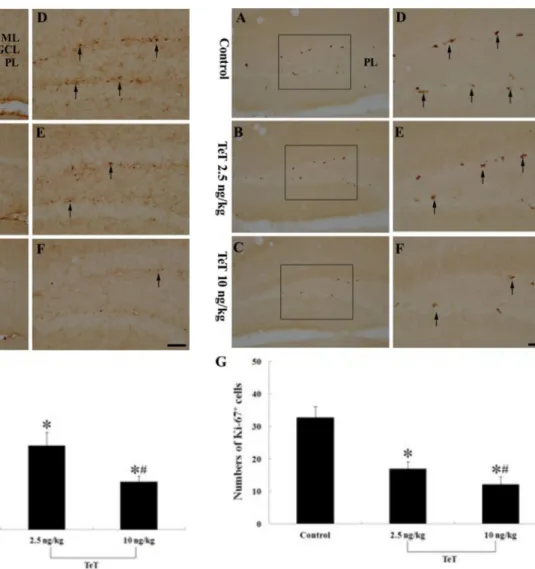
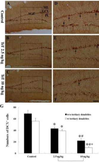
In the present study, we investigated the effect of Tetaus toxin (TeT) on cell proliferation and neuroblast differentiation using specific markers: 5-bromo-2-deoxyuridine (BrdU) as an exogenous marker for cell proliferation, Ki-67 as an endogenous marker for cell proliferation and doublecortin (DCX) as a marker for neuroblasts in the mouse hippocampal dentate gyrus (DG) after TeT treatment. Mice were intraperitoneally administered 2.5 and 10 ng/kg TeT and sacrificed 15 days after the treatment. In both the TeT-treated groups, no neuronal death occurred in any layers of the DG using neuronal nuclei (NeuN, a neuron nuclei maker) and Fluoro-Jade B (F-J B, a high-affinity fluorescent marker for the localization of neuronal degeneration). In addition, no significant change in glial activation in both the 2.5 and 10 ng/kg TeT-treated-groups was found by GFAP (a marker for astrocytes) and Iba-1 (a marker for microglia) immunohistochemistry. However, in the 2.5 ng/kg TeT-treated-group, the mean number of BrdU, Ki-67 and DCX immunoreactive cells, respectively, were apparently decreased compared to the control group, and the mean number of each in the 10 ng/kg TeT-treated-group was much more decreased. In addition, processes of DCX-immunoreactive cells, which projected into the molecular layer, were short compared to those in the control group. In brief, our present results show that low dosage (10 ng/kg) TeT treatment apparently decreased cell proliferation and neuroblast differentiation in the mouse hippocampal DG without distinct gliosis as well as any loss of adult neurons.
Key words: Exotoxin, neuronal damage, neurogenesis, sub-granular zone, granule cell
Received 15 May 2013; Revised version received 30 June 2013; Accepted 16 July 2013
#