Clinical Feasibility and Usefulness of CT Fluoroscopy-Guided Percutaneous Transhepatic Biliary Drainage in
Emergency Patients with Acute Obstructive Cholangitis
Objective: To evaluate the feasibility of CT fluoroscopy (CTF)-guided percuta- neous transhepatic biliary drainage (PTBD) in emergency patients with acute obstructive cholangitis.
Materials and Methods: The study included 28 patients admitted to the emer- gency center due to obstructive jaundice and found to require urgent biliary drainage, as well as judged to have a suitable peripheral bile duct for a CTF-guid- ed puncture (at least 4 mm in width). Prior to the CTF-guided puncture, a CT scan was performed to evaluate bile duct dilatation and the underlying causes of biliary obstruction. If the patient was judged to be a suitable candidate, a CTF-guided PTBD was performed in the same CT unit without additional fluoroscopic guid- ance. Technical feasibility of the procedure was investigated with the evaluation of overall success rate and causes of failure.
Results: A hepatic puncture was attempted at the left lobe in 23 patients and right lobe in five patients. The procedure was successful in 24 of 28 patients (86%) Successful biliary puncture was achieved on the first attempt in 16 patients, the second attempt in five patients, and the third attempt in three patients. The causes of failure included guide wire twisting in one patient, biliary puncture failure in two patients, and poor visualization of the guide wire in one patient. There were no significant procedure-related complication.
Conclusion: The CTF-guided PTBD is technically feasible and highly success- ful in patients judged to have a suitable indication. Moreover, although the proce- dure is unfamiliar and inconvenient to interventionalists, it has economical advan- tages in that it saves time and manpower. We believe this method can be used in the emergency patients requiring urgent biliary drainage as an alternative for the fluoroscopy-guided PTBD.
ince the first clinical introduction by Katada et al. (1, 2) in 1994, real time CT fluoroscopy (CTF) has been increasingly used in the field of interven- tional radiology. Previous reports have described advantages of CTF in the biopsy, drainage, and percutaneous placement of needles or other interventional instruments into the target locations (3-10). CTF had also previously been applied to percutaneous transhepatic biliary drainage (PTBD) and the results were already reported (11-13). However, these results were not purely for CTF-guided PTBD, but for PTBD using the combined guidance of CTF and mobile C-arm fluoroscopy. CTF was used only for the puncture of suitable peripheral bile ducts, and the rest of work was completed under the guidance of C-arm fluoroscopy. Generally, patients with Ji Hyung Kim, MD1, 2
Index terms :
Bile ducts, percutaneous drainage Computed tomography (CT) Fluoroscopy
Computed tomography (CT) fluoroscopy
DOI:10.3348/kjr.2009.10.2.144
Korean J Radiol 2009;10:144-149 Received August 18, 2008; accepted after revision November 7, 2008.
1Department of Radiology, Sam Anyang Hospital, Kyungki-do 430-733, Korea;
2Department of Radiology, Konyang University Hospital, Daejon 305-718, Korea
This article is not related to funding from any organizations or companies.
Address reprint requests to : Ji Hyung Kim, MD, Department of Radiology, Sam Anyang Hospital, 613-8 Anyang 5-dong, Manan-gu, Anyang, Kyungki-do 430-733, Korea.
Tel. (8231) 467-9265 Fax. (8231) 449-0151 e-mail: [email protected]
S
obstructive jaundice require two steps to undergo pallia- tive PTBD. A CT scan or sonography is first performed to evaluate the degree of biliary dilatation and its underlying causes. Next, the patients are moved to intervention unit for the procedure. We believe that these steps could be
simplified using CTF, which would be beneficial for the patients and clinical staff. The purpose of this study is to evaluate the clinical feasibility of CTF-guided PTBD as a means of a simplification in the emergency work steps of patients requiring urgent biliary drainage.
A B
C D
Fig. 1. 65-year-old male patient with distal CBD cancer.
A-C. CT scan images show dilated intrahepatic and extrahepatic ducts. It is possible to trace advancement course of guide wire and catheter on CT images (white arrows).
D-F. Monitoring CT fluoroscopy images, segment 3 IHD is punctured and guide wire is subsequently inserted, finally to CBD (dotty arrows).
E F
MATERIALS AND METHODS
We performed CTF-guided PTBD on five cases as part of a pilot trial before the main study to familiarize ourselves with the procedure. The main study had been prospec- tively performed for a total of 11 months after the pilot trial. The study subjects that were patients hospitalized at the emergency center due to acute cholangitis by biliary obstruction. They clinically complained of fever, abdomi- nal pain at the right upper quadrant, and jaundice. The laboratory tests performed on them revealed elevated serum bilirubin levels and leukocytosis. A CT scan (Somatom plus 4, Siemens Medical Systems, Erlangen, Germany) was performed to evaluate bile duct dilatation and underlying causes. Among them, we selected 28 patients (12 M:16 F, age range 48-82 yrs old, mean 62 yrs old) according to following inclusion criteria; 1) Urgent biliary drainage was clinically required. 2) Taking into
account a rough resolution of the real-time CTF image, the width of the peripheral bile duct should be at least 4 mm or more for the CTF-guided puncture. 3) Patients and their families had to have sufficient understanding of the procedure to allow a CTF-guided PTBD to be performed.
Following the diagnostic CT scanning, selected patients underwent CTF-guided PTBD in the CT room without moving to the intervention unit. Using the C.A.R.E vision system (Siemens Medical Systems, Erlangen, Germany), real-time CTF was acquired in continuous mode (80-90 kVp, 75 mAs, 6 frames per sec, 8 mm thickness). The details of the procedure were as follows; 1) evaluation of the anatomy of the dilated bile ducts and the underlying causative lesions on conventional contrast enhancement CT, 2) determination of the suitable puncture location for the bile duct and the imaginary tracing of the bile duct through which a guide wire and catheter will pass. 3) sterile preparation for the skin, draping, and local anesthe- sia at the puncture site with 2% lidocaine, 4) percutaneous
G H
Fig. 1. 65-year-old male patient with distal CBD cancer.
G. Tip of guide wire is confirmed to be placed in CBD on scanography image.
H, I. Drainage catheter is inserted over guide wire monitoring CT fluoroscopy images (arrows).
J. Cholangiography image is acquired using scanography after finishing procedure.
I J
bile duct puncture with an 18 or 21 G needle under real- time monitoring with CTF, 5) using CTF as a monitoring method, instrument insertion through bile duct; guide wire (0.018” wire in 21 G needle / 0.035” wire in 18 G needle), dilator, and 8 Fr drainage catheter in order, 6) after placing a drainage catheter in the appropriate location (common bile duct [CBD] in the distal obstruction and hilar duct in the proximal obstruction), contrast media infusion through the catheter to facilitate performing a CT topography. The success rate, number of needling for successful bile duct puncture, and technical limitations of the procedure were evaluated with measurement of time spent for the procedure.
RESULTS
The cause of biliary obstruction was CBD stone in 16 patients, distal CBD cancer in eight patients, and hilar cholangiocarcinoma in four patients. Bile duct puncture was attempted at the left lobe in 23 patients and at the right lobe in five patients. CTF-guided PTBD was successful in 24 of 28 patients (86%) (Fig. 1). Successful bile duct puncture
was achieved on the first trial in 16 patients, second trial in five patients and third trial in three patients. The causes of failure included bile duct puncture failure in two patients, guide wire (0.018”) twisting in one patient, and poor visual- ization of the guide wire due to pre-injected contrast media in one patient (Fig. 2). All of them occurred in the left lobe approach. Failure of the bile duct puncture was due to poor respiratory control of the patients. Twisting of the guide wire occurred during its advancement through the bile duct.
In this case, a guide wire was lodged somewhere in the bile duct and could no longer be manipulated. The four patients which failed to successfully undergo the complete
procedure were moved to the intervention unit and PTBD was successfully completed in three patients. One patient also failed even in the intervention unit due to poor respira- tory control. The total elapsed time from bile duct puncture to drainage catheter insertion was 5 to 12 minutes in successful cases. There were no significant procedure- related complications in all patients.
A B
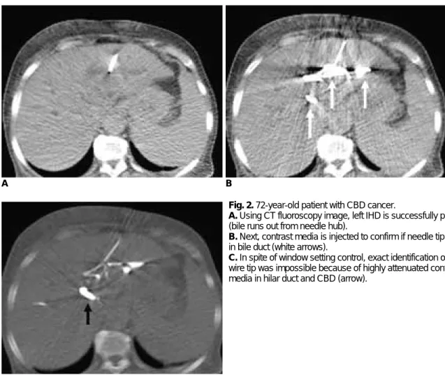
Fig. 2. 72-year-old patient with CBD cancer.
A. Using CT fluoroscopy image, left IHD is successfully punctured (bile runs out from needle hub).
B. Next, contrast media is injected to confirm if needle tip is located in bile duct (white arrows).
C. In spite of window setting control, exact identification of guide wire tip was impossible because of highly attenuated contrast media in hilar duct and CBD (arrow).
C
DISCUSSION
CT fluoroscopy is a recently introduced imaging tool, which facilitates the acquisition of a real-time monitoring of sectional CT images (1, 2). It has been reported that CTF has clinical advantages with regards to biopsy, fluid drainage, local placement of needle for drug injection or other interventional procedures, and radio-frequency ablation (3-10). In spite of the disputes corresponding to differences in opinion regarding the radiation dose (14, 15), the clinical application of CTF has further been extended (16-20). According to previous studies, CTF has the potential to deliver a higher radiation dose and exposure, which can be compensated for by adopting the lowest current, using lead drapes on the patients or needle holder, and a decrease of procedure time by quick needle targeting (5, 21).
To our knowledge, a CTF-guided PTBD has been attempted in only three published studies (11-13). Patients with malignant biliary obstructions were enrolled in the studies, which involved a planned stent-assisted recanaliza- tion following a temporary PTBD. Therefore, the purpose of CTF guiding was the puncturing of suitable peripheral duct that had an obtuse angle with the central bile duct.
After puncturing the bile duct, the rest of the procedures were performed under the guidance of mobile C-arm fluoroscopy. The studies concluded that CTF guiding had its advantages, which involved the visualization of the most suitable bile duct for puncture and reduced time and number of puncture trial as compared with conventional fluoroscopy guided PTBD.
We attempted CTF guided PTBD in the patients who needed urgent biliary drainage. All procedures, from the bile duct puncture to the drainage catheter placement, were performed under CTF guidance without C-arm fluoroscopy. The purpose of the trial was to evaluate the technical feasibility of CTF-guided PTBD and the possibil- ity of one step PTBD in the CT unit immediately after diagnostic CT scanning without moving patients into the intervention unit. The procedure was technically feasible for most patients, however a little modification was needed in the technique based on what was described in previous reports (11-13). A right lobe puncture was preferred in previous studies to ensure the appropriate tracts for stent delivery. It was a reasonable decision and did not result in problems in terms of PTBD, because the next steps were performed under C-arm fluoroscopy guidance. However, one step CTF-guided PTBD is very difficult through the right lobe, because there is limited space for instrument manipulation between the punctured body wall and the CT
gantry. In the five cases performed during the pilot trial prior to the main study, we performed the right lobe approach in three cases, as described in previous reports.
The result indicated that the investigator suffered from limited space in terms of device manipulation. The space between the right lobe and laterally located CT gantry is too narrow and uncomfortable for the operator to go on the rest steps of only CTF guided PTBD. It may cause contamination and limitation of device manipulation. After changing the puncture target to the left lobe in the two remaining cases of the pilot trial, it was much easier to manipulate instruments and helpful for successful trial.
Of the 28 patients included in the main study, only five underwent the procedure through the right lobe intrahep- atic duct (IHD). It was possible in those patients because they had a slim build, securing sufficient space for the procedure between the right abdominal wall and CT gantry. Also, the left lobe puncture has an important advantage in only CTF-guided PTBD. Because segments 5 or 6 IHD is located vertically in the right lobe, it is not easy to monitor the whole course on cross sectional images. On the other hand, segmental ducts in the left lobe have a transverse course, which is easier to trace on CTF.
Contrast media injection should be also carefully consid- ered for a trial to be successful. If it is injected immediately after bile duct puncture, it may disturb visualization of guide wire in the bile duct. We injected contrast media after placing a drainage catheter to confirm location of catheter tip.
The present study is not for a comparison of a CTF-only guided PTBD and fluoroscopy-guided PTBD. Fluoroscopy- guided PTBD is definitely the standard method and we are also adopting it in our practice. However, although the clinical status is very grave and symptoms are very severe to tolerate, patients have to wait to undergo a PTBD until the angiography unit is available. Our belief is that if the CTF-guided PTBD is achieved in the CT room, it is comfortable for the patients and helpful to shorten emergency care steps. Of course, if a combination with the C-arm fluoroscopy is possible in the CT unit, it would be very convenient. However, it is usually difficult to arrange a C-arm as a fixed device in CT unit in the aspect of space and economical efficiency. Bringing the C-arm every time would also be troublesome and it might be sometimes impossible.
We evaluated the feasibility and usefulness for the one step CTF guided PTBD. For the best possible success rate, the proper assessment of suitability for this method must be decided upon beforehand. We have to consider both the clinical and anatomical conditions, as described in the inclusion criteria. In particular, the compliance of patients
should be carefully considered. Although patients have a large dilated peripheral duct width, patients with respira- tory difficulty, uncontrolled motion, severe coughing, and unclear consciousness should be excluded from the indica- tion. Conclusively, although it is not familiar to interven- tional radiologists, performing one step CTF-guided PTBD is technically feasible with a high success rate in patients determined to be suitable candidates for this procedure.
Also, it would be helpful for shortening clinical steps and reducing patient waiting time which is critical due the level of pain endured with the condition.
References
1. Katada K, Anno H, Takeshita G, Ogura Y, Koga S, Ida Y, et al.
Development of real-time CT fluoroscopy. Nippon Igaku Hoshasen Gakkai Zasshi 1994;54:1172-1174
2. Katada K, Kato R, Anno H, Ogura Y, Koga S, Ida Y, et al.
Guidance with real-time CT fluoroscopy: early clinical experi- ence. Radiology 1996;200:851-856
3. White CS, Templeton PA, Hasday JD. CT-assisted tranbronchial needle aspiration: usefulness of CT fluoroscopy. AJR Am J Roentgenol 1997;169:393-394
4. Froelich JJ, Saar B, Hoppe M, Ishaque N, Walthers EM, Regn J, et al. Real-time CT fluoroscopy for guidance of percutaneous drainage procedures. J Vasc Interv Radiol 1998;9:735-740 5. Silverman SG, Tuncali K, Adams DF, Nawfel RD, Zou KH,
Judy PF. CT fluoroscopy-guided abdominal interventions:
technique, results and radiation exposure. Radiology 1999;212:673-681
6. Daly B, Krebs TL, Wong-You-Cheong JJ, Wang SS.
Percutaneous abdominal and pelvic interventional procedures using CT fluoroscopy guidance. AJR Am J Roentgenol 1999;173:637-644
7. Kirchner J, Kickuth R, Walz MV, Schilling EM, Laufer U, Liermann D. CTF-guided puncture of an unenhanced isodense liver lesion during continuous intravenous injection of contrast medium. Cardiovasc Intervent Radiol 1999;22:528-530 8. Muehlstaedt M, Bruening R, Diebold J, Mueller A, Helmberger
T, Reiser M. CT/fluoroscopy-guided transthoracic needle biopsy:
sensitivity and complication rate in 98 procedures. J Comput Assist Tomogr 2002;26:191-196
9. Davies RP, Kew J, West GP. Percutaneous jejunostomy using CT fluoroscopy. AJR Am J Roentgenol 2001;176:808-810 10. Ernst RD, Kim HS, Kawashima A, Middlebrook MR, Sandler
CM. Near Real-time CT fluoroscopy using computer automated scan technology in nonvascular interventional procedures. AJR Am J Roentgenol 2000;174:319-321
11. Froelich JJ, Wagner HJ, Ishaque N, Alfke H, Scherf C, Klose KJ. Comparison of C-arm CT fluoroscopy and conventional fluoroscopy for percutaneous biliary drainage procedures. J Vasc Interv Radiol 2000;11:477-482
12. Laufer U, Kirchner J, Kickuth R, Adams S, Liermann D. First experiences in CT-guided percutaneous transhepatic biliary decompression by means of real-time CT fluoroscopy. Abdom Imaging 2001;26:207-209
13. Laufer U, Kirchner J, Kickuth R, Adams S, Jendreck M, Liermann D. A comparative study of CT fluoroscopy combined with fluoroscopy versus fluoroscopy alone for percutaneous transhepatic biliary drainage. Cardiovasc Intervent Radiol 2001;24:240-244
14. Kato R, Katada K, Anno H, Suzuki S, Ida Y, Koga S. Radiation dosimetry at CT fluoroscopy: physician’s hand dose and development of needle holders. Radiology 1996;201:576-578 15. Stoeckelhuber BM, Leibecke T, Schulz E, Melchert UH,
Bergmann-Koester CU, Helmberger T, et al. Radiation dose to the radiologist’s hand during continuous CT fluoroscopy-guided interventions. Cardiovasc Intervent Radiol 2005;28:589-594 16. Murphy K, Baez JC, Cooney B, Kabaish K. CT fluoroscopy-
guided postmyelographic transthecal radiofrequency ablation of a posterior vertebral body osteoid osteoma. J Vasc Interv Radiol 2008;19:291-293
17. Schaefer PJ, Schaefer FK, Heller M, Jahnke T. CT fluoroscopy guided biopsy of small pulmonary and upper abdominal lesions:
efficacy with a modified breathing technique. J Vasc Interv Radiol 2007;18:1241-1248
18. Bladt O, De Wever W. Additional value of CT-fluoroscopic biopsy of pulmonary lesions: a retrospective study of 69 patients. JBR-BTR 2006;89:298-302
19. Hamuro M, Kaminou T, Nakamura K, Matsuoka T, Sakai Y, Morimoto A, et al. Percutaneous ethanol injection under CT fluoroscopy for hypervascular hepatocellular carcinoma follow- ing transcatheter arterial embolization. Hepatogastroenterology 2002;49:752-757
20. Tay VK, Fitridge R, Tie ML. Computed tomography fluoroscopy-guided chemical lumbar sympathectomy: simple, safe and effective. Australas Radiol 2002;46:163-166 21. Carlson SK, Bender CE, Classic KL, Zink FE, Quam JP, Ward
EM, et al. Benefits and safety of CT fluoroscopy in interven- tional radiologic procedures. Radiology 2001;219:515-529