Multidisciplinary correction of anterior open bite relapse and upper airway obstruction
10
0
0
전체 글
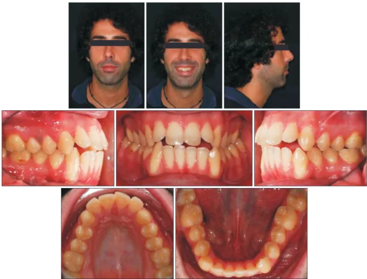
(2) Gracco et al • Anterior open bite and airway obstruction. INTRODUCTION One of the most challenging malocclusions to treat and maintain is open bite. Its etiology involves a com bination of genetic factors, unfavorable growth pattern, and oral habits. Habitual nonnutritive sucking, altered tongue function and posture, and persistent infantile swallowing, for example, can contribute to vertical malocclusion in the growth period.1,2 Several studies have suggested a relationship among nasopharyngeal airway obstruction, mouth breathing, and skeletal and dental malocclusion. Indeed, adenoid hypertrophy and chronic rhinitis may result in mouth breathing and encourage persistent infantile swallowing, which can contribute to anterior open bite.3-6 As such habits can cause relapse following ortho dontic treatment, 7,8 contributing factors should be uncovered when diagnosing patients with anterior open bite. Considering the etiological comple xity, a multidisciplinary approach involving several. health professionals, such as orthodontists, otorh inolaryngologists, orofacial myofunctional therapists, and maxillofacial surgeons, in adult patients, may be necessary. Here, we report the multidisciplinary co rrection of anterior open bite relapse secondary to upper airway obstruction.. DIAGNOSIS AND ETIOLOGY A 27-year-old man presented an anterior open bite relapse (Figures 1-4). He reported that orthodontic treatment was begun when he was 13 years old and lasted 5 years. Although initially satisfied with the outcome, he noticed relapse of anterior open bite one year after the brackets were removed. Intraoral examination showed bilateral Angle Class I molar relationship, slight deviation of the upper midline, proclination of the maxillary lateral incisors, crossbite of the maxillary left canine and second premolar, negative crown inclination of the maxillary posterior teeth, and. Figure 1. Pretreatment photographs. 48. http://dx.doi.org/10.4041/kjod.2015.45.1.47. www.e-kjo.org.
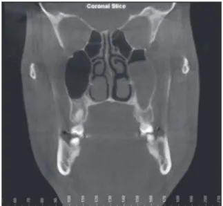
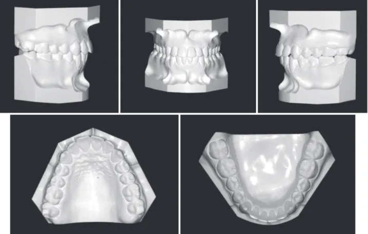
(3) Gracco et al • Anterior open bite and airway obstruction. Figure 2. Pretreatment dental digital models.. Figure 3. Pretreatment panoramic radiograph.. Figure 5. Sagittal cone-beam computed tomography image showing thin mandibular symphysis.. Figure 4. Pretreatment lateral cephalometric radiograph with tracing.. gingival recession at the mandibular canines. Facial analysis showed increased lower facial height with good lip competence and large nasolabial angle. Slight nasal deviation to the right was also apparent when the patient was viewed anteriorly. Gingival exposure while smiling was confined to the maxillary posterior region. Anterior low tongue posture was noted at rest and during swallowing, and the patient reported chronic difficulty in nose breathing. A cone-beam computed tomography (CBCT) of the head was performed to uncover upper airway problems responsible for the breathing difficulty and relapse. The scan showed extremely thin mandibular symphysis, which can markedly affect mandibular incisor inclination during orthodontic treatment (Figure 5). In addition, nasal septum deviation, right turbinate hypertrophy, and left maxillary sinus congestion were visible (Figure. www.e-kjo.org. http://dx.doi.org/10.4041/kjod.2015.45.1.47. 49.
(4) Gracco et al • Anterior open bite and airway obstruction. 6). All these features were thought to contribute to the breathing problem, encourage the improper tongue posture, and thereby cause the relapse. Pretreatment cephalometric analysis confirmed the Class I hyperdivergent skeletal pattern associated with dental anterior open bite and moderate maxillary incisor proclination (Table 1).. TREATMENT OBJECTIVES The treatment objectives were to (1) maintain the bilateral Angle Class I molar relationship; (2) correct the anterior open bite; (3) balance maxillary gingival exposure, ensuring a pleasant smile; (4) minimize mandibular gingival recession during the treatment;. (5) achieve positive crown inclination of the maxillary posterior teeth, creating a wider smile; and (6) ensure stable results overall.. TREATMENT ALTERNATIVES The presence of dental (not skeletal) open bite, lip competence, and good facial and smile esthetics excluded combined orthodontic treatment and orthognathic. Table 1. Summary of the cephalometric analysis Measurement SNA (°). Final. 72.2. 71.7. SNB (°). 75.4. 74.2. ANB (°). −3.2. −2.5. Wits appraisal (mm). −4.1. −9.2. FMA (MP-FH) (°). 27.5. 28.0. MP-SN (°). 43.1. 42.7. Palatal-Mand angle (°). 42.0. 40.3. Palatal-Occ plane (PP-OP) (°). 14.6. 20.1. Mand plane to Occ plane (°). 27.4. 20.2. U-incisor protrusion (U1-APo) (mm). 12.8. 11.8. L1 protrusion (L1-APo) (mm). 9.8. 9.2. 110.6. 103.7. U1-Occ plane (°). 54.8. 56.2. L1-Occ plane (°). 62.4. 75.5. IMPA (°). 90.2. 88.9. U1-palatal plane (°). Figure 6. Coronal cone-beam computed tomography image showing nasal septum deviation, right turbinate hypertrophy, and left maxillary sinus congestion.. Initial. SNA, sella - nasion- A point; SNB, sella - nasion- B point; ANB, A point - nasion - B point; FMA, Frankfort mandibular plane angle; MP, mandibular plane; FH, Frankfort ; Mand, mandibular; Occ, occlusal; U, upper; U1, upper incisor; L1, lower incisor; APo, A point - pogonion; IMPA, lower incisor mandibular plane angle.. Figure 7. Insigna digital set up. 50. http://dx.doi.org/10.4041/kjod.2015.45.1.47. www.e-kjo.org.
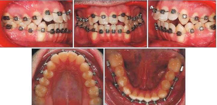
(5) Gracco et al • Anterior open bite and airway obstruction. Figure 8. Photographs of the leveling and aligning phase. surgery as well as first premolar extraction. Although use of tongue spurs or a tongue crib during orthodontic treatment can achieve rapid closure of open bite, this approach was rejected because the tongue posture was judged to be secondary to the breathing problem. To meet all the treatment objectives, we offered the patient a multidisciplinary approach involving or thodontic treatment to correct the malocclusion, otor hinolaryngological intervention to resolve the airway obstruction, and periodontal treatment to correct any gingival recession arising in critical mandibular areas during the treatment.. TREATMENT PROGRESS Damon self ligating brackets with InsigniaTM (Ormco Co., Orange, CA, USA) were bonded to all the teeth (Figures 7, 8). In the digital setup, we prog rammed complete correction of the open bite and, because of the susceptibility to relapse, overcorrection of the overbite, placing the maxillary incisal margin 3 mm below the edge of the mandibular incisors. We also programmed positive crown torque of the maxillary posterior teeth and ensured that all the mandibular teeth would be maintained in the middle of the medullary bone to prevent bone dehiscence and fenestrations, which can cause substantial gingival recession. The brackets were threaded with the following cus tomized archwire sequence: 0.014 inches (in) Damon Copper nickel titanium (Ni-Ti), 0.014 × 0.025 in Damon Copper Ni-Ti, 0.018 × 0.025 in Damon Copper Ni-Ti,. www.e-kjo.org. http://dx.doi.org/10.4041/kjod.2015.45.1.47. Figure 9. Coronal cone-beam computed tomography image showing complete resolution of upper airway ob struction. 0.019 × 0.025 in stainless steel, and 0.019 × 0.025 in titanium molybdenum alloy (TMA). Leveling and alignment were performed using thermal Ni-Ti archwires and completed rapidly because of the previous orthodontic treatment and relapse mainly involved the anterior open bite. When the 0.018 × 0.025 in Damon Copper Ni-Ti archwires were fitted, the patient was also supplied with intermaxillary elastics to facilitate bite closure during the remainder of the active. 51.
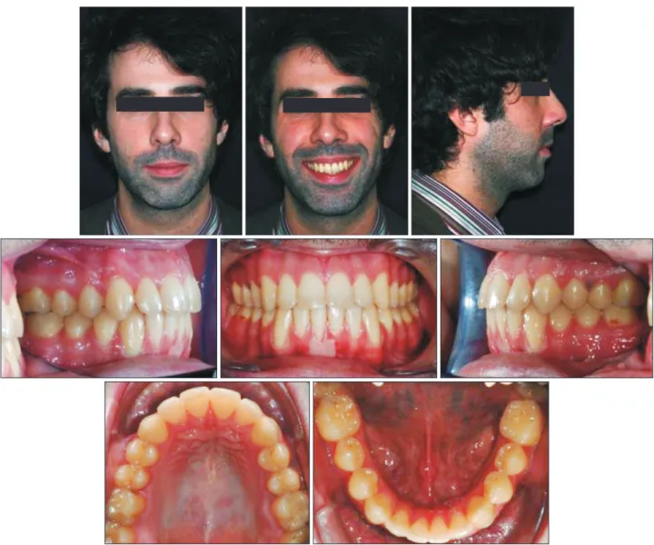
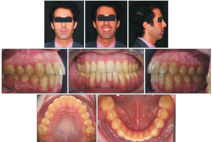
(6) Gracco et al • Anterior open bite and airway obstruction. phase. Bilateral triangular elastics (3/16 in, 6 oz.) were provided for daytime use, and anterior box elastics (5/16 in, 6 oz.) were added for nocturnal wear. After 10 months of orthodontic treatment, we referred the patient to an otorhinolaryngologist for bila teral turbinectomy and maxillary sinus cleaning. The surgery was performed by endonasal endoscopy and prompted immediate improvement in the patient's breathing dynamics. The CBCT scan taken 5 months postoperatively showed complete resolution of upper airway obstruction (Figure 9), and at the subsequent orthodontic appointments, the patient reported that he was finally able to breathe through his nose and his sleep quality had improved. At these appointments, we reminded the patient to continue to maintain correct tongue posture at rest and during swallowing.. RESULTS The orthodontic treatment was concluded at 19 mon ths. The Class I molar and canine relationship was maintained, anterior occlusion was improved, and ideal overbite and overjet was achieved. The treatment brought about mutually protected functional occlusion, with adequate canine disclusion and protrusive guidance. The temporomandibular joints showed no incoordination on opening and closing, and no tem poromandibular joint or muscle problems developed during the retention and postretention periods. The facial profile improved, as did the prominence of the upper lip. The lips remained competent, and the smile was balanced and pleasant. Unfortunately, because of the mandibular anatomy and tooth movement, the existing gingival recession became more pronounced. The periodontist decided to. Figure 10. Post-treatment photographs. 52. http://dx.doi.org/10.4041/kjod.2015.45.1.47. www.e-kjo.org.
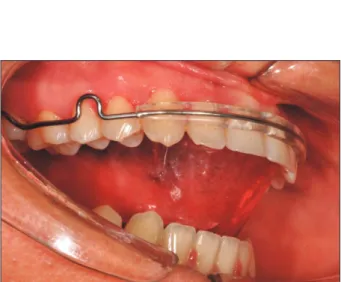
(7) Gracco et al • Anterior open bite and airway obstruction. Figure 11. Post-treatment digital dental models.. Figure 12. Post-treatment panoramic radiograph. improve the periodontal condition of the right man dibular central incisor alone and delay definitive muco gingival surgery for some years to stabilize the gingival conditions. The post-treatment panoramic radiograph showed good root parallelism without root resorption. Posttreatment cephalometric evaluation showed acceptable incisor inclination. Superimposition of the pretreatment and post-treatment cephalometric tracings highlighted the amount of extrusion of the maxillary incisors needed to resolve the open bite and confirmed that the. www.e-kjo.org. http://dx.doi.org/10.4041/kjod.2015.45.1.47. Figure 13. Post-treatment lateral cephalometric radio graph with tracing. mandibular plane angle remained stable (Figures 10-14, Table 1). The patient was instructed to wear a tongue elevator (Figure 15) for retention at night. This device featured. 53.
(8) Gracco et al • Anterior open bite and airway obstruction. Figure 14. Superimposed pretreatment and post-treat ment tracings.. Figure 15. Tongue elevator for retention.. an anterior opening that served as a tactile reminder and a chute that prevented positioning of the tongue between the dental arches. Examination after 3 years 5 months showed stable results. The well-aligned dentition and good overbite were maintained, facial balance was harmonious, and the smile was pleasing with good, stable gingival lines (Figure 16).. DISCUSSION The success of the treatment is mainly ascribable to the thorough diagnosis and multidisciplinary approach, resolving not only the relapse but also the underlying issues. Although the previous orthodontic treatment. 54. lasted 5 years, it did not guarantee stability of results. We believe that it failed predominantly because it was not aimed at tackling the airway obstruction and consequent tongue malpositioning, which were the root causes of the malocclusion. There are many options for correcting the tendency to place the tongue too forward (tongue habit mana gement). A tongue crib or tongue spurs can be used during orthodontic treatment to modify tongue posture, maintain stability of treated open bite, and prevent relapse, by establishing a new engram. 9-11 However, many orthodontists underestimate the importance of upper airway obstruction in oral habits. Anomalies of the nasal septum, turbinates, adenoids, or tonsils and chronic sinusitis can markedly alter respiratory dynamics, compromising dental arch development.4,12,13 Therefore, when poor breathing is suspected, specific investigative tools, such as tomography with a wide field of view, should be used to examine the upper airway. Referral to an otorhinolaryngologist for endoscopic analysis of the airway is also beneficial. Our patient presented with anterior low tongue posture and reported chronic difficulty in nose breathing, so we examined the airway by digital volumetric tomography. The abnormal findings prompted us to refer the patient to the otorhinolaryngologist. The approach selected was orthodontic closure of the open bite using intermaxillary elastics and midcourse surgical turbinectomy, sinus cleaning, and correction of nasal septum deviation. The surgery had an immediate effect on the patient's res piratory difficulty, enabling him to breathe normally and. http://dx.doi.org/10.4041/kjod.2015.45.1.47. www.e-kjo.org.
(9) Gracco et al • Anterior open bite and airway obstruction. Figure 16. Appearance 3 years 5 months after the treatment. position the tongue correctly. However, as open bite presents a high risk of relapse,14,15 we prescribed noc turnal use of a tongue elevator for retention. The good results obtained by this multidisciplinary treatment are still preserved over 3 years later.. CONCLUSION Dental malocclusion often needs to be treated by a multidisciplinary team to arrive at an accurate diagnosis and offer appropriate solutions. In particular, adults pre senting with open bite, especially relapse cases, should be investigated for oral habits and/or airway issues before defining and selecting the best treatment option.. REFERENCES 1. Subtelny JD, Sakuda M. Open bite: diagnosis and treatment. Am J Orthod 1964;50:337-58. 2. Alexander CD. Open bite, dental alveolar protrusion, class I malocclusion: A successful treatment result. Am J Orthod Dentofacial Orthop 1999;116:494-500. 3. Oulis CJ, Vadiakas GP, Ekonomides J, Dratsa J. The. www.e-kjo.org. http://dx.doi.org/10.4041/kjod.2015.45.1.47. effect of hypertrophic adenoids and tonsils on the development of posterior crossbite and oral habits. J Clin Pediatr Dent 1994;18:197-201. 4. Vig KW. Nasal obstruction and facial growth: the strength of evidence for clinical assumptions. Am J Orthod Dentofacial Orthop 1998;113:603-11. 5. Warren DW, Mayo R, Zajac DJ, Rochet AH. Dyspnea following experimentally induced increased nasal airway resistance. Cleft Palate Craniofac J 1996;33:231-5. 6. Fujiki T, Inoue M, Miyawaki S, Nagasaki T, Tanimoto K, Takano-Yamamoto T. Relationship between maxillofacial morphology and deglutitive tongue movement in patients with anterior open bite. Am J Orthod Dentofacial Orthop 2004;125:160-7. 7. Smithpeter J, Covell D Jr. Relapse of anterior open bites treated with orthodontic appliances with and without orofacial myofunctional therapy. Am J Orthod Dentofacial Orthop 2010;137:605-14. 8. Wriedt S, Buhl V, Al-Nawas B, Wehrbein H. Combined treatment of open bite - long-term evaluation and relapse factors. J Orofac Orthop 2009;70:318-26.. 55.
(10) Gracco et al • Anterior open bite and airway obstruction. 9. Giuntini V, Franchi L, Baccetti T, Mucedero M, Cozza P. Dentoskeletal changes associated with fixed and removable appliances with a crib in openbite patients in the mixed dentition. Am J Orthod Dentofacial Orthop 2008;133:77-80. 10. Bosio JA, Justus R. Treatment and retreatment of a patient with a severe anterior open bite. Am J Orthod Dentofacial Orthop 2013;144:594-606. 11. Meyer-Marcotty P, Hartmann J, Stellzig-Eisenhauer A. Dentoalveolar open bite treatment with spur appliances. J Orofac Orthop 2007;68:510-21. 12. Gungor AY, Turkkahraman H. Effects of airway problems on maxillary growth: a review. Eur J Dent. 56. 2009;3:250-4. 13. Jena AK, Singh SP, Utreja AK. Sagittal mandibular development effects on the dimensions of the awake pharyngeal airway passage. Angle Orthod 2010;80:1061-7. 14. Speidel TM, Isaacson RJ, Worms FW. Tongue-thrust therapy and anterior dental open-bite. A review of new facial growth data. Am J Orthod 1972;62:28795. 15. Denison TF, Kokich VG, Shapiro PA. Stability of maxillary surgery in openbite versus nonopenbite malocclusions. Angle Orthod 1989;59:5-10.. http://dx.doi.org/10.4041/kjod.2015.45.1.47. www.e-kjo.org.
(11)
수치

+5
관련 문서
[r]
Objectives: The present study was conducted to determine the relationship between degree of work performance and job satisfaction in NICU nurses.. Methods: The subjects of
Objectives: This study aimed to investigate the relationship between job stress and turnover intention of employed opticians and to investigate the mediating effect of burnout
페놀에 오염된 수 돗물을 마신 수돗물을 마신 시민들은 구토·설사·복통으로 고통을 겪었으며 수돗물로 만든 두부·김치·콩나물 등은 악취 때문에 폐기 처분하는
Effects of phase I periodontal treatment on gingival crevicular fluid levels of matrix metalloproteinase-3 and tissue inhibitor of metalloproteinase-1.
회원국의 영토밖에서 다른 회원국의 , 영토내에서 회원국의 서비스 소비자에게
[r]
등록제 민간자격 운영 사실을 특정한 등록 기관에 비치된 장부에 2. 기재하는 행위로서 등록한 경우에만 민간자격으로
