A Aspergillus Spondylitis involving theCervico-Thoraco-Lumbar Spine in anImmunocompromised Patient: a Case Report
4
0
0
전체 글
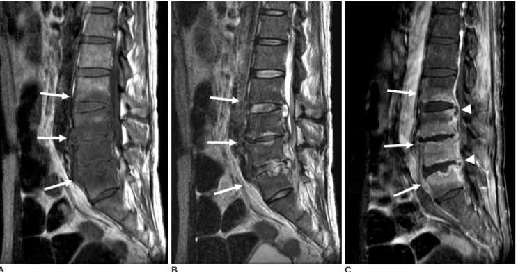
(2) Opportunistic Aspergillus Spondylitis involving Cervico-thoraco-Lumbar Spines. L3 4 and L4 5 levels, and osteolysis was observed at the inferior endplate of L3, the superior and inferior endplates of L4, and the superior endplate of L5. After curettage and debridement of these lesions, an iliac crest bone block was inserted. Histologically, acute inflammation and necrosis with fungal hyphae suggested aspergillus infection. Histopathologically periodic acid-Schiff and Gomori methenamine silver staining revealed aspergillus infection. About three months after he underwent lumbar spine MR imaging, he was readmitted for posterior neck pain. MR images of the cervical and thoracic spine showed changes in bone marrow and endplates at the C4 5 and. A. B. T2 4 levels, the latter of which were similar to the previous findings of lumbar involvement (Figs. 1E G). Increased disc signal and loss of intranuclear cleft were observed in C4 5 and T2 4 discs. Endplate irregularities and disc space narrowing were observed in the involved spine. During surgery on the cervical spine, infected granulation tissue and disc material were found at the C4 5 level with osteolysis at the inferior endplate of C4 and the superior end plate of C5. Debridement and bone grafting were performed, and pathologic reports disclosed acute inflammation with necrosis and osteomyelitis. Histopathological periodic acid-Schiff and Gomori. C Fig. 1. A 46-year-old man with aspergillus spondylitis. A, B. MR image of the lumbar spine showing band-like or diffuse hypointense signals (arrows) in vertebral bodies L2-L5 on T1weighted images (A), whereas these lesions were isointense to slightly hyperintense (arrows) on T2-weighted images (B). Some hypointense signals with intranuclear cleft preservation were evident in L2-3 and L4-5 discs. However, an absence of disc hyperintensity and intranuclear cleft loss were observed in the L3-4 disc. Endplate irregularities were evident in the involved spine, and disc space narrowing was observed in L3-4 and L4-5. C. Band-like or diffuse enhancement (arrows) was observed in involved vertebral bodies with epidural abscesses (arrowheads) on sagittal fat-suppressed contrast-enhanced T1-weighted image. D. Axial fat-suppressed contrast-enhanced T1-weighted image showing relatively well-defined paraspinal abnormal enhancement (arrows).. D Korean J Radiol 8(5), October 2007. 449.
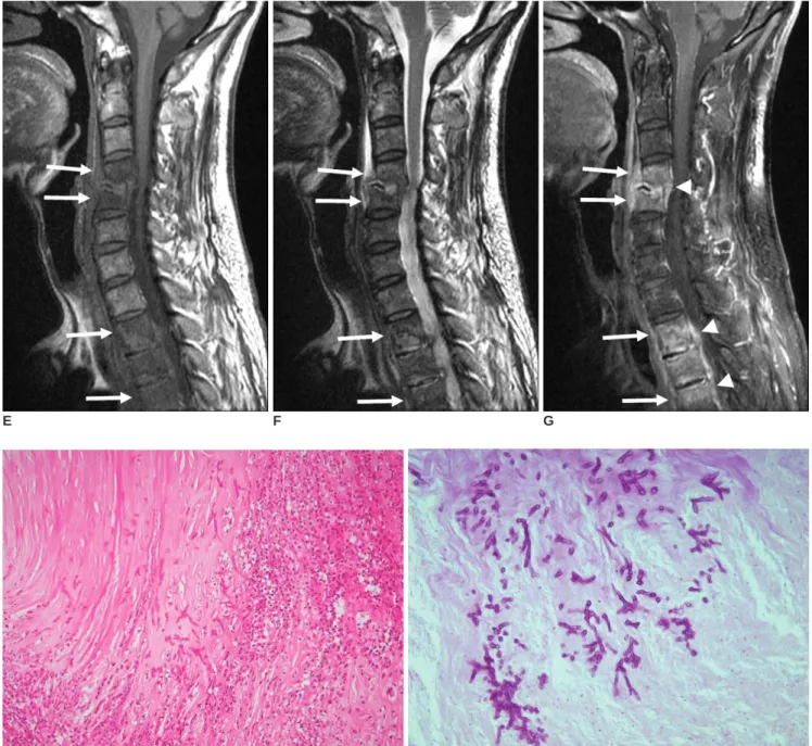
(3) Son et al.. methenamine silver staining findings revealed aspergillus infection (Figs. 1H, I).. DISCUSSION Invasive aspergillosis is a life threatening fungal infection that is associated with a high mortality rate despite. E. F. treatment. Symptoms and signs are nonspecific. In previous reports times between symptom onset and a definite diagnosis were of the order of months (4 7), and in our patient, symptoms persisted for more than two months before pathologic diagnosis. Ordinary laboratory findings are of little help in the diagnosis of aspergillosis (6). According to a previous report by Williams et al. (8), the. G. H I Fig. 1. E, F. MR image of cervical spine showing diffuse hypointense signals (arrows) in C4-5 and T2-4 vertebral bodies on T1-weighted images (E). The lesions were isointense to slightly hyperintense (arrows) on T2-weighted images (F). Increased disc signal and loss of intranuclear cleft were observed in C4-5 and T2-4 discs. Endplate irregularities and disc space narrowing were seen in involved spine. G. Diffuse enhancement was seen in involved vertebral bodies with paraspinal (arrows) and epidural masses (arrowheads) on sagittal fat-suppressed contrast-enhanced T1-weighted images. H. Pathological examination of an intervertebral disc revealed acute inflammation, necrosis and portions of the tissue being invaded by septate hyphae (Hematoxylin & Eosin staining, 200). I. Branching septate hyphae were uniform in width and disposed mainly at acute angles (diastase periodic acid Schiff, 400).. 450. Korean J Radiol 8(5), October 2007.
(4) Opportunistic Aspergillus Spondylitis involving Cervico-thoraco-Lumbar Spines. absence of disc hyperintensity and intranuclear cleft preservation on T2-weighted images are suggestive of nonpyogenic spondylitis. In our case, decreased disc signal intensity was observed in three of six affected discs and intranuclear clefts were preserved in two of six affected discs. The minimal hyperintensity or isointensity of vertebrae on T2-weighted images observed in our case is consistent with the findings of previous reports (8, 9) on Candida or Aspergillus spondylitis. Aspergillus spondylitis is often confused with tuberculous spondylitis (3, 10). In a previous case report (3), MR showed involvements of three consecutive vertebral bodies and a well-defined paraspinal mass, and as a result, tuberculous spondylitis was initially considered. Finally, the patient was pathologically confirmed as having Aspergillus spondylitis. Based on our experiences, the following MR findings favor tuberculous spondylitis rather than pyogenic spondylitis; a well-defined paraspinal abnormal signal rather than an ill-defined paraspinal abnormal signal, a thin and smooth abscess wall rather than a thick and irregular abscess wall, the presence of a paraspinal or an intraosseous abscess, subligamentous spread over at least three vertebral levels, multiple vertebral body involvement, and thoracic spine involvement (5). In the present case, four consecutive vertebral bodies were involved and an abnormal paraspinal signal was relatively well-defined on the initial lumbar spine MRI, which indicated tuberculous spondylitis rather than pyogenic spondylitis. However, no paraspinal or intraosseous abscess was evident despite the extensive involvements of four consecutive vertebral bodies and an abnormal paraspinal signal. Because based on our experiences, about 95% (15/20) of tuberculous spondylitis cases have a concomitant paraspinal or intraosseous abscess (5), the absence of a paraspinal or intraosseous abscess appears to be unusual in tuberculous spondylitis. However, as South Korea is still an endemic area for tuberculosis, and initial diagnosis of tuberculous spondylitis was made despite some inconsistent MR findings. In retrospect, we. Korean J Radiol 8(5), October 2007. should have considered a fungal infection, such as, Candida or aspergillus spondylitis in this immunocompromised patient. In conclusion, aspergillus spondylitis should be considered in the differential diagnosis of immunocompromised patients with MR findings resembling those of tuberculous spondylitis.. References 1. Morgenlander JC, Rossitch E Jr, Rawlings CE 3rd. Aspergillus disc space infection: case report and review of the literature. Neurosurgery 1989;25:126-129 2. Park KU, Lee HS, Kim CJ, Kim EC. Fungal discitis due to Aspergillus terreus in a patient with acute lymphoblastic leukemia. J Korean Med Sci 2000;15:704-707 3. Park SB, Kang MJ, Whang EA, Han SY, Kim HC. A case of fungal sepsis due to aspergillus spondylitis followed by cytomegalovirus infection in a renal transplant recipient. Transplant Proc 2004;36:2154-2155 4. Dagirmanjian A, Schils J, McHenry M, Modic MT. MR imaging of vertebral osteomyelitis revisited. AJR Am J Roentgenol 1996;167:1539-1543 5. Jung NY, Jee WH, Ha KY, Park CK, Byun JY. Discrimination of tuberculous spondylitis from pyogenic spondylitis on MRI. AJR Am J Roentgenol 2004;182:1405-1410 6. Chi CY, Fung CP, Liu CY. Aspergillus flavus epidural abscess and osteomyelitis in a diabetic patient. J Microbiol Immunol Infect 2003;36:145-148 7. Stratov I, Korman TM, Johnson PD. Management of Aspergillus osteomyelitis: report of failure of liposomal amphotericin B and response to voriconazole in an immunocompetent host and literature review. Eur J Clin Microbiol Infect Dis 2003;22:277283 8. Williams RL, Fukui MB, Meltzer CC, Swarnkar A, Johnson DW, Welch W. Fungal spinal osteomyelitis in the immunocompromised patient: MR findings in three cases. AJNR Am J Neuroradiol 1999;20:381-385 9. Munk PL, Lee MJ, Poon PY, O’Connell JX, Coupland DB, Janzen DL, et al. Candida osteomyelitis and disc space infection of the lumbar spine. Skeletal Radiol 1997;26:42-46 10. van Ooij A, Beckers JM, Herpers MJ, Walenkamp GH. Surgical treatment of aspergillus spondylodiscitis. Eur Spine J 2000;9:7579. 451.
(5)
수치
관련 문서
Diffuse pain in lumbar muscles;some radiation to buttocks Localized lumbar pain: similar findings to lumbar strain Leg pain often worse than back
Autho: Effectiveness of surgery for lumbar stenosis and degenerative spondylolisthesis in the octogenarian population: analysis of the Spine Patient Outcomes Research
Second, to see a similarity factor among the relation between a factor which forms peer group and the attitudes toward school life, in case of the
Time series of vertical cross section of potential vorticity and wind vector calculated by Case 1 along the A-A' line indicated at Fig.. Same
Purpose: This study was aimed to compare the functional outcomes of the continuous and discontinuous fractures, retrospectively, in multiple thoracolumbar
Lumbar discogenic radiculopathy (LDR) is one of the most common spinal disorders which can lead to high medical expenses for the patient, occupational impairment,
The struggle was successful, and in January 1926 the first of four papers on “Quantization as an Eigenvalue
출처 : IAEA 발표 자료(Comprehensive inspection exercise at bulk handling facilities, “U-235 Enrichment measurements by gamma-ray spectroscopy”) 13. Uranium
