C Hyaline Vascular-Type Castleman DiseasePresenting as an Esophageal SubmucosalTumor: Case Report
4
0
0
전체 글
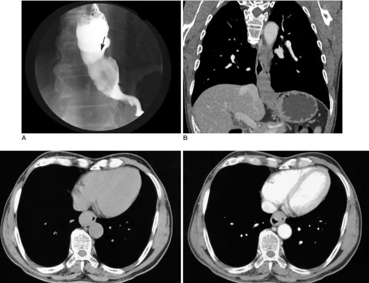
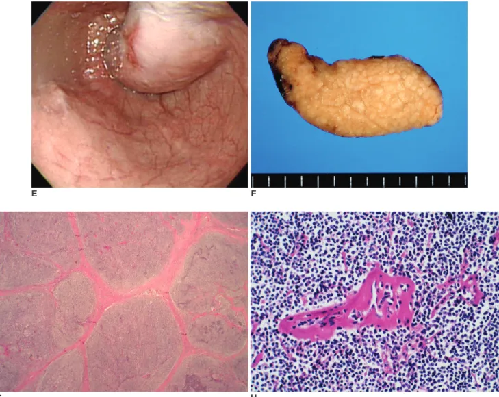
(2) Kim et al.. lesion was located in the submucosal layer of the esophagus. The specimen was reviewed by two pathologists (with 10 and 25 years of experience in pulmonary and lymphoreticular pathology, respectively) and they agreed with the diagnosis of Castleman disease. The excised mass showed a multilobulated mass with whitish color and a smooth margin (3 3 7 cm) at the distal esophagus (27 34 cm from incisor). On sectioning, the mass had a yellowish gray multinodular appearance (Fig. 1F). Microscopically, the esophageal mass revealed nodular lymphoid areas, a marked expansion of the mantle zone, and small, relatively inconspicuous germinal centers (Fig. 1G). The follicles show marked vascular proliferation with hyalinization (Fig. 1H). The final diagnosis of this tumor was Castleman disease of the hyaline vascular type, lymphoid subtype.. A. DISCUSSION Castleman disease is a disease of unknown etiology, and also referred to as angiofollicular lymph node hyperplasia, angiomatous lymphoid hamartoma, and giant mediastinal lymph node hyperplasia. Based on histological differences, the disease has been divided into two variants, and the hyaline vascular variant comprise 91% of cases and is usually asymptomatic. Typically this variant appears as a hypervascular mass with strong enhancement on CT. The plasma cell variant of Castleman disease is often concomitant with systemic manifestations, such as fever, anemia, hypergammaglobulinemia, and increases in acute phase proteins (3). Pathologically, the hyaline vascular type Castleman. B. C D Fig. 1. A 62-year-old man with esophageal Castleman disease. A. Spot radiograph from a single-contrast esophagogram reveals a 6-cm filling defect (arrows) arising from the lateral esophageal wall. B. Coronal reformation CT image shows a definite extent of the disease. C, D. Axial CT images, before (C) and after contrast injection (D), display focal thickening in the anterior wall of the lower esophagus, with circumferential luminal air, and the intramural mass shows homogeneous enhancement.. 74. Korean J Radiol 7(1), March 2006.
(3) Hyaline Vascular-Type Castleman Disease Presenting as an Esophageal Submucosal Tumor. disease is characterized by small hyaline vascular germinal centers and interfollicular capillary proliferation. However, the proportion of the two components - abnormal lymphoid follicles and increased interfollicular vascularity may vary, from cases featuring predominantly large mantle zones with inconspicuous germinal centers (“lymphoid variant”) to cases with a predominantly vascular component and fibrosis (4). The most common locations of Castleman disease are the thorax (63%), abdomen (11%), and axilla (4%), although extrathoracic sites have been reported in the neck, axilla, shoulder, orbit, pelvis, pancreas, leptomeninges, and retroperitoneum (3, 5 8). There are some reports of gastrointestinal involvement in castleman disease, such as gastric or colonic erosion,. E. gastric outlet obstruction, intestinal obstruction or, rectal cancer (1, 2). Most cases are caused by pressure from abdominal, retroperitoneal lymph node enlargement. To our knowledge, Castleman disease of the esophagus has not been previously reported in the literature. There were two reports of mediastinal Castleman disease with esophageal varices, which formed in response to copious blood flow (because of increased blood drainage) from the angiomatous tumor to the esophageal veins (9, 10). In our case, esophagography and CT manifestations were similar to those considered characteristic of leiomyoma. It seems to be difficult to diagnose submucosal Castleman disease without any histological findings. In conclusion, the imaging findings of Castleman disease localized in the esophagus were homogeneous well. F. G H Fig. 1. E. Endoscopy reveals a smoothly bulging mass with smooth surface and focal reddish coloration on the distal esophagus. F. Macroscopy of the excised tumor demonstrates a multilobulated mass with whitish color and smooth margin without hemorrhage or necrosis. G, H. Microscopically, the esophageal mass reveals nodular lymphoid areas, a marked expansion of the mantle zone, and small, relatively inconspicuous germinal centers (H & E staining, 10). The follicles show marked vascular proliferation with hyalinization (H & E staining, 200). Korean J Radiol 7(1), March 2006. 75.
(4) Kim et al.. enhancing mass at CT. From a clinical standpoint, establishing an accurate preoperative diagnosis is difficult, and Castleman disease should be included in the differential diagnosis of asymptomatic, well-enhancing esophageal mass.. References 1. Zamir A, Prasher G, Moukarzel AA, Zeien L, Feldman F. Castleman’s disease: a rare cause of hematemesis. J Pediatr Gastoenterol Nutr 1999;28:112-115 2. Wengrower D, Libson E, Okon E, Goldin E. Gastrointestinal manifestation in Castleman’s disease. Am J Gastroenterol 1990;85:79-82 3. Kaneko T, Takahashi S, Takeuchi T, Goto T, Kitamura T. Castleman’s disease in the retroperitoneal space. J Urol 2003;169:265-266 4. Kojima M, Nakamura S, Iijima M, Murayama K, Sakata N, Masawa N. Lymphoid variant of hyaline vascular Castleman’s disease containing numerous mantle zone lymphocytes with clear cytoplasm. APMIS 2005;113:75-80. 76. 5. Seo BK, Oh YW, Cho KR, Lee NJ, Kim JH, Kim IS, et al. Imaging findings of Castleman’s disease localized in the axilla: a case report. Korean J Radiol 2002;3:136-139 6. Park KS, Choi YJ, Song KS. Hyaline-vascular type Castleman’s disease involving both orbits. Acta Ophthalmol Scand 2002;80:537-539 7. Gaunt GA, Gostout BS, Remstein E, Cliby WA. Pelvic Castleman disease presenting as vaginal occlusion. Obstet Gynecol 2002;100:1082-1085 8. Sotrel A, Castellano-Sanchez AA, Prusmack C, Birchansky S, Brathwaite C, Ragheb J. Castleman’s disease in a child presenting with a partly mineralized solitary meningeal mass. Pediatr Neurosurg 2003;38:232-237 9. Serin E, Ozer B, Gumurdulu Y, Yildirim T, Barutcu O, Boyacioglu S. A case of castleman’s disease with “downhill” varices in the absence of superior vena cava obstruction. Endoscopy 2002;34:160-162 10. Shirakusa T, Iwsaki A, Okazaki M. Downhill esophageal varices caused by benign giant lymphoma. Scand J Thorac Cardiovasc Surg 1988;22:135-138. Korean J Radiol 7(1), March 2006.
(5)
수치
관련 문서
※ Accessory spleen의 유무 확인 또는 Diaphragmatic rupture를 동반한 trauma후에 chest를 보기 위 한 검사일
식도의 통과검사는 환자가 삼킨 bolus가 식도를 통하여 위(stomach)로 내려가는 시간을 측정함으로써 식도 의 기능을 평가하는데 이용.. ④ 컴퓨터를
P.( Kidney
Kim, Hwa-Jung, Hyung-Kyu Nam, Wee-Haeng Hur, Jung-Youn Lee, Jae-Woong Hwang, Ji-Yeon Lee, Yu-Seong Choi, Dong-Won Kim, Jin Young Park. National
Han Soo Kim, Jeong Min Park, Byung No Lee, Myung Gook Moon, Jang Ho Ha, Nam Ho Lee, Young Soo Kim, Chang Goo Kang, Hyung Ki Cha, Moon Sik Chae, Jeong Ho Moon, Kyung Min Oh,
Sanguk Nam, Gyeahyung Jeon,
1) Kim JH, Lee CS, Moon C, Kwak YG, Kim BN, Kim ES, et al. Co-Infection of Scrub Typhus and Human Granulocytic Anaplasmosis in Korea, 2006. Korean Med Sci. Case Report:
The study carried out small group cooperative learning as an experimental group and lecture-type learning as a control group for 12 weeks and compared
