HK-2 세포에서 indoxyl sulfate로 유도된 세포증식억제에 대한 밀몽화의 효과
박형권1․주성민1․강민수1․전병훈1,2* 1 : 원광대학교 한의과대학 병리학교실, 2 : 한국전통의학연구소
Effect of Buddleja officinalis on the Indoxyl Sulfate-induced Inhibition of Cell Proliferation in HK-2 Cells
Hyoung Kwun Park1, Sung Min Ju1, Min Soo Kang1, Byung Hun Jeon1,2* 1 : Department of Pathology, College of Korean Medicine, Wonkwang University,
2 : Research Center of Traditional Korean Medicine, Wonkwang University
The flowers and leaves buds ofBuddleja officinalis are used to treat eye troubles, hernia, gonorrhea and liver troubles in Asia. The present study investigated the effect of aqueous extract of Buddleja officinalis (ABO) on indoxyl sulfate (IS)-induced inhibition of cell proliferation in HK-2 cells. The HK-2 cells were incubated with 5 mM IS 5 mM in the absence or presence of ABO (2, 5, 10, 20 μg/ml) for 24 hr. The effect of ABO on IS-treated HK-2 cells was investigated by using MTT assay, flow cytometric analysis and Western blot. IS-induced proliferation inhibition was recovered dose-dependently by treatment with ABO. ABO reduced S and G2/M phase cell cycle arrest induced by IS.
Recovery effect of ABO on inhibition of cell proliferation by IS is related to the decrease of p21 expression. Taken together, these results suggest that recovery effect of ABO on proliferation of IS-treated HK-2 cells be associated with cell cycle progression through down-regulation of p21 levels.
Key words : Indoxyl sulfate, Buddleja officinalis, HK-2 cells
* 교신저자 : 전병훈, 전북 익산시 신용동 344-2, 원광대학교 한의과대학
․E-mail : [email protected], ․Tel : 063-850-6835
․접수 : 2012/07/26 ․수정 : 2012/08/13 ․채택 : 2012/08/16
서 론
Indoxyl sulfate (IS)는 일종의 요독 물질로 장내에서 트립토 판이 장내세균에 의해 indole로 변하고, 이것이 간에서 IS의 형태 로 바뀌어 생성된다. 신장기능에 이상이 생겨 IS가 소변으로 정 상적 배출되지 않고 혈청과 신세뇨관에 축적이 되면 만성신부전 (chronic kidney disease; CKD)을 유발시킨다1-5). 또한, IS는 만성 신부전 환자에게서 심근섬유모세포, 심근세포, 내피세포, 혈관평 활근세포의 기능장애와 대동맥 석회화 및 동맥경화와 같은 유해 한 효과를 나타내고 있다6-12).
최근 보고된 연구에 의하면, IS는 신근위세뇨관세포에서 세 포노화 및 신섬유화를 유도 및 세포증식을 억제한다13,14). IS에 의 한 증식억제는 CDK활성억제 단백질인 p21발현과 관련되어 있
다15). 무혈청배지에서 IS를 처리한 신근위세뇨관세포는 증식억제 및 p21 단백질 발현이 증가되었고, 혈청이 포함된 배지에 다시 배양하였을 경우 세포증식이 회복되지 않았다.
밀몽화(Buddlejase Flos)는 馬錢科 (Loganiaceae)에 낙엽관목인 밀몽화(Buddlejase officinalis Maxim)의 말린 꽃이나 꽃봉오리로 氣味가 味甘, 平, 微寒, 無毒하고 祛風散熱, 明目退翳하는 효능이 있어 多淚羞明, 眼生翳膜, 視物昏花 등에 사용되어 왔다16,17). 또 한 밀몽화에 연구로는 항균작용, 항산화작용, 신경세포 보호효 과, aldose reductase 활성억제 등이 보고되었다18-22).
본 연구에서는 다양한 약리작용을 나타내는 밀몽화 물추출 물이 IS에 의한 사람 신세뇨관 세포주인 HK-2 세포의 증식억제 에 미치는 효과에 대하여 유의한 결과를 얻었기에 보고하는 바 이다.
재료 및 방법
1. 밀몽화 물추출물 제조
밀몽화(Buddlejase officinalisMaxim)는 원광대학교 한방병원 (익산, 전북)에서 구입한 후 정선하여 사용하였고, 1 kg (Voucher specimen DH)을 건조시킨 후 분쇄기를 이용하여 분말로 하였으 며, 삼각 플라스크에 밀몽화 300 g과 증류수 1.5 L를 넣고 냉각기 를 설치한 후 120 분간 가열하였다. 추출된 전탕액을 3,000 rpm 으로 30 분간 원심 분리 후 여과지 (Whatmann No. 3)를 이용하 여 여과하였다. 여과된 밀몽화 전탕액을 냉동건조기를 이용하여 동결 건조시킨 후 사용 전까지 -20℃에 보관하였다. 수율은 약 9%였다.
2. 시약 및 항체
Indoxyl sulfate, 3-(4,5-dimethylthiazol-2-yl)-2,5-diphenyl tetrazolium bromide (MTT), Propidium iodine (PI), Ribonuclease A (RNase A), Protease inhibitor cocktail은 Sigma-Aldrich Chemical (St. Louis, MO, USA)로부터 구입하였 다. Anti-p21 항체들은 Santa Cruz Biotechnology, INC. (Santa Cruz, CA, USA)로부터 구입하였다. Anti-ERK, anti-phospho-ERK 항체들은 Cell Signaling Technology INC.
(Beverly, MA, USA)로부터 구입하였다.
3. 세포배양
HK-2 세포는 American Type Culture Collection (ATCC, Rockville, MD, USA)로부터 분양받았고, 10% fetal bovine serum (FBS, Invitrogen, Burlington, ON, Canada), 100 U/ml penicillin, 100 μg/ml streptomycin이 포함된 DMEM/F12 배지 (Invitrogen, Burlington, ON, Canada)를 사용하여 37℃와 5%
CO2상태에서 배양하였다.
4. 세포생존률검사
세포생존률은 MTT 방법을 사용하여 측정하였다. MTT 검사 는 미토콘드리아의 숙신산 탈수소효소에 의해 MTT가 formazan 으로 환원되는 원리를 기초로 하고 있다. HK-2 세포는 1×10⁵cells/ml로 12 well 플레이트에 24 시간 전배양 후, 5 mM IS와 밀몽화 물추출물을 일정한 농도로 증가시켜 24 시간 처리하 였다. 처리 후에, 5 mg/ml MTT용액 100 μl를 각각의 well에 첨 가하였고, 4 시간 배양하였다. 비수용성의 MTT-formazan 결정 들은 등량의 solubilization 용액 (10% SDS/0.01 N HCl)을 첨가 하여 용해시켰고, 5% CO2와 37℃ 조건하에서 12 시간 배양하였 다. Formazan의 양은 SpectraMAX 250 microplate spectrophotometer (Molecular Devices, Sunnyvale, CA, USA)를 이용하여 570 nm에서 측정하였다. 세포생존률은 아래의 식으로 계산하였다.
cell viability = (mean OD of treated cells/mean OD of control cells)×100
5. 세포주기분석
HK-2 세포는 5 mM 농도의 IS와 밀몽화 물추출물을 2, 5, 10, 20 μg/ml 농도로 24 시간 처리한 후, 세포 모두를 수확하여
PBS (pH 7.4)로 세척하였다. 세척한 세포는 냉각된 70% 에탄올 로 4℃에서 1 시간 고정하였다. 고정된 세포는 PBS (pH 7.4)로 다시 세척하고 1 ml의 PI (10 μg/ml)/RNase A (100 μg/ml)용액 으로 재 부유시켜 37℃에서 암실조건으로 1 시간 배양하였다.
DNA 양은 FACS-Calibur(BD Biosciences, CA, USA)를 사용하여 측정하였다. 세포주기분석은 고정한 후, 염색한 세포로부터 DNA 양을 측정하여 결정하였다.
6. Western blot analysis
세포는 얼음 냉장된 PBS (pH 7.4)로 세척하고 1% protease inhibitor 혼합액이 포함된 RIPA buffer로 부유 시킨 후 얼음 위 에서 30분간 용해하였다. 세포용해액들은 4℃에서 14,000 rpm으 로 15분간 원심분리 하였고, 단백질 농도는 Bradford 검사법을 이 용하여 측정하였다. 총 40 μg의 단백질 샘플을 SDS-PAGE 젤로 분리하였고 40V에서 3 시간 nitrocellulose 막 위로 단백질을 전이 시켰다. 단백질이 전이된 막은 5% BSA가 포함된 Tris-buffered saline+Tween-20 (20 mM Tris-HCl, pH7.6, 150 mM NaCl, 0.05%
Tween-20)으로 차단시킨 후, 1차 항체를 결합시켰다. 면역 활성 은 peroxidase가 붙어있는 anti-rabbit 또는 anti-mouse 2차 항체 를 사용하여 SuperSignal West Pico Chemiluminescent (Pierce, Rockford, IL, USA)에 의해 탐지하였다.
7. 통계처리
모든 데이터의 결과는 마이크로소프트 오피스 엑셀 (EXCEL) 2010으로 통계처리하여 mean±standard deviation (SD) 로 기록하였다.
결 과
1. IS 억제된 HK-2 세포증식에 대한 밀몽화의 효과
IS는 HK-2 세포의 증식을 억제시킨다14,15). IS에 의해서 억제 된 HK-2 세포의 증식에 대한 밀몽화 물추출물의 효과를 조사하 기 위해 5 mM 농도의 IS와 밀몽화 물추출물을 2, 5, 10, 20 μ g/ml 농도로 동시에 24 시간 처리 한 후, MTT assay로 이용하여 세포생존율을 측정하였다. 밀몽화 물추출물은 IS에 의해서 억제 된 세포증식을 농도의존적으로 증가시켰다(Fig. 1). IS 와 20 μ g/ml 농도의 밀몽화 물추출물 동시처리군은 IS 단독 처리군과 비교하여 약 12% 정도 세포생존율을 증가시켰다. 또한 밀몽화 물추출물의 농도 증가에 따른 세포독성도 보이지 않았다.
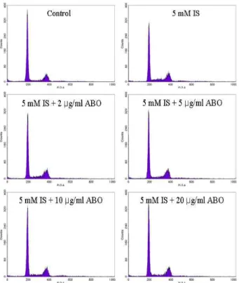
2. IS 처리된 HK-2 세포주기변화에 대한 밀몽화의 효과 IS로 유도된 HK-2 세포 증식억제와 이에 따른 세포주기변화 에 대하여 밀몽화 물추출물의 효과를 조사하였다. 이를 위해 유 세포 분석을 통하여 세포주기를 분석하였다. HK-2 세포에 5 mM 농도의 IS와 밀몽화 물추출물을 2, 5, 10, 20 μg/ml 농도로 동시에 24 시간 처리 한 후, PI 염색하여 세포주기를 분석하였다 (Fig. 2 and Table 1). IS 단독 처리군은 대조군과 비교하여 G0/G1기가 73.76%에서 59.69% 감소하였고, S기는 6.43%에서
13.39%로 증가하였으며, G2/M기는 24.56% 증가시켰다. 밀몽화 물추출물 20 μg/ml 농도 처리군에서는 IS에 의해서 감소된 G0/G1기는 69.42% 증가시켰고, 증가된 S기 및 G2/M기는 각각 7.72%와 20.37%로 감소시켰다. 모든 처리군에서 Sub-G1기의 변 화는 큰 차이가 없었다.
Fig. 1. Effect of ABO on cell viability in IS-treated HK-2 cells. The cells were incubated with IS (5 mM) in the absence or presence of ABO (2, 5, 10, 20 μg/ml) for 24 hr. Cell viability was measured by MTT assay. Value are means
± SD, N = 3.
Fig. 2. Effect of ABO on cell cycle progression in IS-treated HK-2 cells.The cells were incubated with IS (5 mM) in the absence or presence of ABO (2, 5, 10, 20 μg/ml) for 24 hr. The cells were fixed and stained with PI. The DNA content was analyzed by flow cytometry.
Table 1. Effects of ABO on the distribution of cell cycle in IS-treated HK-2 cells
Cell cycle
population (%) 5 mM IS ABO (μg/ml) -
0 +
0 +
2 +
5 +
10 +
20
Sub-G1 1.80 2.68 2.52 2.49 2.10 2.68
G0/G1 73.76 59.69 63.18 65.04 67.27 69.42
S 6.43 13.39 11.22 9.82 8.47 7.72
G2/M 18.11 24.56 23.38 22.89 21.47 20.37
3. IS를 처리한 HK-2 세포에서 p21 단백질발현 증가에 대한 밀 몽화의 효과
CDK 활성억제인자인 p21 단백질은 세포주기조절과 연관되
어 있다14,15). IS에 의해서 증가된 p21 단백질 발현량에 밀몽화 물
추출물의 효과를 조사하기 위해 HK-2 세포에 5 mM 농도의 IS와 밀몽화 물추출물을 2, 5, 10, 20 μg/ml 농도로 동시에 24 시간 처 리 후, p21 단백질의 발현정도를 조사하였다. IS에 의해서 증가된 p21 단백질 발현은 밀몽화 물추출물의 투여로 농도의존적으로 감소되었다(Fig. 3).
A
B
Fig. 3. Effect of ABO on expression of p21 in IS-treated HK-2 cells. The cells were treated with IS (5 mM) in the absence or presence of ABO (2, 5, 10, 20 μg/ml) for 24 hr. Whole cell lysates were subjected to SDS-PAGE followed by Western blot analysis with anti-p21 antibody. Intensity of p21 protein bands (B).
고 찰
신장독성 물질은 신기능장애를 지닌 환자에 축적이되는 화 합물로 다양한 세포와 조직에 유해한 효과를 나타낸다23). 신부전 은 요독 물질의 잔류를 통해 요독증을 유발시키고, 특히 신장질 환의 말기환자에게서 나타난다. 신기능의 작은 장애조차 요독소 의 축적이 원인이 되고 만성신부전의 초기단계에서 세포와 조직 을 악화시킨다. 그러므로 요독소는 신장손상과 만성신부전의 진 행을 촉진시키는 원인이 될 수 있다.
IS는 만성신부전 동물모델에서 신장기능을 악화시키 대표적 인 요독 물질이다4). IS는 트립토판이 장내세균에 의해서 변환된 indole이 장에서 흡수되어 혈류를 통해 간으로 전달되고, 간에서 변환되어 생성된다. 그 후 IS는 담즙으로 분비되어 장에 도착하 고 최종적으로 장간순환 (Enterohepatic circulation, 腸肝循環)에 들어간다15). IS는 마우스 in vivo 실험에서 신근위세뇨관세포의 생존율을 감소시키고, 신근위세뇨관세포에서 산화적스트레스를 통해 plasminogen activator inhibitor-1(PAI-1)을 증가시킨다4,5). 최근 보고된 연구에 의하면 IS는 신근위세뇨관세포에서 세포노
화 및 신섬유화를 유도 및 세포증식을 억제한다고 보고되었다.
본 연구에서는 IS에 의해 억제된 HK-2 세포의 증식에 대한 밀몽화 물추출물이 미치는 효과를 세포생존률 측정과 유세포분 석을 통하여 확인할 수 있었다. 밀몽화 물추출물은 IS에 의해서 감소된 세포증식을 농도의존적으로 증가시켰다. 밀몽화 물추출 물 20 μg/ml 농도에서 IS에 의해서 감소된 세포증식을 IS 단독 처리군 비교하여 12% 정도 세포증식을 증가시켰다. 유세포분석 에서도 밀몽화 물추출물은 IS 단독처리군과 비교하여 G0/G1 phase가 10% 정도 감소하였고, S phase는 6%, G2/M phase는 4% 정도 감소시켰다. 이 결과들은 밀몽화 물추출물이 IS에 의해 서 증가된 S phase와 G2/M phase를 감소시키고 G0/G1 phase 를 증가시킴으로써 세포주기를 진행시켜 세포증식을 증가시키는 것을 보여주고 있다.
세포주기정지는 세포증식에 있어서 중요한 역할을 수행한 다. 세포주기는 cyclin-dependent kinases(CDKs)에 의해 양성적 으로 조절되지만24), CDK활성을 억제하는 CDK 억제제(CKIs)에 의해 음성적으로도 조절된다25). 현재 포유동물세포의 CKIs는 2 가지 종류가 있다. 하나는 CIP/KIP 패밀리로 p21, p27, p57등이 있고, 모든 CDK활성에 광범위하게 작용을 한다25,26). 또 다른 종 류는 INK4 패밀리인 p15, p16, p18, p19로 CDK4와 CDK6의 활 성을 조절한다25). 이러한 세포주기조절 기전은 세포증식을 조절 하는 기본이 된다. IS는 HK-2 세포에서 p21 단백질 발현을 증가 시키고, p21 단백질 증가가 IS로 유도된 HK-2 세포의 증식을 억 제시키는 것으로 보고되어 있다14,15). 밀몽화 물추출물은 IS에 의 해서 증가된 p21 단백질 발현을 농도의존적으로 감소시켰다. 이 결과는 밀몽화 물추출물에 의한 p21 단백질 발현 감소가 IS의 세 포증식억제에 대한 증식회복효과에 관련되어 있다는 것을 보여 주고 있다.
결 론
밀몽화 물추출물은 IS에 의해 억제된 HK-2 세포의 증식을 증가시켰고, S 및 G2/M phase 증가를 감소시켰다. 또한, 밀몽화 물추출물은 IS에 의해 증가된 p21 단백질 발현을 농도의존적으 로 감소시켰다. 이상 결과를 종합하면, 밀몽화 물추출물의 증식 회복효과는 IS에 의해 증가된 세포주기조절 억제인자인 p21 단 백질 발현 감소를 통하여 이루어졌다고 생각된다.
감사의 글
이 논문은 2011년도 정부[교육과학기술부]의 재원으로 한국 연구재단의 지원을 받아 수행된 연구임[NO. 2010-0029469].
참고문헌
1. Miyazaki, T., Ise, M., Seo, H., Niwa, T. Indoxyl sulfate increases the gene expression of TGF-β1, TIMP-1 and pro- α (I) collagen in uremic rat kidneys. Kidney Int. Suppl. 63:
S15-S22, 1997.
2. Niwa, T., Ise, M. Indoxyl sulfate, a circulating uremic toxin, stimulates the progression of glomerular sclerosis. J. Lab.
Clin. Med. 124: 96-104, 1994.
3. Niwa, T., Ise, M., Miyazaki, T. Progression of glomerular sclerosis in experimental uremic rats by administration of indole, aprecursor of indoxyl sulfate. Am. J. Nephrol. 14:
207-212, 1994.
4. Enomoto, A., Takeda, M., Tojo, A., Sekine, T., Cha, S.H., Khamdang, S., Takayama, F., Aoyama, I., Nakamura, S., Endou, H., Niwa, T. Role of organic anion transporters in the tubular transport of indoxyl sulfate and the induction of its nephrotoxicity. J. Am. Soc. Nephrol. 13: 1711-1720, 2002.
5. Motojima, M., Hosokawa, A., Yamato, H., Muraki, T., Yoshioka, T. Uremic toxins of organic anions up-regulate PAI-1 expression by induction of NF-κB and free radical in proximal tubular cells. Kidney Int. 63: 1671-1680, 2003.
6. Lekawanvijit, S., Adrahtas, A., Kelly, D.J., Kompa, A.R., Wang, B.H., Krum, H. Does indoxyl sulfate, a uraemic toxin, have direct effects on cardiac fibroblasts and myocytes? Eur. Heart J. 31: 1771-1779, 2010.
7. Tumur, Z., Shimizu, H., Enomoto, A., Miyazaki, H., Niwa, T. Indoxyl sulfate upregulates expression of ICAM-1 and MCP-1 by oxidative stress-induced NF-kappaB activation.
Am. J. Nephrol. 31: 435-441, 2010.
8. Muteliefu, G., Enomoto, A., Niwa, T. Indoxyl sulfate promotes proliferation of human aortic smooth muscle cells by inducing oxidative stress. J. Ren. Nutr. 19: 29-32, 2009.
9. Shimizu, H., Hirose, Y., Nishijima, F., Tsubakihara, Y., Miyazaki, H. ROS and PDGF-β receptors are critically involved in indoxyl sulfate actions that promote vascular smooth muscle cell proliferation and migration. Am. J.
Physiol. Cell Physiol. 297: C389-C396, 2009.
10. Adijiang, A., Goto, S., Uramoto, S., Nishijima, F., Niwa, T.
Indoxyl sulphate promotes aortic calcification with expression of osteoblastspecific proteins in hypertensive rats. Nephrol. Dial. Transplant 23: 1892-1901, 2008.
11. Muteliefu, G., Enomoto, A., Jiang, P., Takahashi, M., Niwa, T. Indoxyl sulphate induces oxidative stress and the expression of osteoblastspecific proteins in vascular smooth muscle cells. Nephrol. Dial. Transplant 24: 2051-2058, 2009.
12. Taki, K., Tsuruta, Y., Niwa, T. Indoxyl sulfate and atherosclerotic risk factors in hemodialysis patients. Am. J.
Nephrol. 27: 30-35, 2007.
13. Shimizu, H., Bolati, D., Adijiang, A., Enomoto, A., Nishijima, F., Dateki, M., Niwa, T. Senescence and dysfunction of proximal tubular cells are associated with
activated p53 expression by indoxyl sulfate. Am. J. Physiol.
Cell Physiol. 299: C1110-1117, 2010.
14. Shimizu, H., Bolati, D., Adijiang, A., Muteliefu, G., Enomoto, A., Nishijima, F., Dateki, M., Niwa, T.. NF-κB plays an important role in indoxyl sulfate-induced cellular senescence, fibrotic gene expression, and inhibition of proliferation in proximal tubular cells. Am J. Physiol. Cell Physiol. 301: C1201-1212, 2011.
15. Kawakami, T., Inagi, R., Wada, T., Tanaka, T., Fujita, T., Nangaku, M. Indoxyl sulfate inhibits proliferation of human proximal tubular cells via endoplasmic reticulum stress. Am. J. Physiol. Renal Physiol. 299: F568-576. 2010.
16. 중화인민공화국위생부약전위원회. 중화인민공화국약전, p 291, 1995.
17. 강소신의학원. 중약대사전, 서울, 정담, pp 1904-1906, 1998.
18. Blondelle, S.E., Houghten, R.A. Designs of model amphipathic peptides having potent antimicrobial activities. Biochem. 31: 12688-12694, 1992.
19. Piao, M.S., Kim, M.R., Lee, D.G., Park, Y., Hahm, K.S., Moon, Y.H., Woo, E.R. Antioxidative constituents from Buddleja officinalis. Arch. Pharm. Res. 26: 453-457, 2003.
20. Lee, D.H., Ha, N., Bu, Y.M., Choi, H.I., Park, Y.G., Kim,
Y.B., Kim, M.Y., Kim, H. Neuroprotective effect of Buddleja officinalis extract on transient middle cerebral artery occlusion in rats. Biol. Pharm. Bull. 29: 1608-1612, 2006.
21. 김호철. 밀몽화의 신경보호 효능에 관한 연구, 경희대학교 동 서의학대학원, 2004.
22. Matsuda, H., Cai, H., Kudo, M., Tosa, H., Iinuma, M. Study on anti-cataract drugs from natural sources. II. Effects on Buddlejae flos on in vitro aldose reductase activity. Biol.
Pharm. Bull. 18: 463-466, 1995.
23. Raff, A.C., Meyer, T.W., Hostetter, T.H. New insights into uremic toxicity. Curr. Opin. Nephrol. Hypertens 17:
560-565, 2008.
24. Covacci, V., Bruzzese, N., Sgambato, A., Di Francesco, A., Russo, M.A., Wolf, F.I., Cittadini, A. Magnesium restriction induces granulocytic differentiation and expression of p27Kip1 in human leukemic HL-60 cells. J. Cell Biochem.
70: 313-322, 1998.
25. Sherr, C.J., Roberts, J.M. Inhibitors of mammalian G1 cyclin-dependent kinases. Genes Dev. 9: 1149-1163, 1995.
26. Brugarolas, J., Chandrasekaran, C., Gordon, J.I., Beach, D., Jacks, T., Hannon, G.J. Radiation-induced cell cycle arrest compromised by p21 deficiency. Nature 377: 552-557, 1995.