Inhibitory Effect of NAD(P)H:Quinone Oxidoreductase 1 on the Activation of Macrophages
Ji Hong, Peng Zhang, I Na Yoon and Ho Kim*
Division of Life Science and Chemistry, College of Natural Science, Daejin University, Pocheon, Gyeonggido 487-711, Korea Received March 22, 2017 /Revised October 16, 2017 /Accepted October 16, 2017
We previously reported that NAD(P)H:quinone oxidoreductase 1 (NQO1)-knockout (KO) mice ex- hibited spontaneous inflammation in the gut. We also found that NQO1-KO mice showed highly in- creased inflammatory responses compared with NQO1-WT control mice when subjected to DSS-in- duced experimental colitis. In a Clostridium difficile toxin-induced mouse enteritis model, NQO1-KO mice were also sensitive compared with NQO1-WT mice. Moreover, numerous studies have shown that NQO1 is functionally associated with immune regulation. Here, we assessed whether NQO1 de- fects can alter macrophage activation. We found that peritoneal macrophages isolated from NQO1-KO mice produced more IL-6 and TNF-a than those isolated from NQO1-WT mice. Moreover, the dicu- marol-induced inhibition of NQO1 significantly increased IL-6 and TNF-a production in peritoneal macrophages isolated from NQO1-WT mice, as well as in the cultured mouse macrophage cell line, RAW264.7. These results indicate that NQO1 may negatively regulate the activation of macrophages.
Knockout or chemical inhibition of NQO1 markedly reduced the expression of IκB (inhibitor of NFκB) in both mouse peritoneal macrophages and RAW264.7 cells. Finally, RAW264.7 cells treated with dicu- marol exhibited morphological changes reflecting macrophage activation. Our results suggest that NQO1 may suppress the NFkB pathways in macrophages, thereby suppressing the activation of these cells. Thus, immunosuppressive activity may be among the many possible functions of NQO1.
Key words : Immunosuppression, IL-6, macrophage, NQO1, TNF-α
*Corresponding author
*Tel : +82-31-539-1855, Fax : +82-31-539-1850
*E-mail : [email protected]
This is an Open-Access article distributed under the terms of the Creative Commons Attribution Non-Commercial License (http://creativecommons.org/licenses/by-nc/3.0) which permits unrestricted non-commercial use, distribution, and reproduction in any medium, provided the original work is properly cited.
Journal of Life Science 2017 Vol. 27. No. 8. 873~878 DOI : https://doi.org/10.5352/JLS.2017.27.8.873
서 론
NAD(P)H:quinone oxidoreductase 1 (NQO1)은 세포질에 존재하며 플라보 단백질로서 quinone 화합물과 그 유도체를 환원시키거나 독성을 낮추는 역할을 하는 것으로 알려져 있다 [1, 5, 11, 17, 19, 20]. 이 과정은 활성산소(reactive oxygen spe- cies)에 의한 세포 독성을 감소시키기도 한다[3, 5, 16]. 한편 종양형성 과정에서 NQO1은 세포를 보호하는 것으로도 알려 져 있다[23, 27, 28]. NQO1의 기능은 또한 면역질환과 연관되 어 있다고 알려져 있다[4]. 손상된 면역반응을 보이는 환자나 자가면역질환 환자들에게서 NQO1 유전자 돌연변이가 높은 빈도로 확인된다[22, 25]. 더욱이 NQO1과 NQO2가 각각 결핍 된 생쥐에서, 말초혈액 속 B세포 수와 배중심(germinal center) 세포들의 면역 활성이 크게 감소되어 있음이 보고되었다[4].
또한 형성된 항체의 양과 활성이 현저하게 감소되어 있음도 확인되었다. NQO1 KO 생쥐에서는 자가면역질환 유발 비율
도 유의하게 증가한다는 사실이 확인되었다[4]. LPS로 자극된 단핵구(monocytes) 속에서도 NQO1의 주요 기능이 면역반응 억제임이 확인되었다[21]. LPS 자극으로 활성화된 대식세포를 억제하는 타이완 버섯(Antrodia salmonea)의 효능 역시 NQO1 발현량 증가와 연관되어 있음이 보고된 바 있다[27]. NQO1을 포함하는 phase II 항산화 효소들이 T세포의 세포자살을 억제 한다는 보고도 있다[8]. 면역기관이 아닌 소화기관 부속 장기 들에서 NQO1 발현량이 높다는 보고가 있지만[14], 상기 결과 들을 고려해볼 때, NQO1이 면역세포의 활성화와 분화과정에 서 중요한 역할을 수행하고 있음을 강하게 제시하고 있다. 우 리는 최근 연구를 통해, NQO1 KO 생쥐의 대장에서 자연발생 적으로 염증이 일어남을 보고하였다[14]. 이 결과 역시 NQO1 의 기능이 면역반응 조절과 밀접하게 연관되어 있음을 보여준 다.
본 연구에서는 NQO1이 대식세포 활성화를 낮추는 억제효 능을 가지는지를 확인해보고자 하였다. 이를 위해, NQO1 KO 생쥐의 복강에서 분리한 복강대식세포의 활성화 정도를 NQO1 WT 생쥐와 비교해 보았다. 또한 정상 생쥐의 복강에서 분리한 복강대식세포에 NQO1 억제제(dicumarol)를 처치한 다음 활성화 정도를 비교하였다. 마지막으로 대식세포주인 RAW264.7 세포에 dicumarol을 처치한 후 상기 변화들을 재 검증 해보았다. 본 연구는 향 후 대식세포 활성화 과정을 조절 하는 NQO1의 역할을 연구하는데 기초가 될 것으로 기대한다.
시약
IκB와 β-actin 항체 등은 Santa Cruz biotech (USA)으로부 터 구매하였다. Dicumarol과 Avertin 마취제 등은 Sigma Aldrich (USA)에서 구매하여 사용하였다. IL-6와 TNF-α ELISA (enzyme-linked immunosorbent assay) kit는 R&D System (USA)에서 구매하여 사용하였다[13].
세포배양
생쥐의 macrophage-like cell line인 RAW264.7 세포는 ATCC (USA)에서 구매하여 사용하였고 Dulbecco’s modified Eagle’s medium (DMEM) 배양액(invitrogen, USA)에 10%
FBS (Invitrogen, USA)을 첨가하여 CO2 배양기에서 배양하였 다[7]. Confluence 상태에서 배지와 비부착세포를 제거하고 새 로운 배지를 가하여 24시간 배양한 후, rubber policeman으로 세포를 모아 3회 수세하고 생존 세포의 수를 세었다. Well 당 1×10⁶의 세포수로 24 well에 분주한 후, 2시간 이상 배양하여 plate에 부착시킨 다음 실험에 사용하였다.
생쥐 복강 대식세포 분리(isolation of peritoneal mac- rophages)
NQO1 WT 생쥐와 NQO1 KO 생쥐를 경추탈골하여 도살시 킨 다음, 복강에 따뜻한 생리식염수(PBS) 10 ml를 넣어 복강세 포를 수집하였다. 수집한 세포를 4℃에서 1,300 rpm으로 10분 간 원심분리하고 DMEM 배지로 2회 세척 후, 배양 용기에 분주하여 CO2 incubator에서 배양시키고, 2시간 후에 부착되 지 않은 세포를 제거하였다. 배지는 DMEM 배지를 사용하였 으며, 배지에 10% FBS와 penicillin-streptomycin (100 units/
ml, 100 μg/ml)을 첨가하여 사용하였다[13]. 본 연구는 대진대 학교 동물실험위원회(Animal Care and Use Committee of Daejin University)의 방침 및 범규에 따라 진행되었다(승인번 호: 2013-03).
IL-6와 TNF-α 정량
IL-6와 TNF-α 양은 ELISA assay로 측정하였다. 생쥐로부터 분리한 복강대식세포에 dicumarol을 처치한 다음 배양액을 분리하였다. 그 후 ELISA plate의 각 well에 동일한 양의 배양 액을 100 μl씩 넣어주고 2시간 동안 상온에서 반응시켰다. 3회 수세한 다음, 일차 항체를 이용하여 1시간 동안 상온에서 반응 시켰다. 추가로 수세한 다음, avidin-horseradish peroxidase (avidin-HRP)를 이용하여 30분 동안 반응하였다. 최종 수세 후 15분간 발색시킨 다음 반응 중지용액을 첨가하여 반응을 끝냈다. 마지막으로 Microplate reader (model 3550; Bio-Rad, Mississauga, Canada)를 이용 450 nm 파장에서 흡광도를 측 정하였다. 배양액 ml당 IL-6와 TNF-α양을 결정하였다[13].
RAW264.7 세포에 dicumarol을 처치 한 후 세포들은 SDS sample buffer (150 mM NaCl, 50 mM Tris-HCl, pH8.0, 0.5 mM EDTA, 1% Nonidet P-40)를 첨가하여 용해시켰다. 단백 질 정량은 BCA protein assay kit (Thermo Scientific, USA)를 이용하여 실시하였다. SDS-PAGE를 실시하여 분리한 단백질 은 transfer 과정을(120 V, 1시간) 통해 nitrocellulose mem- brane으로 옮겼다. 일차 항체들을 이용해 4℃에서 밤새 항원 항체 반응을 수행한 다음 이차 항체를 이용하여 1시간 동안 상온에서 추가 반응시켰다. LumiGlo 시약(New England Biolabs, USA)을 이용하여 발색시킨 후 Fusion FX (Vilber Lourmat, France)를 이용하여 현상하였다[15].
Statistical analysis
모든 실험은 3회 반복으로 행하여 표준오차로 나타내었고 SIGMA-STAT professional statistics software program (Jandel Scientific Software, USA)의 t-test를 이용하여 유의성 을 검정하였다.
결과 및 고찰
NQO1 KO 생쥐 대식세포의 과잉 활성화
이전 연구에서, 우리는 NQO1 KO 생쥐 대장에서 염증반응 이 나타남을 확인하였다[14]. 이 결과는 NQO1이 면역반응에 대한 억제효능이 있음을 간접적으로 보여준다고 할 수 있다.
이를 확인하기 위해 NQO1 WT 생쥐와 NQO1 KO 생쥐의 복 강에서 대식세포를 각각 분리한 다음, IL-6와 TNF-α와 같은 염증성 사이토카인[7] 양을 측정하였다. Fig. 1에서처럼, IL-6 (A)와 TNF-α (B) 분비량이 NQO1 WT 생쥐에서보다 NQO1 KO 생쥐에서 증가되어 있음을 확인하였다. TNF-α의 경우, 약 31% 이상 증가하는 것으로 확인되었다. 이 결과는 NQO1 기 능이 대식세포의 활성화를 억제하는 경로들과 연관되어 있음 을 보여준다.
우리는 이전 연구에서 NQO1 KO 생쥐에서 관찰되는 대장 염증이 점막을 구성하는 상피세포들의 밀착연접 감소에 기인 함을 보고하였다[14]. 그러나 NQO1 KO 생쥐에서 분리한 대 식세포가 과잉 활성화되어 있다는 본 연구결과를 감안할 때, NQO1 KO 생쥐 대장에서 관찰된 염증반응이 점막 상피세포 장벽기능 소실과 더불어 대식세포의 과잉 활성화에 기인한 복합적인 원인이라고 추측할 수 있다.
Dicumarol에 의한 NQO1 억제는 생쥐 복강대식세포를 활 성화시킴
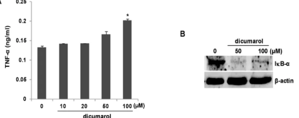
다음으로 연구자 등은 NQO1 WT 생쥐의 복강에서 분리한 대식세포에 NQO1 억제제인 dicumarol을 24시간 동안 처치한 다음 TNF-α 분비량을 측정하여 대식세포의 활성화를 평가하
Fig. 1. Peritoneal macrophages from NQO1-knockout (KO) mice produce much more IL-6 and TNF-α than those of NQO1 wild-type (WT) mice. Peritoneal macrophages (106) isolated from NQO1 WT and NQO1 KO mice were cultured in DMEM supplemented with 10% FBS for 24 hr. Culture supernatants were harvested, and ELISA was used to assess the levels of IL-6 (A) and TNF-α (B). The bars represent the mean ± SEM of three independent experiments (*, p<0.05 vs. NQO1 WT mice).
Fig. 2. Inhibition of NQO1 by dicumarol activates peritoneal macrophages. (A) Peritoneal macrophages (106) were isolated from NQO1 WT mice and treated with the indicated concentrations of dicumarol, and TNF-α levels in culture supernatants were measured by ELISA. The bars represent the mean ± SEM of three independent experiments (*, p<0.005 vs. untreated cells).
(B) Peritoneal macrophages (106) were isolated from NQO1 WT mice and treated with dicumarol (0, 50, or 100 μM) for 12 hr. Cell lysates were resolved by SDS-PAGE, and blots were probed with antibodies against IκB-α and β-actin. The pre- sented results are representative of three independent experiments.
였다. Fig. 2A에서처럼, dicumarol이 처치된 복강대식세포에 서 TNF-α 분비량이 농도 의존적으로 증가함을 확인하였다.
50 μM 농도에서 TNF-α 분비량이 유의하게 증가하였으며 100 μM 농도에서 최고 농도를 나타내었다. 지금까지 IL-6와 TNF- α를 조절하는 대표적인 전사개시인자로서 NFκB가 잘 알려져 있으며, 이 단백질의 활성화 과정이 IκB에 의해 조절된다는 사실도 명확하다[6]. 특히 IκB의 활성조절이 단백질 변성과정 에 의존적임도 잘 알려져 있다[10]. 따라서 dicumarol 처치로 활성화되는 대식세포에서 NFκB가 주요 경로임을 확인해보고 자 하였다. 이를 위해, NQO1 WT 생쥐에서 분리한 복강대식 세포에 dicumarol을 12시간 동안 처치하고 단백질을 분리한 다음 IκB 항체를 이용 immunoblot analysis를 수행하였다.
Fig. 2B에서처럼, dicumarol 처치는 세포 속 IκB 단백질을 완 전하게 감소시키는 것으로 확인되었다. 앞서 결과처럼, dicu- marol 50 μM 농도에서부터 IκB 단백질 감소가 나타났다. 이상 의 결과는 dicumarol에 의해 NQO1가 억제되면 NFκB 전사인
자가 작동하여 대식세포의 활성이 증가했음을 보여준다.
NQO1의 억제로 인해 NFκB가 활성화되는 기작은 세포 속 활성산소 생성과정과 연관될 것으로 추측된다. 왜냐하면 NQO1 의 주요 기능 중 하나가 활성산소를 제거하는 항산화 효능이 기 때문이다[14, 23, 25]. 따라서 NQO1 억제로 인해 증가한 세포 속 활성산소가 IκB의 파괴경로를 촉진시켜 단백질 감소 로 이어졌다고 생각된다. 실제 많은 연구들이 활성산소가 IκB 의 감소 과정에서 필수적이라고 보고하였다[2, 6]. 예를 들어, LPS에 의한 대식세포 활성화에서 NFκB 경로가 반드시 요구 되며 이 과정이 세포 속 활성산소 양에 의존적으로 조절됨이 보고되었다[2].
Dicumarol에 의한 NQO1 억제는 대식세포주(RAW264.7) 를 활성화시킴
다음으로 대식세포주인 RAW264.7 세포에 dicumarol을 24 시간 동은 처치한 다음 TNF-α 분비량을 측정하였다. Fig. 3A
Fig. 3. Dicumarol treatment activates Raw264.7 macrophages.
(A) RAW264.7 cells (106) were treated with dicumarol (0, 50, 100 μM) for 24 hr, and TNF-α levels in culture supernatants were measured by ELISA. The bars repre- sent the mean ± SEM of three independent experiments (*, p<0.005 vs. untreated cells). (B) RAW264.7 cells (106) were treated with dicumarol (0, 50, or 100 μM) for 24 hr. Cell lysates were resolved by SDS-PAGE, and blots were probed with antibodies against IκB-α and β-actin.
The presented results are representative of three in- dependent experiments. (C) Light microscopic images of RAW264.7 cells treated with dicumarol. The pre- sented images are representative of three independent experiments.
에서처럼, dicumarol이 처치된 대식세포에서 TNF-α 생성량이 유의하게 증가함을 확인하였다. 생쥐 복강에서 분리한 대식세 포에서의 결과와 마찬가지로, dicumarol 50 μM 농도에서 TNF-α 분비량이 유의하게 증가하기 시작하였으며 100 μM 농 도에서 최고 농도를 나타내었다. Dicumarol 처치에 의한 IκB 단백질 감소현상도 농도 의존적으로 나타남을 확인하였다 (Fig. 3B). 비활성형 대식세포의 일반적인 형태로 알려진 둥근 원형 모양이[12] dicumarol 처치로 인해서 많은 돌기를 포함하 는 확장된(spreading) 구조의 활성형[12]으로 변한다는 사실 도 확인하였다(Fig. 3C).
들에 의해 조절되는 것으로 알려져 왔다. 그 이유는 이들 단백 질들이 세포골격계의 하나인 미세섬유 형성에 주요하게 관여 하는 인자이기 때문이다. 실제로 이들 단백질이 결핍된 대식 세포에서, 활성형의 대식세포 형태 발달과 세포이동이 현저하 게 감소된다고 알려져 있다[9]. 특이하게도, Rho 가족 단백질 에 의한 세포골격계 조절과정이 활성산소에 의해 영향을 받는 다는 것이 확인되었다[26]. 이상의 결과들을 고려하면, NQO1 결핍으로 축적된 활성산소가 대식세포의 형태 결정에 관여하 는 Rho 가족 단백질의 활성화를 유도하여 대식세포의 모양변 화를 초래했을 가능성이 있다. 더욱이 대식세포의 경우, 탐식 과정 후 섭취한 미생물 파괴를 위해 과산소(superoxide anion) 와 같은 활성산소를 많이 필요로 한다[18, 24]. 결국 NQO1 결핍은 더 많은 활성산소 축적으로 이어져서 급격한 대식세포 형태 변화를 초래하였다고 추측할 수 있다.
감사의 글
이 논문은 2017학년도 대진대학교 학술연구비 지원에 의한 것임.
References
1. Berger, F., Ramirez-Hernandez, M. H. and Ziegler, M. 2004.
The new life of a centenarian: signalling functions of NAD (P). Trends Biochem. Sci. 29, 111-118.
2. Diao, Y., Xin, Y., Zhou, Y., Li, N., Pan, X., Qi, S., Qi, Z., Xu, Y., Luo, L., Wan, H., Lan, L. and Yin, Z. Extracellular polysaccharide from Bacillus sp. strain LBP32 prevents LPS-induced inflammation in RAW 264.7 macrophages by inhibiting NF-kappaB and MAPKs activation and ROS production. Int. Immunopharmacol. 18, 12-19.
3. Hwang, J. H., Kim, D. W., Jo, E. J., Kim, Y. K., Jo, Y. S., Park, J. H., Yoo, S. K., Park, M. K., Kwak, T. H., Kho, Y.
L., Han, J., Choi, H. S., Lee, S. H., Kim, J. M., Lee, I., Kyung, T., Jang, C., Chung, J., Kweon, G. R. and Shong, M. 2009.
Pharmacological stimulation of NADH oxidation amelio- rates obesity and related phenotypes in mice. Diabetes 58, 965-974.
4. Iskander, K., Li, J., Han, S., Zheng, B. and Jaiswal, A. K.
2006. NQO1 and NQO2 regulation of humoral immunity and autoimmunity. J. Biol. Chem. 281, 30917-30924.
5. Jaiswal, A. K. 2000. Regulation of genes encoding NAD(P) H:quinone oxidoreductases. Free Radic. Biol. Med. 29, 254- 262.
6. Kang, J., Tae, N., Min, B. S., Choe, J. and Lee, J. H. 2012.
Malabaricone C suppresses lipopolysaccharide-induced in- flammatory responses via inhibiting ROS-mediated Akt/
IKK/NF-kappaB signaling in murine macrophages. Int.
Immunopharmacol. 14, 302-310.
7. Kim, H., Kokkotou, E., Na, X., Rhee, S. H., Moyer, M. P., Pothoulakis, C. and Lamont, J. T. 2005. Clostridium difficile
toxin A-induced colonocyte apoptosis involves p53-depend- ent p21(WAF1/CIP1) induction via p38 mitogen-activated protein kinase. Gastroenterology 129, 1875-1888.
8. Kim, H. J. and Nel, A. E. 2005. The role of phase II anti- oxidant enzymes in protecting memory T cells from sponta- neous apoptosis in young and old mice. J. Immunol. 175, 2948-2959.
9. Konigs, V., Jennings, R., Vogl, T., Horsthemke, M., Bachg, A. C., Xu, Y., Grobe, K., Brakebusch, C., Schwab, A., Bahler, M., Knaus, U. G. and Hanley, P. J. Mouse macrophages completely lacking Rho subfamily GTPases (RhoA, RhoB, and RhoC) have severe lamellipodial retraction defects, but robust chemotactic navigation and altered motility. J. Biol.
Chem. 289, 30772-30784.
10. Lawrence, T., Bebien, M., Liu, G. Y., Nizet, V. and Karin, M. 2005. IKKalpha limits macrophage NF-kappaB activation and contributes to the resolution of inflammation. Nature 434, 1138-1143.
11. Long, D. J., Iskander, K., Gaikwad, A., Arin, M., Roop, D.
R., Knox, R., Barrios, R. and Jaiswal, A. K. 2002. Disruption of dihydronicotinamide riboside:quinone oxidoreductase 2 (NQO2) leads to myeloid hyperplasia of bone marrow and decreased sensitivity to menadione toxicity. J. Biol. Chem.
277, 46131-46139.
12. McWhorter, F. Y., Wang, T., Nguyen, P., Chung, T. and Liu, W. F. 2013. Modulation of macrophage phenotype by cell shape. Proc. Natl. Acad Sci. USA 110, 17253-17258.
13. Nam, H. J., Oh, A. R., Nam, S. T., Kang, J. K., Chang, J.
S., Kim, D. H., Lee, J. H., Hwang, J. S., Shong, K. E., Park, M. J., Seok, H. and Kim, H. 2012. The insect peptide CopA3 inhibits lipopolysaccharide-induced macrophage activation.
J. Pept. Sci. 18, 650-656.
14. Nam, S. T., Hwang, J. H., Kim, D. H., Park, M. J., Lee, I.
H., Nam, H. J., Kang, J. K., Kim, S. K., Hwang, J. S., Chung, H. K., Shong, M., Lee, C. H. and Kim, H. 2014. Role of NADH: quinone oxidoreductase-1 in the tight junctions of colonic epithelial cells. BMB. Rep. 47, 494-499.
15. Nam, S. T., Seok, H., Kim, D. H., Nam, H. J., Kang, J. K., Eom, J. H., Lee, M. B., Kim, S. K., Park, M. J., Chang, J.
S., Ha, E. M., Shong, K. E., Hwang, J. S. and Kim, H. 2012.
Clostridium difficile toxin A inhibits erythropoietin receptor- mediated colonocyte focal adhesion through inactivation of Janus Kinase-2. J. Microbiol. Biotechnol. 22, 1629-1635.
16. Oh, G. S., Kim, H. J., Choi, J. H., Shen, A., Choe, S. K., Karna, A., Lee, S. H., Jo, H. J., Yang, S. H., Kwak, T. H., Lee, C.
H., Park, R. and So, H. S. 2014. Pharmacological activation of NQO1 increases NAD levels and attenuates cispla- tin-mediated acute kidney injury in mice. Kidney Int. 85, 547-560
17. Palming, J., Sjoholm, K., Jernas, M., Lystig, T. C., Gummes- son, A., Romeo, S., Lonn, L., Lonn, M., Carlsson, B. and Carlsson, L. M. 2007. The expression of NAD(P)H:quinone
oxidoreductase 1 is high in human adipose tissue, reduced by weight loss, and correlates with adiposity, insulin sensi- tivity, and markers of liver dysfunction. J. Clin. Endocrinol.
Metab. 92, 2346-2352.
18. Park, J. B. 2003. Phagocytosis induces superoxide formation and apoptosis in macrophages. Exp. Mol. Med. 35, 325-335.
19. Pollak, N., Dolle, C. and Ziegler, M. 2007. The power to reduce: pyridine nucleotides--small molecules with a multi- tude of functions. Biochem. J. 402, 205-218.
20. Radjendirane, V., Joseph, P., Lee, Y. H., Kimura, S., Klein- Szanto, A. J., Gonzalez, F. J. and Jaiswal, A. K. 1998. Disrup- tion of the DT diaphorase (NQO1) gene in mice leads to increased menadione toxicity. J. Biol. Chem. 273, 7382-7389.
21. Rushworth, S. A., MacEwan, D. J. and O'Connell, M. A.
2008. Lipopolysaccharide-induced expression of NAD(P)H:
quinone oxidoreductase 1 and heme oxygenase-1 protects against excessive inflammatory responses in human mono- cytes. J. Immunol. 181, 6730-6737.
22. Siegel, D., Anwar, A., Winski, S. L., Kepa, J. K., Zolman, K. L. and Ross, D. 2001. Rapid polyubiquitination and pro- teasomal degradation of a mutant form of NAD(P)H:qui- none oxidoreductase 1. Mol. Pharmacol. 59, 263-268.
23. Siegel, D., Yan, C. and Ross, D. 2012. NAD(P)H:quinone oxi- doreductase 1 (NQO1) in the sensitivity and resistance to antitumor quinones. Biochem. Pharmacol. 83, 1033-1040.
24. Stanley, A., Thompson, K., Hynes, A., Brakebusch, C. and Quondamatteo, F. 2014. NADPH oxidase complex-derived reactive oxygen species, the actin cytoskeleton, and Rho GTPases in cell migration. Antioxid. Redox Signal. 20, 2026- 2042.
25. Traver, R. D., Horikoshi, T., Danenberg, K. D., Stadlbauer, T. H., Danenberg, P. V., Ross, D. and Gibson, N. W. 1992.
NAD(P)H:quinone oxidoreductase gene expression in hu- man colon carcinoma cells: characterization of a mutation which modulates DT-diaphorase activity and mitomycin sensitivity. Cancer Res. 52, 797-802.
26. van Wetering, S., van Buul, J. D., Quik, S., Mul, F. P., Anthony, E. C., ten Klooster, J. P. Collard, J. G. and Hordijk, P. L. 2002. Reactive oxygen species mediate Rac-induced loss of cell-cell adhesion in primary human endothelial cells.
J. Cell Sci. 115, 1837-1846.
27. Yang, H. L., Lin, S. W., Lee, C. C., Lin, K. Y., Liao, C. H., Yang, T. Y., Wang, H. M., Huang, H. C., Wu, C. R. and Hseu, Y. C. 2015. Induction of Nrf2-mediated genes by Antrodia salmonea inhibits ROS generation and inflammatory effects in lipopolysaccharide-stimulated RAW264.7 macrophages.
Food Funct. 6, 230-241.
28. Zhu, H., Jia, Z., Zhang, L., Yamamoto, M., Misra, H. P., Trush, M. A. and Li, Y. 2008. Antioxidants and phase 2 en- zymes in macrophages: regulation by Nrf2 signaling and protection against oxidative and electrophilic stress. Exp.
Biol. Med. (Maywood) 233, 463-474.
초록:NQO1 (NAD(P)H:quinone oxidoreductase 1)에 의한 대식세포 활성화 억제
훙지․장펑․윤이나․김호*
(대진대학교 자연과학대학 생명화학부 생명과학전공)
본 연구는 대식세포 활성화 과정에서 NQO1의 역할을 확인하는 것이다. 대식세포의 활성화 정도는 배양액으로 분비하는 IL-6와 TNF-α 양을 측정하여 평가하였다. 먼저 NQO1 WT 생쥐와 NQO1 KO 생쥐에서 각각 분리한 복강대식세포의 활성화 정도를 비교해 보았다. 특이하게도 NQO1 KO 복강대식세포가 NQO1 WT에 비해서 더 높게 활성화되어 있었다. 또한 일반 생쥐의 복강대식세포에 NQO1 억제제(dicumarol)을 처치한 경우에도 강한 활성이 유도됨을 확인하였다. Dicumarol을 처치한 RAW264.7 (대식세포주)에 서도 강한 활성화가 관찰되었다. 이 는 NQO1이 대식세포의 활성화 과정을 억제하는 경로와 연관되어 있음을 보여준다. 더욱이 dicumarol을 처치하 여 NQO1의 기능을 억제시킨 다양한 대식세포에서 IκB 단백질이 유의하게 감소한다는 사실을 확인하였다. 대식 세포 활성화 과정을 매개하는 주요 신호분자가 NFκB이며 이 분자에 대한 억제자가 IκB라는 사실들을 감안할 때, NQO1의 기능이 IκB 단백질변성 억제와 연관되어 있으며 이를 통해 대식세포의 활성화를 차단했을 가능성이 있 다. 본 연구는 향 후 대식세포 활성화 과정을 조절하는 NQO1의 역할을 규명하는데 있어서 중요한 기초 결과가 될 것이다.