The root extract of Paeonia lactiflora Pall inhibits the oxidative damage via its anti-oxidant activity
Ji Young Yun
1#, Jin Boo Jeong
1, Hyun Ji Eo
1, Kun Woo Kwon
1, Se Chul Hong
2Hyung Jin Jeong
1, Jin Suk Koo
1*1 : Medicinal Plant Resources Major, Andong National University, Andong, 760749, Korea 2 : International Ginseng & Herb Research Institute, Geumsan, 312804, Korea
ABSTRACT
Objectives : Reactive oxygen species (ROS) have been associated with pathogenic processes including carcinogenesis through direct effect on DNA directly and by acting as a tumor promoter. Therefore, it has been regarded that ROS may be a major target for cancer prevention. The root of Paeonia lactiflora pall (PL), a traditional Chinese herb, has been a component of effective prescriptions for treatment of liver disease. Also, there are some reports about the antioxidant activities of the extracts from PL. However, little has been known about the effects of PL against oxidative damage. This work aimed to elucidate the anti-oxidant effects of Paeonia lactiflora pall (PL) in the non-cellular system and cellular system.
Methods : Antioxidant activities of PL were evaluated by hydroxyl radical scavenging assay and Fe
2+chelating assay. Anti-oxidative effect of PL was evaluated by φX-174 RF I plasmid DNA cleavage assay in non-cellular system. In addition, DNA migration assay, expression level of phospho-H2AX, MTT assay and lipid peroxidation assay were performed for evaluate the anti-oxidative effect of PL in cellular system.
Results : PL had a dose-dependent hydroxyl radical scavenging and Fe
2+chelating capacity. In addition, PL inhibited oxidative DNA and cell damage induced by hydroxyl radical in non-cellular system and cellular system.
Conclusion : Taken together, P. lactiflora pall may be possible for the application to a potential drug for treating the oxidative diseases such as cancer.
Key words : Anti-oxidant activity, Oxidative DNA damage, Oxidative cell damage, Paeonia lactiflora Pall
Introduction 1)2)
Reactive oxygen species (ROS) have been associated with pathogenic processes including carcinogenesis through direct effect on DNA directly and by acting as a tumor promoter
1). Radicals have been demonstrated to be initiators of the oxidative process
2)and to be involved in the development of disease such as cancer
3). Catalase, superoxide dismutase, glutathione and uric acid are examples of antioxidants produced by organisms under normal conditions as part of defense system against ROS-mediated cellular injury.
However, if this defense system is challenged or
overwhelmed by excessive generation of ROS, redox imbalance or oxidative stress may occur. This can result in damage to the organism and disease initiation
4). ROS have also been shown to play an important role in carcinogenesis by damaging DNA and acting as tumor promoters
1). The health promoting effect of antioxidants from plants is thought to arise from their potential effects on the reactive oxygen/nitrogen species. In addition, antioxidants have been widely used in food industry to prolong the shelf life. However, there is widespread agreement that some synthetic antioxidants such as butylhydroxyanisole and butylhydroxytoluene need to be replaced with natural antioxidants due to
* Corresponding author : Jin Suk Koo, Medicinal Plant Resources Major, Andong National University, Andong, Korea. 760749.
·Tel : +82 54 820 5464 ·Fax: +82 54 820 6252 ·E-mail : [email protected]
# First authors : Ji Young Yun, Medicinal Plant Resources Major, Andong National University, Andong, Korea. 760749.
·Tel: +82 54 820 7753 ·E-mail: [email protected]
·접수:2012년 9월 18일 ·수정:2012년 11월 3일 ·채택:2012년 11월 6일
their potential health risks and toxicity
5).
Medicinal plants have been used to treat various human diseases in the East for centuries because of their good therapeutic performance and low toxicity
5). The use of traditional medicine is widespread and plants still represent a large source of natural antioxidants that might serve as leads for the development of novel drugs.
Paeonia lactiflora pall (PL) root, a traditional Chinese herb, has been a component of effective prescriptions for treatment of liver disease
6). Also, there are some reports about the antioxidant activities of the extracts from PL
5). However, little has been known about the functional role of PL against oxidative damage.
Therefore, it is important to understand the inhibitory mechanism of the extracts from PL against oxidative damage since these is a hallmark of many tumors and ROS-mediated signaling and genomic instability contributing to the initiation and progression of cancer
Materials and Methods
1. Sample preparation
Paeonia lactiflora pall (PL) root, a traditional Chinese herb, was kindly provided by the Bonghwa Alpine Medicinal Plant Experiment Station, Korea.
One kilogram of the root of PL was extracted with 2L of 80% methanol with shaking for 24 hours. After 24 hours, Extract with 80% methanol was filtered, concentrated to approximately 400 ml volume using by a vacuum evaporator, and fractioned with ethyl acetate in a separating funnel. Aqueous fraction (32 g, yield rate: 3.2%) after fractionation with ethyl acetate was freeze-dried and kept at -70
oC until use.
2. Hydroxyl radical scavenging activity
Hydroxyl radical scavenger ability was measured according to a literature procedure
7)with a few modifications. Hydroxyl radical was generated from Fenton reaction between 1.5 mM FeSO4 and 6 mM H
2O
2(1.4:1, v/v) at 37
oC for 30 min before the assay and detected by their ability to hydroxylate salicylate. The reaction mixture (1 ml) contained 760 ㎕ of hydroxyl radical, 40 ㎕ of varying concentrations of the extracts (0.125, 0.25, 0.5, 1 and 2 mg/ml) and 300 ㎕ of sodium salicylate (20 mM). After a reaction for 30 min at 37
oC, the absorbance of the hydroxylated salicylate complex was measured at 562 nm. Hydroxyl radical scavenger ability was calculated from the log-dose inhibition curve. All determination was carried out in triplicate. Ascorbic acid was used as a positive control.
3. Fe
2+chelating activity
This assay was measured according to a literature procedure
8)with a few modifications. The reaction mixture (800 ㎕) contained 120 ㎕ of 2 mM FeCl
2, 40 ㎕ of varying concentrations of the extracts (0.125, 0.25, 0.5, 1 and 2 mg/ml) and 640 ㎕ of distilled water. The mixture was shaken vigorously and left at room temperature for 5 min.
After 5 min, 200 ㎕ of 5 mM ferrozine was added and mixed. The absorbance of the Fe
2+-ferrozine complex was measured at 562 nm. Fe
2+-chelating activity assay was calculated from the log-dose inhibition curve. All determination was carried out in triplicate. Deferoxamine was used as a positive control.
4. φX-174 RF I plasmid DNA cleavage assay
Conversion of the supercoiled form of plasmid DNA to the open-circular and further linear forms has been used as an index of DNA damage
9). For DNA cleavage assay by hydroxyl radical and ferrous iron, reaction mixtures (90 ㎕) contained 10 ㎕ of φ X-174RF I plasmid DNA, 4 ㎕ of varying concentrations of the extracts (0.25, 0.5, 1 and 2 mg/ml), 76 ㎕ of hydroxyl radical generated from Fenton reaction between 250 ㎕ of 1.5 mM FeSO
4and 175 ㎕ of 6 mM H
2O
2. The mixtures were incubated at 37 oC for 30 min. For DNA cleavage assay by ferrous iron, The mixtures contained 4 ㎕ of varying concentrations of the extracts, 12 ㎕ of 2 mM FeCl
2, 64 ㎕ of distilled water and 10 ㎕ of φX-174 RF I plasmid DNA. The mixtures were incubated at 37 oC for 30 min. After the reaction for 30 min, 10 ㎕ of a solution containing 50% glycerol (v/v), 40 mM EDTA and 0.05% bromophenol blue was added to stop the reaction and the reaction mixtures was electrophoresed on 1% agarose gel. The DNA in the gel was visualized and photographed under ultraviolet light after ethidium bromide staining.
5. Cell culture
NIH 3T3 cell, mouse fibroblast cell line was purchased from the Korean Cell Line Bank (Seoul, Korea). Cell was cultured in Dulbeco’s modified Eagle’s medium (DMEM) containing 100 U/ml of penicillin, 100 lg/ml of streptomycin and 10% fetal bovine serum. The cells were incubated in an atmosphere of 5% CO
2at 37 oC. In all experiments, cells were grown to 80–90% confluence and subjected to no more than 20 cell passages.
6. Intracellular DNA migration assay
NIH3T3 cells (2 × 10
6) were cultured in 6-well
plates for 24 hours at 37 oC in an incubator with a humidified atmosphere of 5 % CO
2.The cells were treated with the varying concentration of the extracts (0.5, 1 and 2 mg/ml) for 30 min and then added with the total concentration of 1 mM FeSO
4and 1 mM H
2O
2for 24 hour. Each cell was harvested and after the centrifugation at 1250 rpm, the supernatant was discarded. Each cell was resuspended with 20 ㎕ of lysis buffer (50 mM Tris-HCl, pH 8.0, 10 mM EDTA, 0.5% SDS and 0.5 mg/ml proteinase K) by pipetting cells to ensure complete lysis and then incubated at 55
oC for 60 min. After the centrifugation, 5 ㎕ of RNase A was added to the supernatant and each cell was incubated at 55
oC for another 60 min. After 60 min, each cell was spun briefly to remove any further cell debris and collect the supernatant. Each lysate was heated at 70
oC for a few minutes and mixed with 10 ㎕ of loading buffer (50% glycerol (v/v), 40 mM EDTA and 0.05% bromophenol blue). the reaction mixtures was electrophoresed on 2% agarose gel, and the DNA in the gel was visualized and photographed under ultraviolet light after ethidium bromide staining.
7. Lipid peroxidation assay
This assay was carried according to literature procedure
10)with some modification. NIH3T3 cells were cultured in a 6-well plate at 2×10
6cells/well for 24 hours. Twenty four hours after plating, the cells treated with the varying concentrations of extract (0.5, 1 and 2 mg/ml) for 30 min. After 30 min, the total concentration of 1 mM H
2O
2and 1 mM FeSO
4was added to the plate and incubated for 24 hours. The cells were then washed with cold phosphate-buffered saline (PBS), harvested, and homogenized in an ice-cold 1.15% KCl. One hundred microliters of the cell lysates was mixed with 0.1 ml of 8.1% sodium dodecylsulfate, 0.75 ml of 20% acetic acid (adjusted to pH 3.5), and 0.75 ml of 0.8%
thiobarbituric acid (TBA). The mixture was made up to a final volume of 4 ml with distilled water and heated to 95 oC for 2 hours. After cooling to room temperature, 2.5 ml of an n-butanol/pyridine mixture (15:1, v/v) was added and the mixture was shaken.
After centrifugation at 1000 g for 10 min, the supernatant fraction was isolated and the absorbance was measured spectrophotometrically at 532 nm.
8. MTT assay
NIH3T3 cells (5×10
3cells/well) were cultured in 96-well plate at 37
oC for 24 hours. After 24 hours, the varying concentrations of the extracts (0.5, 1 and
2 mg/ml) were treated to each well, and then incubated at 37
oC for 30 min. After 30 min, the total concentration of 1 mM H
2O
2and 1 mM FeSO
4was applied to each well and then incubated at 37 oC for 24 hours. After 24 hours, 50 ㎕ of MTT solution (1 mg/ml) was treated to each well for 4 hours, the supernatant was removed, and then 100 ㎕ of DMSO was treated to each well. The observance was measured with a microplate reader at 570 nm.
9. Western blot of phospho-H2A.X
NIH3T3 cells (2×10
6cells/well) were cultured in 6-well plate at 37
oC for 24 hours. After 24 hours, the varying concentrations of the extracts (0.5, 1 and 2 mg/ml) were treated to each well, and then incubated at 37
oC for 30 min. After 30 min, the total concentration of 1 mM H
2O
2and 1 mM FeSO
4was applied to each well and then incubated at 37 oC for 24 hours. After 24 hours, each cell was harvested for the analysis. The cells were lysed with lysis buffer (50 mM Tris-HCl pH 7.4, 150 mM NaCl, 1 mM EDTA, 1 mM EGTA, 10 ㎍/ml aprotinin, 10 ㎍/ml leupeptin, 5 mM phenylmethylsulfonyl fluoride [PMSF], and 1 mM DTT) containing 1 % Triton X-100. Insoluble debris was removed by centrifugation at 15,000 g for 15 min three times. The protein extracted from the cells was separated on 15 % Tris-HCl ready gel (Bio-rad, Hercules, CA) following the manufacturer’s instruction.
Gels were transblotted on to PVDF membranes for western blot analysis. The membranes were blocked for non-specific binding for 90 min in block buffer (5 % non-fat milk and 0.1 % Tween 20 in 1×Tris-buffered saline[TBS]) and then washed with 1×TBS solution (0.1% Tween 20 in 1×TBS). After washing with 1×TBS solution, the membrane was incubated with phospho-H2A.X at 1:1000 dilutions in antibody dilution buffer (5% BSA, 1×TBS, 0.1% Tween 20) with gentle shaking at 4 oC for 16 hours and then washed with 1×TBS solution. After washing, the membranes were incubated with Phototope-HRP Western Blot Detection System, Anti-rabbit IgG, HRP-linked Antibody as the secondary antibody at 1:1000 dilutions in antibody dilution buffer (5% non-fat milk and 0.1% Tween 20 in 1TBS) for 1 h and then washed again. After washing, the membranes were treated with the detection agent (Amersham Biosciences) and immediately developed in Polaroid film.
10. Immuno-staining for phospho-H2A.X foci
NIH3T3 cells (5 × 10
4) were cultured in 6-well plates with the cover glass for 24 hours at 37 oC.
After 24 hours, the varying concentrations of water
extract (0.5 and 2 mg/ml) were treated to each well, and the cells were incubated at 37
oC for 30 min.
After 30 min, the total concentrations of 1 mM H
2O
2and 1 mM FeSO
4were applied to each well and the cells were incubated at 37
oC for 24 hours. After 24 hours, the cells were fixed with 2% formaldehyde for 30 min and then washed with 1×PBS for 5 min three times. PBS with 10% fetal bovine serum (PBS/FBS) was then added to block non-specific binding for an hour. After washing, anti phospho-H2A.X polyclonal anti-body was diluted in 0.1% saponin/PBS/FBS solution at 1:500 dilutions for an hour, and then washed with 1×PBS for 5 min three times. Subsequent antibody, Alexa-Flour 488 goat anti-rabbit IgG, and DAPI were used against phospho-H2A.X antibody and nuclei, respectively and the cells were incubated in the dark for an hour. Then, the cells were washed with 1×PBS and mounted with antifade mounting medium. Mounted slides were viewed under a fluorescence microscope using a 60×oil immersion objective. The excitation wavelengths for DAPI and phospho-H2A.X were 359nm and 494nm, respectively.
11. Statistical analysis
All results were expressed as the mean ±the standard deviation of triplicate analysis. Statistical analysis was performed with the Student’s unpaired t-test, with statistical significance set at *, P < 0.05.
Results
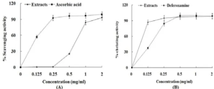
1. Antioxidant activities of PL extract
Antioxidant activities of ethyl acetate and aqueous fractions were evaluated by hydroxyl radical scavenging assay and Fe
2+chelating assay. In hydroxyl radical scavenging assay (Fig. 1A), aqueous fraction removed hydroxyl radical by 26% at 0.5 mg/ml, 84% at 1 mg/ml and 93% at 2 mg/ml. Ascorbic acid used as the positive control removed it by 57% at 0.125 mg/ml, 93% at 0.25 mg/ml, 97% at 0.5 mg/ml and 1 mg/ml, and 100% at 2 mg/ml. Although hydroxyl radical scavenging activity of the extract was lower than that of ascorbic acid, it showed a strong Fe
2+chelating activity.
In Fe
2+chelating assay (Fig. 1B), aqueous fraction chelated Fe
2+ion by 87% at 0.125 mg/ml, 95% at 0.25 mg/ml, 97% at 0.5 mg/ml and 1 mg/ml and 98% at 2 mg/ml. Deferoxamine used as the positive control chelated it by 37% at 0.125 mg/ml, 84% at 0.25 mg/ml and 99% at 0.5 mg/ml~2mg/ml.
Fig. 1. Hydroxyl radical scavenging activity (A) and Fe2+ chelating activity (B) of PL extract. The absorbance values were converted to scavenging activity or chelating activity (%) and the data plotted as the mean of replicate scavenging effects or chelating effects (%) values ± 1 S.D. (n=3) against extract concentration in mg extract per ml reaction volume. Ascorbic acid and deferoxamine were used as the positive control.
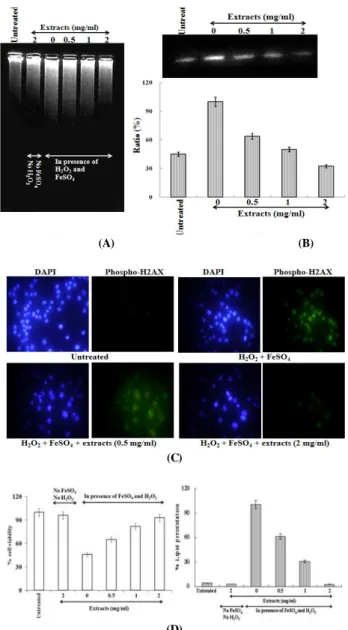
2. Protective effect of PL extract against oxidative damage in non-cellular system
The plasmid DNA cleavage assay using φX-174 RF I plasmid DNA was used as an initial approach toward determining whether aqueous fraction with antioxidant activity could inhibit oxidative DNA damage induced by hydroxyl radical or Fe
2+ion. Conversion of the supercoiled form (SC) of plasmid DNA to the open-circular form (OC) and further linear form (LC) has been used as an index of DNA damage
9).
It had no inhibitory effect on DNA damage induced by hydroxyl radical at the low concentrations (0.25 mg/ml and 0.5 mg/ml) while aqueous fraction (1 mg/ml and 2 mg/ml) inhibited DNA damage (Fig. 2A).
But, aqueous fraction (0.25 mg/ml ~ 2 mg/ml) inhibited DNA damage induced by Fe
2+ion(Fig.2B).
For the elucidation of the main mechanism for
inhibitory effect on DNA damage at the low
concentration (0.5 mg/ml) (Fig. 2C), the treatments
were divided into three groups. In the first group,
water extract was reacted with hydroxyl radical and
DNA together for 30 min at 37
oC (lane c). In the
second group, it was firstly reacted with Fe
2+ion for
10 min at the room temperature and then re-reacted
with H
2O
2and DNA together for 30 min at 37
oC
(lane d). And in the third group, it was firstly reacted
with H
2O
2at 10 min at the room temperature and
then re-reacted with Fe
2+ion and DNA together for
30 min at 37
oC (lane e). In this result, the
simultaneous reaction of the extract with hydroxyl
radical (lane c) and the pre-reaction with H
2O
2(lane
e) had no inhibitory effect on DNA damage, while the
pre-reaction with Fe
2+ion inhibited the oxidative DNA
damage by 90% (lane d). This result suggests that the
extract may protect DNA from the oxidative damage
by inhibiting the generation of hydroxyl radical
through the blocking of Fenton reaction via Fe
2+ion
chelation.
3. Protective effect of PL extract against oxidative damage in cellular system
The inhibitory effect of the extract against oxidative DNA damage was evaluated by intracellular DNA migration and the expression level of phospho-H2A.X in the cellular system. DNA migration assay is a sensitive biomarker of the DNA damage. The extract inhibited DNA migration induced by hydroxyl radical in a dose-dependent manner (Fig. 3A). In the expression level of phospho-H2A.X, a sensitive marker for breaks of double stranded DNA
11), the treatment of the extract in presence of H
2O
2and FeSO
4reduced the expression of phospho-H2A.X by 32% at 0.5 mg/ml, 53% at 1 mg/ml and 72% at 2 mg/ml, compared with the cells treated with H
2O
2and FeSO
4in absence of the extract (Fig. 3B). This result was confirmed by changes of phospho-H2A.X foci into the nuclear in immuno-staining (Fig. 3C). Therefore, the results of the present study indicate that PL can inhibit the oxidative DNA damage induced by excessive ROS. Protective effect of the extract against oxidative cell damage was evaluated by MTT assay and lipid peroxidation assay. In MTT assay (Fig. 3D), hydroxyl radical generated from Fenton reaction between H
2O
2and FeSO
4induced the oxidative cell death by 54%, compared with the untreated cells.
However, the treatment of water extract in presence of H
2O
2and FeSO
4reduced the oxidative cell death, compared with the cell treated with H
2O
2and FeSO
4in absence of the extract. It is thought that this result correlates with the inhibitory effect of the extract against lipid peroxidation. In the lipid peroxidation assay (Fig. 3E), the extract inhibited the lipid peroxidation by 39% at 0.5 mg/ml, 69% at 1 mg/ml and 97% at 2 mg/ml compared with the cells treated with H
2O
2and FeSO
4. From this result, it is thought that the extract from PL displays a significant protective capability against the oxidative cell damage.
Fig. 2. Protective effect of PL extract against oxidative DNA damage in non-cellular system. (A) treatment of hydroxyl radical
in presence of aqueous fraction; (B) treatment of Fe2+ ion in presence of aqueous fraction; (C) Lane a was the normal DNA without hydroxyl radical and aqueous fraction. Lane b was treated with hydroxyl radical alone without the aqueous fraction. In the lane c, aqueous fraction was reacted with hydroxyl radical and DNA together for 30 min at 37 oC. In the lane d, aqueous fraction was firstly reacted with Fe2+ ion for 10 min at the room temperature, and then re-reacted with H2O2 and DNA together for 30 min at 37 oC. In the lane e, aqueous fraction was firstly reacted with H2O2 at 10 min at the room temperature, and then re-reacted with Fe2+ ion and DNA together for 30 min at 37 oC.
In this assay, aqueous fraction was treated with 0.5 mg/ml. In this assay, Hydroxyl radical was generated from Fenton reaction between H2O2 and FeSO4. Iron was generated from FeCl2.
(A) (B)
(C)
(D)
Fig. 3. Protective effect of PL extract against oxidative DNA and cell damage in the cellular system. The protective effect was evaluated by intracellular DNA migration assay (A), western blot for the level of phospho-H2AX expression (B), immuno-staining for the formation of phospho-H2AX foci (C), MTT assay (D) and lipid peroxidation assay (E). In western blot, each well was loaded with 25 ㎍ protein. % ratio of the expression level of phospho-H2A.X was calculated by the density using the software Un-SCAN-IT gel Version 5.1 (Silk Scientific, Inc.). In MTT assay and lipid peroxidation assay, the absorbance values were converted to cell viability (%) and inhibitory effects (%), and the data plotted as the mean of replicate inhibitory effects (%) values
± 1 S.D. (n=3).