파라-쿠마린산의 자외선B 차단 효과
송 교 선*⋅부 용 출*,**,†
*경북대학교 의학전문대학원 분자의학교실 세포기질연구소, **(주)루비크라운 (2012년 5월 23일 접수, 2012년 8월 14일 수정, 2012년 8월 22일 채택)
UVB-Shielding Effects of para-Coumaric Acid
Kyosun Song* and Yong Chool Boo*,**,†
*Department of Molecular Medicine and Cell and Matrix Research Institute, Kyungpook National University School of Medicine, Daegu 700-422, Korea.
**Ruby Crown Co. Ltd.
(Received May 23, 2012; Revised August 14, 2012; Accepted August 22, 2012)
요 약: 최근 연구에서 잠재적 피부 색소침착 경감제로서 파라-쿠마린산(PCA)의 주목되는 특성이 발견되었다. 본 연구의 목적은 이 물질의 자외선 차단 효과를 탐구하는 것이다. 자외선에 노출된 HaCaT 세포의 생존율에 대한 PCA의 영향을
in vitro에서 조사하고, 자외선 흡수 스펙트럼이 유사한 방향성 아미노산 대사물들의 작용과 비교하였다.
In vivo시험으 로는 PCA 크림(1.5 %)과 크림 베이스를 SKH-1 무모 쥐의 등 피부에 도포하고 UVB에 의한 염증 반응으로 나타나는 피부색(홍반) 및 두께 변화(부종)를 측정하였다. 크림 도포-자외선 조사는 2일 간격으로 총 12회 반복하였다. HaCaT 세포를 UVB에 노출시켰을 때 광량 의존적으로 세포 생존율이 감소하였다. 자외선 노출(10 mJ cm
-2)에 의한 세포 생존율 감소는 100 µM의 PCA, cinnamic acid, urocanic acid, 그리고 indole acrylic acid에 의해 각각 39, 27, 39, 31 %가 억제되 었다. 무모 쥐의 등 부위에 도포된 PCA크림(10 µg cm
-2) 은 자외선(150 mJ cm
-2)- 노출 피부의 색 지수, 즉 L*, a* 및 b* 값, 그리고 두께의 변화를 각각 59, 50, 58, 53 %씩 억제하였다. 본 연구의 결과는 PCA의 멜라닌 생성 억제 작용을 밝힌 선행 연구와 함께 PCA가 자외선에 노출된 피부의 색소 이상 침착과 염증 반응을 막아줄 수 있음을 시사하였다.
Abstract: Recent studies have uncovered attractive properties of para-coumaric acid (PCA) as a potential skin hy- whitening agent. The purpose of the current study was to examine its UVB-shielding effects. Effects of PCA on the viability of HaCaT cells exposed to UVB were assessed
in vitroin comparison with other aromatic amino acid metabolites that have similar UV absorption spectra. For
in vivotest, PCA cream (1.5 %) and cream base were topically applied to the dorsal skin of SKH-1 hairless mice and the inflammatory responses due to UVB exposure were monitored by changes in skin color (erythema) and thickness (edema). The cream application-UVB exposure regimen was repeated every other day for a total of 12 sessions. When HaCaT cells were irradiated with UVB, there was a dose-dependent decline in cell viability. The cell viability decline due to UVB exposure (10 mJ cm
-2) was sig- nificantly prevented by 100 µM PCA, cinnamic acid, urocanic acid, or indole acrylic acid by 39, 27, 39, or 31 %, respectively. Topical application of PCA cream onto the dorsal skin of hairless mice (10 µg cm
-2) attenuated the changes of color parameters, L*, a*, b* values, and thickness of the UVB (150 mJ cm
-2)-exposed skin by 59, 50, 58, and 53 %, respectively. The current study, together with the previous studies that demonstrated the anti- melanogenic effects of PCA, suggested that PCA may prevent not only dyspigmentation but also inflammatory re- actions in the UVB-exposed skin.
Keywords: para-coumaric acid, ultraviolet radiation, keratinocytes viability, hairless mouse, inflammatory response 1)
† 주 저자 (e-mail: [email protected])
1. Introduction
Even though melanin is essential for photoprotection of the skin, abnormal melanin accumulation due to UV or inflammation can cause an aesthetic problem observed in the cases of melasma, freckles, and senile lentigines [1,2]. Thus, various pharmacological agents have been developed to control unwanted skin pigmentation [3-5], but their in vivo efficacies remain controversial [6].
Nonetheless, considering the essential role of melanin in the protection against UV-induced skin damage, the ideal skin hypopigmenting agents should not only inhibit melanin synthesis / accumulation but also provide UV-shielding effects. In this regard, phenolic compounds of the plants are attractive for their UV-shielding as well as antimelanogenic properties.
Trans-para-coumaric acid (PCA) is a common secondary metabolite in plants. It has a potent antioxidant activity, which has been demonstrated in cultured cells [7] and animal models [8,9]. Recent studies including those of our laboratory have revealed attractive properties of PCA as a potential hypopigmenting agent. It is a specific and potent inhibitor of human tyrosinase [10,11] and its antimelanogenic effects have been verified in murine melanoma cells [10,12], human epidermal melanocytes [10,13], 3-dimensional human skin equivalents [14], animal skins [15] and human skins [16]. There have been preliminary observations suggesting that PCA may be useful as a natural UVB-shielding agent. For example, PCA has been observed to mitigate the cytotoxicity of UVB in human epidermal melanocytes [13]. Long-term pre-application of PCA in the form of cream has also been observed to attenuate erythema induction in animal and human skin exposed to UV [15,16]. However, UVB-shielding effect of PCA has been relatively less investigated compared to its antimelanogenic effect. Thus, the present study was aimed at the assessment of the UVB-shielding effect of PCA in vitro in HaCaT human keratinocytes and in vivo in SKH-1 hairless mice.
PCA, trans-cinnamic acid (CA), trans-urocanic acid (UCA) and trans-indole acrylic acid (IAA) are known to be formed from L-tyrosine, L-phenylalanine, L-histidine,
and L-tryptophan respectively. These reactions are catalyzed by L-tyrosine ammonia lyase, L-phenylalanine ammonia lyase, L-histidine ammonia lyase (also called histidase) and L-tryptophan ammonia lyase that are distributed in prokaryotes, plants or animals [17,18]. Of these compounds, UCA has attracted an attention in dermatology and cosmetics fields as a natural sunscreen since it was identified as the major acid-soluble, UV- absorbing compound in the stratum corneum [19,20].
The photon energy absorbed by UCA is effectively dissipated through the reversible isomerization to cis-urocanic acid [21,22]. However, UCA's reputation as an ideal natural sunscreen was shattered by the observations that cis-urocanic acid mediated the immunosuppressive effect of UV [23], and UCA increased the photocarcinogenic risk in hairless mice [24]. In contrast, Barresi and colleagues observed that histidase-deficient mice were more susceptible to UV-induced DNA damage and apoptosis than wild type mice [25]. Thus it remains inconclusive whether topically applied UCA has a beneficial or detrimental role in the skin exposed to UV radiation [26].
The present study addressed the specific issue of UVB-shielding by PCA. The effects of PCA on UVB-induced cytotoxicity were assessed in vitro in HaCaT human keratinocytes. Other aromatic acid metabolites including CA, UCA and IAA were also tested for comparative purposes. The UVB-protective effect of PCA formulated in the form of cream was also examined in vivo in SKH-1 hairless mice exposed to UVB. The present study demonstrated that PCA may be useful for topical applications to mitigate the harmful effects of UVB.
2. Materials and Methods
2.1. Materials
PCA, CA and IAA were purchased from Sigma-Aldrich (St. Louis, MO, USA). UCA was purchased from Santa Cruz Biotechnology (Santa Cruz, CA, USA).
2.2. UV spectrophotometry
Test materials were dissolved in phosphate buffered
saline (PBS) so that the final concentration was 10, 30, and 100 µM. The transmittance spectra were recorded in the 200 ~ 400 nm range using a Shimadzu UV-1650PC spectrophotometer (Shimadzu Corporation, Kyoto, Japan).
2.3. In vitro Experiments
2.3.1. Cell Culture
HaCaT cells were grown in DMEM/F-12 medium (GIBCO-BRL, Grand Island, NY, USA) supplemented with a 10 % fetal bovine serum, 100 U mL
-1penicillin, 0.1 mg mL
-1streptomycin, 0.25 µg mL
-1amphotericin B, and 10 mg mL
-1hydrocortisone. The cells were cultured at 37 ℃ in a humidified atmosphere containing 5 % CO
2and 95 % air.
2.3.2. UVB Irradiation and Cell Viability Test
Cells were seeded on a six-well plate at a density of 2 × 10
5cells per well and grown in the culture medium for 48 h to reach 80 % confluency. The cells were washed twice with PBS and exposed to UVB radiation in PBS that contained the test material. The UVB treatment was conducted in a cell culture hood using a UVB lamp (Model UVB-18, ULTRA*LUM. Inc., Claremont, CA, USA) that emits radiation in the range of wavelengths from 280 to 340 nm, with the maximum emission wavelength of 300 nm. The intensity of radiation was determined using UV Light meter (Model UV 340A, Lutron Electronic Enterprise Co. Taipei, Taiwan). The UV intensity at the culture plate position was set at 80 µW cm
-2and the duration of irradiation was changed to provide the specified doses of UV radiation (5, 10, and 15 mJ cm
-2). After irradiation, cells were fed with the growth medium and incubated for 2 days. Cell viability was assayed using 3-[4,5-dimethylthiazol-2-yl]-2,5-diphenyltetrazolium bromide (MTT) [7,27]. Percent inhibition of a test sample against the UVB-induced viability loss was calculated as (C - B) / (B - A) × 100 where A is the viability of the non-irradiated vehicle control group, and B and C are the viabilities of the UVB-irradiated test (C) and vehicle control (B) groups.
2.4. Preparation of PCA Cream
The cream containing PCA as the active ingredient was manufactured by Dasso & Company Co. (Inchon, Korea) [15]. The cream base had the following volumetric composition : moisturizer & emollient 18 %, antioxidant 0.1 %, viscosity increasing agent 3 %, wax 2.5 %, emulsifying agent 5 %, preservative 0.5 %, and water 60.9 %. PCA was dissolved in propylene glycol and incorporated into a cream so that the final content in the cream was 1.5 % (w/v). The molar concentration of the PCA in the PCA cream was 91 mM. The transmittance spectra of the PCA cream and cream base diluted in ethanol were recorded in the 200 ~ 400 nm range as above.
2.5. Construction of a Mouse-housing Device for UVB Irradiation of Dorsal Skin Sites
This device was designed for convenient and accurate UVB irradiation on the dorsal skin areas of the mice without anesthesia usage. The device was constructed with Plexiglas (3 mm thick), by putting together one roof board, one ground board, and 6 lateral boards, hence allowing simultaneous housing of five mice. For UVB exposure of the mice dorsal skin areas, 5 windows (20 mm × 20 mm) were drilled through the roof board.
Three small slits (20 mm × 4 mm) on the animal head space and 2 slits on the tail space of each block were also drilled through the roof board, and inserted with black barriers (12 mm × 35 mm) for the immobile positioning of the mice at a specific position. The walls and roof were made black so that UVB could be irradiated only to the designated sites.
2.6. In vivo Experiments
2.6.1. Animals
Animal experiments were performed in accordance
with the guideline of Kyungpook National University,
intramural Animal Use and care Committee. Five-
week-old female SKH-1 hairless mice were purchased
from Orient Bio, INC. (Gyeonggido, Korea). The animals
were maintained under controlled environmental
conditions (23 ± 1 ℃, 55 ± 5 % humidity, 12 h light/dark
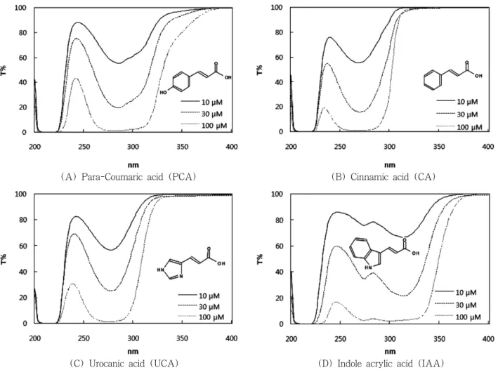
(A) Para-Coumaric acid (PCA) (B) Cinnamic acid (CA)
(C) Urocanic acid (UCA) (D) Indole acrylic acid (IAA)
Figure 1. Transmittance spectra of PCA, CA, UCA, and IAA. The chemical structures of these compounds are also shown.
cycle) with free access to water and an ad libitum standard laboratory diet (Superfeed Co, Wonju, Kangwon, Korea).
2.6.2. UVB Irradiation of SKH-1 Hairless Mice
After an acclimation period, mice were randomly divided into 3 groups of 10 ∼ 11 animals. Cream base or PCA cream (100 µL) was topically applied to a designated site (32 mm × 32 mm) on the dorsal skin of the animal, 30 min before every exposure to UVB. Mice were irradiated with a UVB lamp (Model G8T5E UVB lamp, Sankyo Denki Co., Hiratsuka, Kanagawa, Japan). This lamp emits UV radiation in the range of wavelengths from 280 to 360 nm, with the maximum wavelength of 306 nm. Mice were irradiated at 0.54 mW cm
-2for 277 sec to provide 150 mJ cm
-2of
UVB radiation which corresponds 3 × minimal ery- thema dose (MED) for the hairless mouse. In this step, mice were kept in a home-made housing device which was placed at a specific distance from the UVB source.
The roof windows of the device allowed reproducible irradiation of the dorsal skin areas (20 mm × 20 mm) with downward UVB radiation from a lamp. The cream application-UVB exposure was repeated every other day for a total of 12 sessions.
2.6.3. Measurement of Skin Color and Thickness
The skin color and thickness as indices of inflammation
were measured before the cream application-UVB
exposure protocol every other day. The color of the skin
sites was measured using a spectrophotometer CR-10
(Konica Minolta Sensing, Inc., Sakai, Osaka, Japan) in
(A)
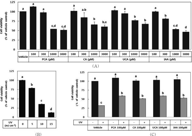
(B) (C)
Figure 2. Effects of PCA, CA, UCA, and IAA on the viability of HaCaT cells exposed to UVB. Cells were incubated for 48 h in a growth medium that contained a test material at the indicated concentration (A). Cells were exposed to UVB at the indicated dose in PBS (B). Cells were irradiated with 10 mJ cm-2 UVB in PBS that contained 100 µM of a test material or vehicle only (C). Cell viability was assayed using MTT. Data are presented as % of vehicle control without UVB irradiation (Means ± SEM, n = 3). Data marked not sharing the same letters (a, b, c, or d on the bars) are statistically different from each other (p < 0.05). For example data marked with b, c are not statistically different from those marked with b or c, but different from those marked with a or d.
which the colors are described by L* (lightness), a*
(red to green ratio), and b* values (blue to yellow ratio) according to the Commission International de l’Eclairage color system. The double skin fold thickness was measured along the midline of a uniform dorsal skin sites using a digital caliper (Mitutoyo Co., Hiratsuka, Kanagawa, Japan). Percent inhibition of PCA cream against the UVB-induced changes of skin parameters was calculated as (C - B) / (B - A) × 100 where A is the value for the non-irradiated cream base group, and B and C are the values for the UVB-irradiated PCA cream (C) and cream base (B) groups.
2.7. Statistical Analysis
Data were presented as the means ± SEM of three
or more independent experiments. Significant differences among the groups were determined by one-way ANOVA. Duncan’s multiple-range test was done if differences were identified between the groups at p <
0.05.
3. Results
In the first set of experiments, the UVB-shielding
effects of PCA were assessed in vitro in HaCaT cells,
a spontaneously immortalized cell line of human
keratinocytes. Other aromatic acid metabolites including
CA, UCA and IAA were also tested for comparative
purposes. The transmittance spectra of PCA, CA, UCA
and IAA at 10, 30 and 100 µM were shown in Figure 1.
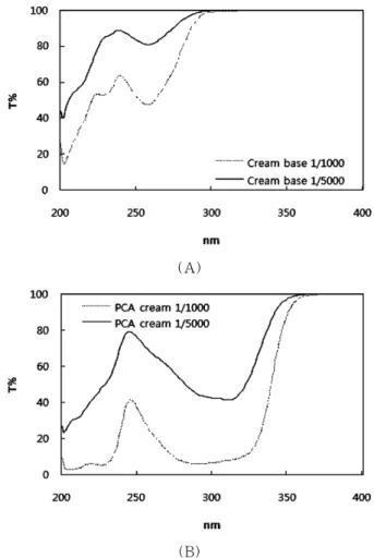
(A)
(B)
Figure 3. Transmittance spectra of PCA cream and cream base used in this study. The transmittance spectra of cream base (A) and PCA cream (B) diluted in ethanol were recorded in the 200 ∼ 400 nm range.
(A)
(B)
Figure 4. A mouse-housing device used in this study.
The housing device has roof windows for the irradiation of dorsal skin sites of the mice (A). The device with mice inside is also shown (B).
All these compounds were observed to have similar spectral properties, implicating their potential utility as UV-protective agents. CA and UCA had very similar spectral properties, and PCA and IAA appeared to shield a little wider range of UVB.
The potential UVB-protective effects of PCA, CA, UCA and IAA were examined using HaCaT cells. All these compounds appeared to be non-toxic to HaCaT cells up to 100 µM (Figure 2(A)). At 300 µM, the cytotoxicity was on the order of IAA > CA > UCA = PCA. The effect of UVB irradiation on cell viability was then examined. As expected, there was a gradual decline in cell viability with increasing doses of UVB from 5 to 15 mJ cm
-2(Figure 2(B)). Cells were irradiated with UVB (10 mJ cm
-2) in the absence and
presence of a test compound (100 µM) to compare the cell protective effects of PCA, CA, UCA and IAA. As shown in Figure 2C, At 100 µM, PCA, CA, UCA, and IAA attenuated the UVB (10 mJ cm
-2)-induced viability loss of HaCaT cells by 39, 27, 39, and 31 %, respectively. PCA and UCA were a little more protective compared to CA and IAA although the difference was not statistically significant.
For the in vivo assessment of the skin protective effects of PCA, a cream formulation containing 1.5 % PCA and cream base were prepared. The PCA cream had strong UVB absorption compared to cream base (Figure 3). HPLC profiles of the PCA cream and cream base has been shown previously [15].
The UVB-shielding effects of the PCA cream was
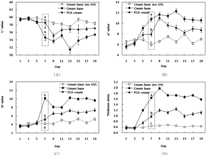
(A) (B)
(C) (D)
Figure 5. Effects of PCA cream on the inflammatory responses in mouse skins exposed to UVB. SKH-1 hairless mice were treated with 100 µL of cream base or PCA cream on a designated dorsal skin sites, 30 min before UVB irradiation at 150 mJ cm-2. The cream application-UVB exposure session was repeated every other day, starting from day 3. Skin color and thickness were measured before cream application. Color changes of the irradiated skin sites are represented by L* (lightness, (A)), a*(red to green ratio, (B)), and b* values (blue to yellow ratio, (C)). Edema is represented by skin thickness of the irradiated dorsal skin (D). Data represent the Means ± SEM (n = 10 ~ 11).
The data from day 7 (dotted box) were analyzed for statistical significance. Data marked with different letters (a, b, or c) are statistically different from each other (p < 0.05). For example, data marked with b are not statistically different from those marked with b, but different from those marked with a or c.
assessed in comparison with the cream base in mice exposed to UVB repetitively. During UVB exposure, mice were housed in a device specially designed for this purpose (Figure 4). Control mice were treated with the cream base and entered the device, but not irradiated with UVB. The inflammatory response of the mouse skin was assessed by changes in the skin color and thickness. As shown in Figure 5(A)∼(C), repetitive UVB irradiation altered the mouse skin color parameters
monitored with a spectrometer. The L* value decreased while a* and b* values increased due to exposure to UVB irradiation. UVB irradiation also increased dorsal skin thickness (Figure 5(D)). Compared to the cream base, the PCA cream attenuated the changes in the skin color parameters, L*, a*, b* values, and thickness of the UVB-exposed skin by 59, 50, 58, and 53 %, respectively (Figure 5).
Typical skin images of the non-irradiated control
(A) Cream base (no UV) (B) Cream base + UVB (C) PCA cream + UVB Figure 6. Protective effect of PCA cream against the UVB-induced skin injury in mice. SKH-1 hairless mice were treated as in Figure 5. Pictures of dorsal skin sites of 5 representative mice were taken on day 7.