유해물질 노출로 인한 분자·생화학적 바이오마커와 담수 어류에 대한 현장 적용성
김정곤·박예나·김우근*·김지원*·이성규*·최경호† 서울대학교 보건대학원, *안전성평가연구소 환경독성연구센터
(2010. 9. 6. 접수/2010. 10. 1. 수정/2010. 10. 25. 채택)
Molecular/biochemical Biomarkers for Exposure to Hazardous Chemicals in the Water Environment and their
Application to Freshwater Fish
Jungkon Kim · Yena Park · Woo-Keun Kim* · Jiwon Kim* · Sung-Kyu Lee* · Kyungho Choi†
School of Public Health, Seoul National University, Seoul, Korea
*Ecotoxicology Research Center, Korea Institute of Toxicology, Daejeon, Korea (Received September 6, 2010/Revised October 1, 2010/Accepted October 25, 2010)
ABSTRACT
As concerns regarding water pollution grow, the need increases for a fast and accurate assessment of ecological risk.
In this context, many studies have been conducted to identify biomarkers which can sensitively indicate exposure to and effects of various contaminants in a water environment. However, the utility of most such biomarkers in the real water environment is not yet validated. In this paper, we conducted a thorough review of publications that were related to developing or evaluating molecular and biochemical biomarkers of freshwater fish in ecological risk assessment, and evaluated whether these biomarkers of interest could link to the effects on higher biological levels, such as his- topathology and above. Biomarkers of interest included those associated with metabolism, oxidative stress, reproduction and endocrine disruption, genotoxicity, and defense against heavy metal exposure. We found that, when used alone, most molecular and biochemical biomarkers are not sufficient to understand the effects of toxic substances in higher biological levels, due to defense or acclimation mechanisms of organisms. Moreover, some biomarkers respond not only to hazardous substances but also to the changes in water quality and disease outbreak. Molecular and biochemical biom- arkers may be most useful in understanding the potential biological effects of toxic compounds when used in parallel with relevant endpoints of higher biological levels.
Keywords: aquatic ecosystem, risk, metabolic enzyme, oxidative stress, endocrine disruption, genotoxicity, metallothionein
I. 서 론
인간활동으로 인하여 다양한 유해화학물질이 물환경 으로 유입되고 있다. 그러나 이들 유해화학물질의 노출 로 인해 초래되는 생태계 영향의 기전과 심각성에 대 해서 정확히 파악하는 것은 쉽지 않다. 환경오염물질의 영향을 평가하기 위해서 많은 연구가 수행되어 왔으나 대부분의 연구가 실험실 내에서 이루어져 현장에서의
상황을 적절히 반영하지 못하는 경우가 많다. 게다가 유해인자 노출로 인한 영향을 분절적으로 평가하는 경 우가 대부분이어서 실제의 생태계 영향을 정확히 이해 하기가 어려운 실정이다.
실제 환경에서 나타나는 생태계 영향은 대부분 오랜 기간 동안 지속적으로 발생되었던 노출의 결과로 초래 되는 것이며, 일단 발생한 생태계 영향을 되돌려 회복 시키는 것이 극히 어려운 경우가 많다. 이러한 사태의 발생을 미연에 방지하기 위해서는 개체, 개체종, 또는 군집 수준의 부정적인 영향이 나타나기 전에 유해인자 의 생물학적 영향을 조기에 감지할 수 있는 바이오마 커를 활용하는 것이 필요하다. 독성학 분야에서 바이오
†
Corresponding author : School of Public Health, Seoul National University
Tel: 82-2-880-2738, Fax: 82-2-745-9104
E-mail : [email protected]
마커는 외인성물질의 노출, 이로 인한 생물체의 영향, 그리고 생물체의 감수성을 나타내는, 다양한 수준에서 관찰 또는 측정 가능한 지표라고 정의될 수 있다.1-4)
일반적으로 개체와 같은 상위 단계에서의 영향은 그 아래의 생물학적 수준, 예를 들면 분자·생화학적, 세 포 또는 조직수준에서 일어나는 변화를 거친 다음에야 발생한다. 하위 단계의 생물학적 변화는 상위단계에서 의 변화에 비해 훨씬 더 신속하면서도 독성영향의 생 물학적 기전을 설명하는데 용이하다는 장점이 있다. 그 러므로 바이오마커를 적절히 이용하면 개체군 혹은 생 태계 수준의 영향을 예측하는 조기경보지표로 활용할 수 있다.5,6)
그러나 이러한 바이오마커를 이용한 대부분의 연구는 특정한 생물학적 수준에 국한되어 분절적으로 이루어 져 왔다. 또한, 이들 연구에서 관찰된 바이오마커의 반 응이 실제로 생물 개체에 어떤 의미를 갖고 있는 것인 지에 대해 충분히 이해하기가 어렵다는 단점이 있다.
분자·생화학적 바이오마커를 적절한 조기경보 바이오 마커로 활용하면 물환경으로 유입되는 유해물질의 효 과적인 관리에 도움이 될 것으로 판단된다. 따라서 실 제 환경에서의 생태계 영향을 이해하기 위해 분자·생 화학적 바이오마커의 반응을 활용하는 것이 적절한지 파악할 필요가 있다.
물환경에 유해물질의 노출과 영향을 지시하는 바이오 마커는 매우 다양한데, 그 가운데서도 대사관련 효소 (대사효소), 산화적인 스트레스 요인(항산화효소, 지질과 산화, DNA 손상 등), 스트레스 단백질(heat-shock protein), 중금속 노출 인자(metallothionein, MT), 혈액 학적 요인(혈구용적, 헤모글로빈, 단백질, 당 등), 생식 및 내분비계 요인(vitellogenin, VTG), 신경학적 요인 (acetylcholinesterase, AChE), 유전독성학적 요인, 생리 학적 그리고 형태학적 요인 등이 대표적이다. 이러한 바이오마커는 물환경 유해인자의 독성영향 기전에 대 한 정보를 제공하고 잠재적인 생태계 영향을 제시한다 는 측면에서 활용도가 크다.
이 연구는 지금까지 생태독성학 및 생태위해성평가 분야에서 개발되어 활용되어 온 주요 분자·생화학적 수준의 바이오마커가 생태위해성관리 측면에서 어떤 효 용을 갖는지 평가하기 위한 목적으로 수행되었다. 이를 위해 생태독성학 연구에서 활용되는 주요 바이오마커 의 종류와 기능에 대해서 요약하고, 이 바이오마커를 현장에서 포획된 담수어종에 적용하여 수행된 여러 가 지 분자·생화학적 수준의 바이오마커의 분석 사례들을 검토하였다. 이를 통하여 이러한 바이오마커가 생태위 해성 평가를 위해 적절히 활용될 수 있는지 알아보았다.
II. 연구방법
생태독성학 및 생태위해성평가 분야에서 활용되는 바 이오마커에 대한 보고를 탐색하기 위하여 상용 데이터 베이스를 활용하였다. 학술지 데이터베이스 “ISI Web of Science”를 이용하여 오염 현장과 대조지역의 담수 어류를 대상으로 분자·생화학적 바이오마커 혹은 이 것과 조직병리학적 수준 이상의 지표를 병행하여 조 사한 사례를 검색하였다(2010년 5월 검색). “fish”,
“biomarker” 및 “field”의 검색어를 사용하여 검색한 결 과 모두 198 건의 논문을 파악할 수 있었다. 이 가운 데 현장에서 포획한 담수어류를 대상으로 분자·생화학 적 바이오마커 혹은 조직 수준 이상의 지표를 동시에 분석한 사례들을 선별하여 주요 분자·생화학적 바이오 마커 중심으로 조사하였다.
III. 연구결과
1. 대사효소
대사와 관련된 여러 가지 효소 중에서 바이오마커로 가장 많이 활용되어 온 대사효소는 phase I 대사를 담 당하는 cytochrome P450 효소(CYP)이다. CYP은 모 든 생명계에서 발견될 수 있는 헴단백질(hemoprotein) 로 27종의 다양한 superfamily를 포함하고 있다. CYP 에 의해 일어나는 가장 일반적인 반응은 기질에 1개의 산소를 첨가함으로써 기질의 수용성을 증가시키는 것 이다. 이를 mixed function oxygenase (MFO) system 이라고도 하며 거의 모든 동물과 식물에 존재하며 내 인성 물질일 뿐 아니라 외인성 유독물질의 대사에 관 여하는 효소계이다.
CYP은 오염물질 노출에 민감하게 반응하는 경우가 많아서 환경중 오염물질의 노출평가와 유해성평가에 널 리 사용되는 바이오마커 중의 하나이다. 일반적으로 오 염물질에 노출되면 CYP가 유도되고 이에 따라 오염물 질의 대사가 촉진되어 궁극적으로 체외 배출이 용이하 게 된다. 다이옥신과 polychlorinated biphenyls(PCBs) 등의 잔류성 유기오염물질과 β-naphthalene과 같은 방 향족 탄화수소류(polycyclic aromatic hydrocarbons, PAHs) 화합물은 CYP1A를 유도하는 대표적 물질로 알 려져 있다. CYP 유도를 저해하는 물질도 다양하다.
현장 환경 시료에서 CYP은 주로 단백질 발현량이나 효소의 활성도(arylhydrocarbon hydroxylase[AHH], ethyoxyresorufin O-deethylase[EROD]) 등을 측정하는 방법으로 평가된다(Table 1). CYP의 수준은 대개 유기 오염물질의 오염도에 따라 증가하는 경향을 보인다. 특
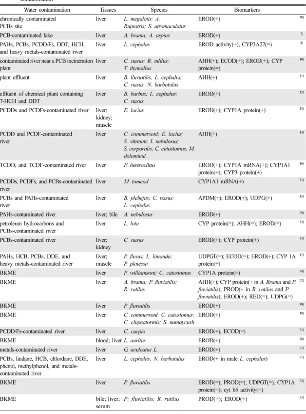
Table 1. Summary of studies which applied metabolic enzymes of freshwater fish as biomarkers to assess the effects of water contamination
Water contamination Tissues Species Biomarkers
chronically contaminated PCBs site
liver L. megalotis; A.
Rupestris; S. atromaculatus
EROD(+) 70)
PCB-contaminated lake liver A. brama; A. aspius EROD(+) 7)
PAHs, PCBs, PCDD/Fs, DDT, HCH, and heavy metals-contaminated river
liver L. cephalus EROD activity(+); CYP3A27(=) 8)
contaminated river near a PCB incineration plant
liver C. nasus; R. mlilus;
T. thymallus
AHH(+); ECOD(+); EROD(+); CYP protein(=)
10)
plant effluent liver B. lluviatilis; L. cephalvs;
C. nasus; N. barbatulus
AHH(+) 11)
effluent of chemical plant containing 7-HCH and DDT
liver B. barbus; L. cephalus;
C. nasus
EROD(+) 12)
PCDDs and PCDFs-contaminated river liver;
kidney;
muscle
E. lucius EROD(+); CYP1A protein(+) 13)
PCDD and PCDF-contaminated river
liver C. commersoni; E. lucius;
S. vitreum; I. nebulosus;
S. corporalis; C. cutostomus; M.
dolomieui
AHH(+) 14)
TCDD, and TCDF-contaminated river liver F. heteroclitus EROD(+); CYP1A mRNA(+); CYP1A1 protein(+); CYP3 protein(+)
16)
PCDDs, PCDFs, and PCBs-contaminated river
liver M. tomcod CYP1A1 mRNA(+) 71)
PCBs and PAHs-contaminated river
liver B. plebejus; C. nasus;
L. cephalus
APDM(+); EROD(+); UDPG(+) 15)
PAHs-contaminated river liver; bile A. nebulosus EROD(+) 29)
petroleum hydrocarbons and PCBs-contaminated river
liver L. lota CYP protein(=); AHH(=); EROD(=) 72)
PCBs-contaminated river liver;
kidney
C. nasus EROD(+); CYP protein(+) 73)
PAHs, HCB, PCBs, DDE, and heavy metals-contaminated river
liver;
muscle
P. flesus; L. limanda;
P. platessa
UDPGT(=); ECOD(=); EROD(+); CYP 1A protein(+)
17)
BKME liver P. williamsoni; C. catostomus CYP1A protein(=) 74)
BKME liver A. brama; P. fluviatilis;
R. rutilus
AHH(+); CYP protein(+ in A. Brama and P.
fluviatilis); PROD(+ in R. rutilus and P.
fluviatilis); EROD(+); RED(=); UDPG(=)
22)
BKME liver P. fluviatilis EROD(+) 18)
BKME liver C. commersonl; C. catostomus;
C. clupeatormis; S. namaycush
EROD(+) 19)
PCDD/Fs-contaminated river liver C. carpio EROD(+), ECOD(=) 21)
BKME blood; liver L. aurltus EROD(+) 26)
metals-contaminated river liver G. aculeatus L. EROD(+) 27)
PCBs, lindane, HCB, chlordane, DDE, phenol, methylphenol, and metals- contaminated river
liver L. cephalus; N. barbatulus EROD(+ in male L. cephalus) 31)
BKME liver P. fluviatilis EROD(=); PROD(=); UDPGT(=); CYP1A
protein(=); cyt b5 activity(=)
32)
BKME bile; liver;
serum
P; fluviatilis; R. rutilus PROD(+); EROD(+) 33)
Table 1. Continued
Water contamination Tissues Species Biomarkers
BKME liver C. cafostomus; P. wlliamson;
L. tola
EROD(+); CYP1A protein(+) 75)
PCDD/Fs and PAHs-contaminated river liver P. oregonensis; O. clarkia;
C. carpio
EROD(+ in C. carpio); CYP protein (+ in C. carpio);CYP1A1 protein(+ in C. carpio); RED(- in C. carpio)
23)
BKME liver A. brama; P. fluviatilis;
R. rutilus
UDPGT(+); EROD(+) 25)
PCBs and PAHs-contaminated river liver A. brama p-Nitroanisole-O-demethylase(+) 28) metals, PAHs, PCBs, lindane, and DDE-
contaminated river
liver B. barbus; L. cephalus;
G. gobio
EROD(-) 30)
PAHs and heavy metals-contaminated river liver feral fish AHH(+) 47)
PCBs, PAHs, and OCPs-contaminated river
liver R. rutilus cyt b5(-); EROD(=); total CYP protein (-); CYP1A protein(=); RED(=)
62)
effluents of a coking plant containing 2,3,7,8- TCDD and PAHs
liver A. nebulosus EROD(-) 63)
oil-contaminated site liver; gill S. trutta AHH(+) 76)
pesticides-contaminated river (diuron, simazine, fenithrothion, methidathion, bromopropylate, tetradifon)
liver P. phoxinus; R. rutilus; N barbatulus; L. soufia;
G. gobio; L. cephalus
EROD(+) 77)
PAHs and PCBs-contaminated river liver P. vetulus; L. bilineata;
P. stellatus
CYP1A protein(+); AHH(+); EROD(+)78)
PCBs and PAHs-contaminated river liver; muscle L. cephalus EROD(+); CYP1A protein(+) 79) urban rivers and a paper mill liver S. trutta; P americanus EROD(+ in liver; - in muscle) 80) HCB, PCBs, and heavy
metals-contaminated river
liver B. graellsii CYP1A mRNA and protein(+);
EROD(+)
68)
complex mixture of organics (chloroform, methylene chloride, toluene, 2-butanone, o-xylene, 2-hexanone, dieldrin, 4,4’-DDE, 4,4’-DDD, PCB-1254) and trace metals (Ag, Cr, Cu, Hg, NI)
liver I. nebulosus CYP protein(-); EROD(-); cyt b5(=) 81)
PCB, TCDD, DDE, and heavy metals-contaminated river
liver C. carpio; M. salmoides;
M. dolomieui; I. furcatus
EROD activity(+) 82)
PCBs and DDE-contaminated river liver; kidney I. nebulosus UDPGT(=); EROD(+) 83)
PCBs, PAHs, OCPs, and PCDD/
Fs-contaminated river
liver A. anguilla UDPGT(+); CYP1A protein(+); CYP3A
protein(=); EROD(+); cyt b5(+); CYP RED(=); PROD(+)
84)
PCBs and Hg-contaminated river liver A. brama EROD(+); ECOD(+) 85)
oil shale processing plant with high PAHs and heavy metals (Cd, Cu, Hg, Pb) contamination
Liver; kidney;
spleen;
intestine
P. fluviatilis; R. rutilus EROD(=); CYP1A protein(=); AHH(-);
UDPGT(=)
86)
heavy metals and OCs-contaminated river liver L. cephalus; G. gobio EROD(+) 87)
PCBs, metals, chlorine, and nutrient- contaminated river
liver L. auritus EROD(+) 88)
urban, industrial or agricultural contamination sites
G. aculeatus L. EROD(+) 89)
BKME liver C. gobio L. AHH(+) 90)
-, inhibition; =, no (significant) response; +, induction; OCs, organochlorine compounds; 7-HCH, hexachlorocyclohexane; PCDDs, polychlorinated dibenzodioxins; PCDFs, polychlorinated dibenzofurans; TCDD, 2,3,7,8-tetrachlorodibenzo-p-dioxin; TCDF, 2,3,7,8- tetrachlorodibenzofuran; ECOD, 7-ethoxycoumarin O-deethylase; UDPGT, uridine diphosphate glucuronyltransferase; RED, cytochrome c- reductase; The numbers in parenthesis represent references.
히 PCBs 또는 다이옥신과 같은 잔류성유기오염물질과 bleached kraft mill effluent(BKME)가 오염된 지역에 서 포획된 담수어류에서는 대사 효소의 활성이 대조지 역에 비해 현저히 증가하였다.7-25) BKME 유출 하류지 역에서 포획된 개체의 경우, 전 개체종에서 CYP 활성 이 높게 나타났으나 해당 제지공장의 조업이 정지된 2주 후에는 CYP의 활성이 감소된 것은 CYP가 BKME 함유 오염물질의 독성을 잘 반영하는 바이오마 커임을 보여준다.19) 지금까지의 조사결과를 종합해 볼 때 CYP1A1 단백질 수준과 효소 활성도는 PAHs, PCBs, dichloro-diphenyl-trichloroethanes(DDTs) 등을 포함한 유기오염물질로 인한 담수 어류의 영향을 모 니터링하기 위한 목적으로 유용한 바이오마커로 판단 된다.17)
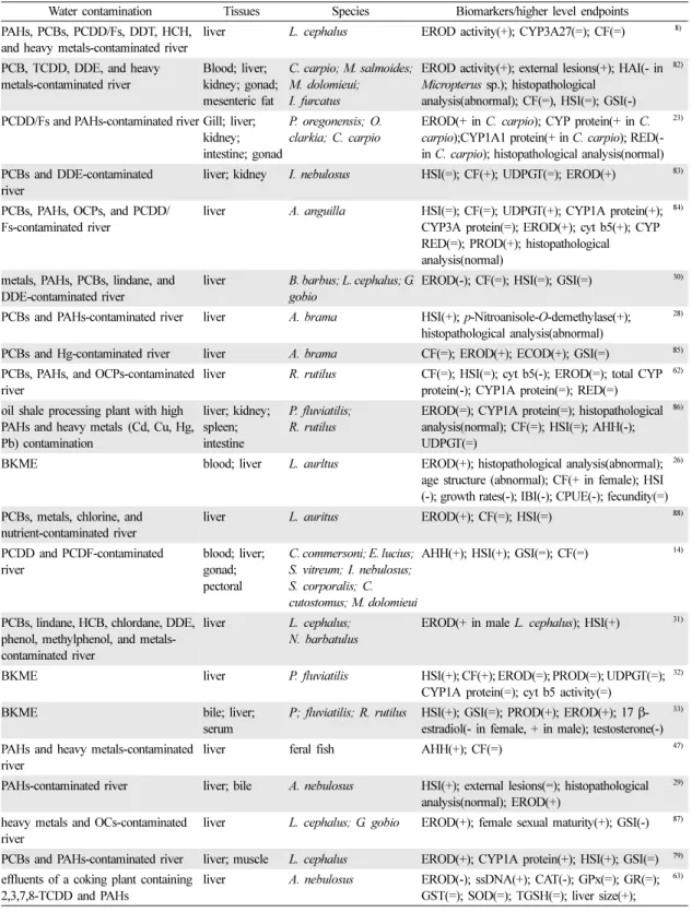
대사효소 바이오마커의 반응과 조직병리학적 수준 이상의 상위 단계 지표를 병행하여 측정한 보고사례는 총 25건이 검색되었다(Table 2). 관련 오염물질은 PCBs가 가장 많았으며 PAHs, BKME, 중금속 순으로 나타났다. 대사효소와 함께 분석된 상위단계 지표로는 hepatosomatic index(HSI), gonadosomatic index(GSI), condition factor(CF) 등의 생리학적 지표가 많았다. 이 외에도 마크로파지 응집, 성 성숙도 및 조직병리학적 분석이 병행된 사례도 있었으며, 개체 연령구조, 성장 률, 개체군 지표 그리고 군집 지표를 대사 효소와 함께 적용한 사례도 2건이 검색되었다.26,27)
PCBs와 PAHs로 오염된 하천에서 포획된 개체에서는 대조지역에 비해 HSI가 유의하게 높게 나타났다. 간의 상대 중량이 증가하였음은 유해물질 대사 및 해독을 위 한 장기 수준의 보상적 노력을 반영하는 것으로, 오염 물질의 노출로 인한 개체의 영향을 반영하는 것으로 여 겨진다.28) Arcand-Hoy와 Metcalfe29)도 PAHs로 오염된 하천의 물고기에서 HSI 및 EROD 활성 증가를 보고한 바 있다. 그러나 위의 두 사례 이외에는 염소계 화합물 과 PAHs의 노출에 의해 여러 바이오마커의 반응이 일 관성 있게 나타난 경우는 발견하기 어려웠다. Van der Oost24)는 PCBs, organochlorine pesticides(OCPs), PAHs 그리고 PCDF/Ds로 오염된 하천에서 포획된 개 체가 대조지역의 생물에 비해 CYP 단백질 수준이 높 은 것으로 보고하였으나, 대조지역과 오염지역의 개체 간의 HSI의 차이는 관찰되지 않았다. 또한 Flammarion 과 Garric30)의 연구에 의하면 PCBs와 PAHs로 오염된 하천의 개체에서 CF, HSI 및 GSI 등의 생리학적 지표 가 지역간 오염도를 비교적 잘 반영하였지만 지역에 따 른 EROD 활성의 차이는 관찰되지 않았다.
염소계 화합물질 및 PAHs의 경우와는 달리 BKME
의 노출에 의한 영향을 평가한 연구에서는 대사효소 수 준의 반응이 비교적 상위 수준 반응을 잘 반영하는 것 으로 보고된다. 대조지역에 비해 BKME가 방류되는 지 점에서 포획된 개체에서 AHH 활성이 10배 그리고 방 류지점부터 95 km 떨어진 하류에서는 5배 높았다. 또 한 BKME에 노출된 개체에서 HSI, 혈구용적, 혈청 단 백질과 당, 그리고 지느러미 비대칭 지표 등은 대사효 소 활성과 동일한 양상으로 증가하였다.14)그 외 다수 의 연구에서도 BKME에 노출된 개체에서 대사효소와 관련된 바이오마커의 수준과 HSI 및 CF 등의 생리학 적 지표가 증가하는 것으로 나타났다.31-33)이러한 사례 들을 종합해 볼 때 대사효소와 관련된 바이오마커는 BKME 오염으로 인한 상위 수준의 영향을 잘 반영하 는 지표인 것으로 보이지만, 그 이외의 오염물질에 대 해서는 상위 단계의 지표와 함께 분석했을 때 활용성 이 증가하는 것으로 판단된다.
2. 산화적 손상 관련 지표
생체 내에서 다양한 과정을 거쳐 생산된 자유라디 칼(free radical)은 단백질, 핵산, 지질 등의 생분자들 을 공격하여 분해시키거나 그 기능을 저해한다. 생체 는 자유라디칼의 악영향을 제어하기 위해 다양한 방 어 메커니즘을 가지고 있다. 자유라디칼의 한 종류인 활성산소종(reactive oxygen species: ROS)은 산소라 디칼 및 이것으로부터 파생된 산소화합물로 최외곽 전 자궤도에 쌍을 이루지 않은 전자를 가지고 있어 반응 성이 높기 때문에 DNA 손상, 단백질 불활성화, 지질 과산화(lipid peroxidation: LPO) 등의 영향을 초래한 다.34) 이에 대처하여 항산화효소는 외인물질의 생전 달 과정 혹은 세포내 대사과정에서 생성된 ROS로부 터 생체를 보호하는 역할을 한다. 대표적인 항산화효 소는 superoxide dismutase(SOD),35) catalase(CAT),36) glutathione peroxidase (GPx),37) 그리고 glutathione S-transferase(GST)34)가 있다. 항산화효소는 특정 ROS 를 기질로 반응한다. 그러므로 특정한 항산화효소의 활 성이 증가했다면 이것이 어떤 ROS의 노출 영향 때문 인지 간접적으로 추정할 수 있다.
물 환경 중에 존재하는 다양한 금속이온은 물고기 에게 산화적 손상을 일으킬 수 있는 것으로 알려져
있다.38,39)실내 노출 연구를 토대로 보면 항산화효소나
산화적 손상 관련 지표들이 다양한 환경오염물질 노출 로 인해 발생하는 산화적 손상을 잘 지시하는 것으로
보인다.40-44) Table 3은 수질오염으로 인한 담수어류의
독성영향을 평가하기 위해 산화적 손상과 관련된 분 자·생화학적 수준의 바이오마커를 적용한 사례를 제시
Table 2. Summary of studies which link biomarkers of metabolic enzymes to the observational endpoints of higher biological levels in freshwater fish
Water contamination Tissues Species Biomarkers/higher level endpoints
PAHs, PCBs, PCDD/Fs, DDT, HCH, and heavy metals-contaminated river
liver L. cephalus EROD activity(+); CYP3A27(=); CF(=) 8)
PCB, TCDD, DDE, and heavy metals-contaminated river
Blood; liver;
kidney; gonad;
mesenteric fat
C. carpio; M. salmoides;
M. dolomieui;
I. furcatus
EROD activity(+); external lesions(+); HAI(- in Micropterus sp.); histopathological
analysis(abnormal); CF(=), HSI(=); GSI(-)
82)
PCDD/Fs and PAHs-contaminated river Gill; liver;
kidney;
intestine; gonad
P. oregonensis; O.
clarkia; C. carpio
EROD(+ in C. carpio); CYP protein(+ in C.
carpio);CYP1A1 protein(+ in C. carpio); RED(- in C. carpio); histopathological analysis(normal)
23)
PCBs and DDE-contaminated river
liver; kidney I. nebulosus HSI(=); CF(+); UDPGT(=); EROD(+) 83)
PCBs, PAHs, OCPs, and PCDD/
Fs-contaminated river
liver A. anguilla HSI(=); CF(=); UDPGT(+); CYP1A protein(+);
CYP3A protein(=); EROD(+); cyt b5(+); CYP RED(=); PROD(+); histopathological analysis(normal)
84)
metals, PAHs, PCBs, lindane, and DDE-contaminated river
liver B. barbus; L. cephalus; G.
gobio
EROD(-); CF(=); HSI(=); GSI(=) 30)
PCBs and PAHs-contaminated river liver A. brama HSI(+); p-Nitroanisole-O-demethylase(+);
histopathological analysis(abnormal)
28)
PCBs and Hg-contaminated river liver A. brama CF(=); EROD(+); ECOD(+); GSI(=) 85)
PCBs, PAHs, and OCPs-contaminated river
liver R. rutilus CF(=); HSI(=); cyt b5(-); EROD(=); total CYP protein(-); CYP1A protein(=); RED(=)
62)
oil shale processing plant with high PAHs and heavy metals (Cd, Cu, Hg, Pb) contamination
liver; kidney;
spleen;
intestine
P. fluviatilis;
R. rutilus
EROD(=); CYP1A protein(=); histopathological analysis(normal); CF(=); HSI(=); AHH(-);
UDPGT(=)
86)
BKME blood; liver L. aurltus EROD(+); histopathological analysis(abnormal);
age structure (abnormal); CF(+ in female); HSI (-); growth rates(-); IBI(-); CPUE(-); fecundity(=)
26)
PCBs, metals, chlorine, and nutrient-contaminated river
liver L. auritus EROD(+); CF(=); HSI(=) 88)
PCDD and PCDF-contaminated river
blood; liver;
gonad;
pectoral
C. commersoni; E. lucius;
S. vitreum; I. nebulosus;
S. corporalis; C.
cutostomus; M. dolomieui
AHH(+); HSI(+); GSI(=); CF(=) 14)
PCBs, lindane, HCB, chlordane, DDE, phenol, methylphenol, and metals- contaminated river
liver L. cephalus;
N. barbatulus
EROD(+ in male L. cephalus); HSI(+) 31)
BKME liver P. fluviatilis HSI(+); CF(+); EROD(=); PROD(=); UDPGT(=);
CYP1A protein(=); cyt b5 activity(=)
32)
BKME bile; liver;
serum
P; fluviatilis; R. rutilus HSI(+); GSI(=); PROD(+); EROD(+); 17 β- estradiol(- in female, + in male); testosterone(-)
33)
PAHs and heavy metals-contaminated river
liver feral fish AHH(+); CF(=) 47)
PAHs-contaminated river liver; bile A. nebulosus HSI(+); external lesions(=); histopathological analysis(normal); EROD(+)
29)
heavy metals and OCs-contaminated river
liver L. cephalus; G. gobio EROD(+); female sexual maturity(+); GSI(-) 87)
PCBs and PAHs-contaminated river liver; muscle L. cephalus EROD(+); CYP1A protein(+); HSI(+); GSI(=) 79) effluents of a coking plant containing
2,3,7,8-TCDD and PAHs
liver A. nebulosus EROD(-); ssDNA(+); CAT(-); GPx(=); GR(=);
GST(=); SOD(=); TGSH(=); liver size(+);
63)
Table 2. Continued
Water contamination Tissues Species Biomarkers/higher level endpoints
metals, PAHs, PCBs, lindane, and DDE-contaminated river
liver B. barbus;
L. cephalus; G. gobio
EROD(-); CF(=); HSI(=); GSI(=) 30)
urban, industrial or agricultural contamination sites
Liver G. aculeatus L. CF(+); HSI(+); EROD(+); GST(+); GPx(+);
GSH(+); TBARS(+)
89)
metals-contaminated river liver G. aculeatus L. EROD(+); HSI(+); GSI(+); CF(+); population disturbance(+)
27)
-, inhibition; =, no (significant) response; +, induction; GSI, gonadosomatic index; HAI, health assessment index; PROD, 7- pentocyresorufin O-dealkylase; NADPH cytochrome P450 reductase; CPUE, catch per unit effort; The numbers in parenthesis represent references.
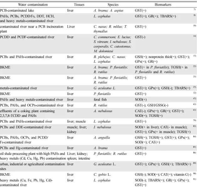
Table 3. Summary of studies which applied oxidative stress parameters of freshwater fish as biomarkers to assess the effects of water contamination
Water contamination Tissues Species Biomarkers
PCB-contaminated lake liver A. brama; A. aspius GST(+) 7)
PAHs, PCBs, PCDD/Fs, DDT, HCH, and heavy metals-contaminated river
L. cephalus GST(+); GR(+); TBARS(=) 8)
contaminated river near a PCB incineration plant
Liver C. nasus; R. mlilus; T.
thymallus
GST(+) 10)
PCDD and PCDF-contaminated river liver C. commersoni; E. lucius;
S. vitreum; I. nebulosus; S.
corporalis; C. cutostomus;
M. dolomieui
GST(-) 14)
PCBs and PAHs-contaminated river liver B. plebejus; C. nasus;
L. cephalus
GSH(=); nonprotein thiol(=); GST(+);
GPx(=); GR(=)
15)
BKME liver A. brama; P. fluviatilis;
R. rutilus
GST(+ in P. fluviatilis); TGSH(+ in P. fluviatilis and R. rutilus)
22)
BKME liver A. brama; P. fluviatilis;
R. rutilus
GST(=) 25)
metals-contaminated river liver G. aculeatus L. GST(+); GPx(+); GSH(-); TBARS(+) 27)
BKME liver P. fluviatilis GST(=) 32)
PAHs and heavy metals-contaminated river liver feral fish SOD(+) 47)
PCBs, PAHs, and OCPs-contaminated river liver R. rutilus GST(-); GSH/GSSG(-) 62)
effluents of a coking plant containing 2,3,7,8-TCDD and PAHs
liver A. nebulosus CAT(-); GPx(=); GR(=); GST(=);
SOD(=); TGSH(=)
63)
PCBs and PAHs-contaminated river liver; muscle L. cephalus GST(=) 79)
PCBs and DDE-contaminated river muscle; liver;
kidney
I. nebulosus SOD(+ in liver); CAT(- in muscle);
GST(+); GPx(+ in muscle); TGSH(+)
83)
PCBs, PAHs, OCPs, and PCDD/
Fs-contaminated river
liver A. anguilla GSH(=); TGSH(=); GST(+); GPx(=);
SOD(=); CAT(+)
84)
PCBs and Hg-contaminated river liver A. brama GST(+) 85)
oil shale processing plant with high PAHs and heavy metals (Cd, Cu, Hg, Pb) contamination
Liver; kidney;
spleen; intestine
P. fluviatilis; R. rutilus GST(+) 86)
urban, industrial or agricultural contamination sites
liver G. aculeatus L. GST(+); GPx(+); GSH(+); TBARS(+) 89)
BKME liver C. gobio L. GSH(-); SOD(=); CAT(=); vitamin C(-) 90)
heavy metals (Cu, Fe, Pb, Hg, Cd)- contaminated river
liver L. cephalus SOD(-); TBARS(=); GR(=); GPx(+);
GST(=)
91)
-, inhibition; =, no (significant) response; +, induction; GR, glutathione reductase; TGSH, total glutathione; The numbers in parenthesis represent references.
하고 있다. Sturve 등45)은 무지개 송어(Oncorhynchus mykiss)를 하수처리장 방류수에 노출시켜 항산화효소의 활성 변화를 관찰한 결과, 방류수 농도 50%에 5일 동 안 노출된 개체에서 CAT의 활성이 대조군에 비해 유 의하게 증가함을 보고하였다. 하지만 하수처리 시설 하 류에서 서식하는 무지개 송어를 대상으로 분석한 항산 화효소(GST, CAT)의 활성과 LPO의 수준은 청정지역 의 대조군에 비해 방류지역의 어류에서 LPO의 수준만 높게 관찰되었을 뿐, 항산화효소의 활성에는 변화가 없 었다.46) 오염된 지역의 담수 어류에 대한 독성영향을 평가하기 위해 산화적 손상과 관련된 바이오마커와 상 위 수준 지표를 동시에 적용한 사례에서도 이와 유사 한 경향이 관찰되었다(Table 4). Roberts Jr 등47)은
PAHs와 중금속으로 오염된 하천의 개체에서 SOD와 CF를 측정한 결과, 대조지역과 오염지역의 개체 모두 에서 SOD 활성이 높게 나타났으나, 물고기 개체의 성 장에는 부정적 영향이 관찰되지 않았다. Sanchez 등27) 도 다양한 오염원에 의해 오염된 지역에서 개체군의 교 란을 발견하였으나 바이오마커와 개체군 반응 간에 뚜 렷한 상관성을 찾지 못하였다.
이와 같이 오염으로 인한 산화적 손상 바이오마커의 반응과 상위 수준의 지표 사이의 반응이 일치하지 않 는 것에는 여러 가지 이유가 있다. 항산화효소의 활성 또는 발현은 산화적 손상을 지시하는 좋은 바이오마커 이지만, 산화적 손상의 유발 요인은 화학물질 이외에도 산소부족, 산소과포화, 열충격, 내인성독소, 질병, 자외
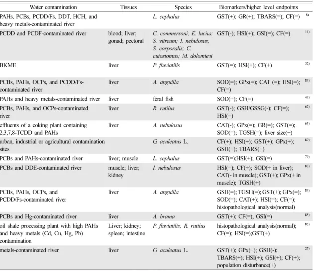
Table 4. Summary of studies which link biomarkers of oxidative stress to the observational endpoints of higher biological levels in freshwater fish
Water contamination Tissues Species Biomarkers/higher level endpoints
PAHs, PCBs, PCDD/Fs, DDT, HCH, and heavy metals-contaminated river
L. cephalus GST(+); GR(+); TBARS(=); CF(=) 8)
PCDD and PCDF-contaminated river blood; liver;
gonad; pectoral
C. commersoni; E. lucius;
S. vitreum; I. nebulosus;
S. corporalis; C.
cutostomus; M. dolomieui
GST(-); HSI(+); GSI(=); CF(=) 14)
BKME liver P. fluviatilis GST(=); HSI(+); CF(+) 32)
PCBs, PAHs, OCPs, and PCDD/Fs- contaminated river
liver A. anguilla SOD(=); GPx(=); CAT (=); HSI(=);
CF(=)
84)
PAHs and heavy metals-contaminated river liver feral fish SOD(+); CF(=) 47)
PCBs, PAHs, and OCPs-contaminated river
liver R. rutilus GST(-); GSH/GSSG(-); CF(=);
HSI(=)
62)
effluents of a coking plant containing 2,3,7,8-TCDD and PAHs
liver A. nebulosus CAT(-); GPx(=); GR(=); GST(=);
SOD(=); TGSH(=); liver size(+)
63)
urban, industrial or agricultural contamination sites
G. aculeatus L. CF(+); HSI(+); GST(+); GPx(+);
GSH(+); TBARS(+)
89)
PCBs and PAHs-contaminated river liver; muscle L. cephalus GST(=);HSI(+); GSI(=) 79) PCBs and DDE-contaminated river muscle; liver;
kidney
I. nebulosus HSI(=); CF(+); SOD(+ in liver);
CAT(- in muscle); GST(+); GPx(+ in muscle); TGSH(+)
83)
PCBs, PAHs, OCPs, and PCDD/Fs-contaminated river
liver A. anguilla GSH(=); TGSH(=); GST(+); GPx(=);
SOD(=); CAT(+); HSI(=); CF(=);
histopathological analysis(normal)
84)
PCBs and Hg-contaminated river liver A. brama GST(+); CF(=); GSI(=) 85)
oil shale processing plant with high PAHs and heavy metals (Cd, Cu, Hg, Pb) contamination
Liver; kidney;
spleen; intestine
P. fluviatilis; R. rutilus histopathological analysis(normal);
CF(=); HSI(=);GST(+)
86)
metals-contaminated river liver G. aculeatus L. GST(+); GPx(+); GSH(-);
TBARS(+); HSI(+); GSI(+); CF(+);
population disturbance(+)
27)
-, inhibition; =, no (significant) response; +, induction; The numbers in parenthesis represent references.
선 등 다양하다.48)한편 생물이 유해인자 또는 독성물 질에 만성적으로 노출되는 경우 이 스트레스에 점진적 으로 적응하여 개체가 산화적 손상 요인에 대해 나타 내는 반응이 달라지는 경우도 있다.49)동일 생물종이라 도 야외에서 생존해 온 야생종이 실내에서 사육된 실 험종보다 자외선에 대한 저항성이 크다는 보고50)는 수 서생물이 처한 환경에 따라 산화적 손상에 대한 방어 기전을 획득할 수 있다는 것을 뒷받침한다. 강화된 방 어기전 덕분에 다른 오염물질에 의해 유발될 수 있는 산화적 손상에 대해서도 보호 효과를 나타낼 수도 있 다. 이러한 복잡성 때문에 상위 수준의 영향평가와 병 행하여 활용될 때 산화적 손상 관련 바이오마커의 효 용성이 증가할 것으로 판단된다.
3. 생식 및 내분비계교란 관련 지표
내분비계장애물질은 화학구조가 호르몬과 유사하여 노출될 경우 생체의 정상적인 호르몬의 작용을 저해하 거나 마치 호르몬처럼 작용하여 불필요한 생리작용을 초래하게 된다. 이러한 외인성 내분비계장애물질들은 주로 에스트로겐과 유사한 작용을 하여 특히 수컷의 생 식기능에 영향을 준다. 어류에서 내분비계장애물질의 영향을 평가하기 위해 자주 사용하는 방법은 생체내 성 호르몬의 변화를 측정하거나 난황단백질(vitellogenin, VTG)의 생성을 측정하는 것이다. VTG는 난황 단백질
의 전구체로서 어류, 양서류, 또는 곤충 등의 비포유류 난생동물의 암컷 혈장에서 주로 발견된다.51,52) VTG는 간에서 생성된 후 혈액을 통해 난소로 이동하여 발달 중인 난모세포로 흡수된 후 알 내부에서 리포비텔린 (lipovitelline)과 포스비틴(phosvitin)으로 전환되는데, 에 스트로겐은 이러한 VTG의 합성과 분비를 촉진시킨다.
암컷 붕어의 VTG 농도는 수컷에 비해 20~40배 높으 며 수컷에서는 10 µg/ml 이하로 측정된다.53) 그러나 외부에서 에스트로겐이나 내분비장애 물질에 노출된 경 우에는 수컷이나 미성숙 암컷에서도 VTG의 합성이 유 발될 수 있어 VTG는 외인성 에스트로겐 유사물질 (estrogenic chemical)의 노출을 지시하는 바이오마커로 널리 활용되고 있다.54,55)
생식 및 내분비계교란 관련 바이오마커를 이용하여 외인성 내분비계장애물질의 노출과 영향을 분석한 사 례는 총 10건이 파악되었으며(Table 5), 바이오마커로 호르몬 변화를 측정한 사례는 4건, VTG 수준을 평가 한 사례는 6건이었다. TCDD와 PCB 등의 잔류성유기 화합물과 17α-ethynylestradiol 및 BKME가 주요 오염 물질로 제시되었다.
BKME가 어류에 초래하는 내분비계 교란작용은 여 러 연구에서 확인되었다. Karels 등33)은 BKME 오염 지역에서 포획한 암컷 농어에서 HSI 수준이 대조지역 에 비해 높게 측정되었다고 보고하였다. 또한 공장 근
Table 5. Summary of studies which applied reproduction and endocrine disruption parameters of freshwater fish as biomarkers
to assess the effects of water contamination
Water contamination Tissues Species Biomarkers
PCDD and PCDF-contaminated river blood C. commersoni; E. lucius;
S. vitreum;/ I. nebulosus;
S. corporalis; C. cutostomus;
M. dolomieui
testosterone(=); 17 β-estradiol(=) 14)
TCDD, and TCDF-contaminated river
liver F. heteroclitus 6β-hydroxyprogesterone activity(+) 16)
BKME liver C. commersonl; C. catostomus testosterone and estradiol(- in female C. catostomus)
19)
BKME blood L. aurltus 17 β-estradiol(-) 26)
metals-contaminated river liver G. aculeatus L. VTG(+) 27)
BKME bile; liver;
serum
P. fluviatilis; R. rutilus VTG mRNA( -); testosterone(-);
17 β-estradiol(- in female, + in male)
33)
PCB, TCDD, DDE, and heavy metals- contaminated river
blood C. carpio; M. salmoides;
M. dolomieui; I. furcatus
VTG(+ in Micropterus sp.) 82)
effluents from a single sewage treatment plant
liver S. trutta VTG mRNA(-); VTG protein(+) 92)
17α-ethynylestradiol (5-6 ng/L)- contaminated lake
liver P. promelas VTG mRNA and protein expression in
males(+)
93)
contaminated river blood L. cephalus VTG(+) 94)
-, inhibition; =, no (significant) response; +, induction; The numbers in parenthesis represent references.
처의 수체에서 포획된 암컷의 혈장 17β-estrodiol 농도 와 VTG 유전자 발현이 감소하였지만 수컷의 17β- estrodiol 농도는 증가하였으며, 암·수 모두에서 testosterone 수준이 감소하였다. 또 다른 사례26)에서는 BKME로 오염된 지역의 개체에서 종의 풍부함과 구성 을 나타내는 IBI(index of biotic integrity) 지표가 낮 게 나타나 물고기 군집의 영양구조가 불균형하게 나타 났다. 오염 지역의 개체군에서 크기와 연령의 구조가 비정상적이었으며, 오염된 지역의 개체에서 다수의 폐 쇄성 난모세포와 17β-estradiol 저하가 관찰되었다. 이 두 사례의 결과를 종합해 볼 때 BKME는 암·수컷 개 체에 내분비계교란을 일으켜 성호르몬 수준을 변화시 키고, 이로 인해 암컷에서 난황전구물질인 VTG 발현 을 감소시켜 생식기능장애와 자손번식 실패를 통해 물고기 개체군에 영향을 주는 것으로 결론지을 수 있 었다.
수질 오염으로 인한 생식 영향 및 내분비계교란을 분 자·생화학적 바이오마커들과 상위 수준 지표로 평가한 사례는 7건을 찾을 수 있었다(Table 6). 이 사례들을 분석한 결과 VTG발현과 성호르몬 변화는 내분비계교
란 물질의 노출에 따른 생식 독성을 잘 반영하는 것으 로 나타났으며, 생식선의 발달이나 양성화 등과 같은 상위 수준의 관측과도 일치하는 반응을 보였다. 하지만 VTG는 암컷의 성장과 생육단계 별로 생성되는 농도가 다르고, 계절적 변이가 크다는 점은 바이오마커로서 VTG의 활용성을 제한하는 특성이다.56)내분비계 교란 물질 오염과 번식장애를 파악하기 위해 VTG를 바이오 마커로 사용하는 경우 개체의 번식주기, 지리적 분포와 생물 다양성의 차이를 고려할 필요가 있다.57)
4. 유전독성 매개변수
일부 유해물질은 직·간접적으로 세포에 염색체 이상 및 DNA 손상을 유발하여 어떤 경우에는 돌연변이, 암, 유전자 이상과 같은 유전독성을 초래할 수 있다.
예를 들면 유해물질 노출로 인해 세포 내에 ROS가 발 생되고 세포 내 redox 균형을 붕괴시켜 산화적 스트레 스를 유발함으로써 이러한 유전독성이 발생할 수 있 다. DNA의 구조적 손상은 유전독성의 바이오마커로 나아가 생태계 이상의 조기 경보로서 활용성이 있다.58) 유전독성 관련 분자·생화학적 바이오마커로 자주 활
Table 6. Summary of studies which link biomarkers of reproduction and endocrine disruption to the observational endpoints of higher biological levels in freshwater fish
Water contamination Tissues Species Biomarkers/higher level endpoints PCB, TCDD, DDE, and heavy
metals-contaminated river
blood; liver;
kidney; gonad;
mesenteric fat
C. carpio; M.
salmoides; M.
dolomieui; I. furcatus
EROD activity(+); external lesions(+); HAI(- in Micropterus sp.); histopathological analysis (abnormal); CF(=), HSI(=); GSI(-); VTG(+ in Micropterus sp.)
82)
BKME blood; liver L. aurltus histopathological analysis(abnormal); age structure (abnormal); CF(+ in female); HSI(-); growth rates (-); IBI(-); CPUE(-); 17 β-estradiol(-); vitellogenic oocytes(=); fecundity(=); atretic oocyte(+)
26)
PCDD and PCDF-contaminated river
blood; liver;
gonad; pectoral
C. commersoni;
E. lucius; S. vitreum;
I. nebulosus; S.
corporalis; C.
cutostomus; M.
dolomieui
HSI(+); CF(=); testosterone(=); 17 β-estradiol(=) 14)
BKME bile; liver; serum P. fluviatilis;
R. rutilus
HSI(+); GSI(=); VTG mRNA( -); Testosterone(-);
17 β-estradiol(- in female, + in male)
33)
17α-ethynylestradiol (5-6 ng/L)- contaminated lake
liver P. promelas feminization of males(+); VTG mRNA and protein expression in males(+); gonad development and intersex in males(+); oogenesis in females(altered);
population effect(extinction)
93)
metals and organics-contaminated river
blood L. cephalus VTG(+); testicular structure(altered) 94)
metals-contaminated river liver G. aculeatus L. VTG(+); HSI(+); GSI(+); CF(+); population disturbance(+)
27)
-, inhibition; =, no (significant) response; +, induction; The numbers in parenthesis represent references.
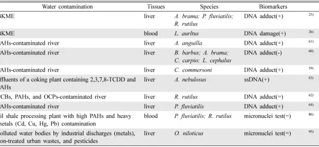
용되는 것에는 DNA 부가물(DNA adduct), DNA 절 단(breakage), comet assay, 염색체 이상(chromosome aberration), 소핵분석(micronucleus; MN), 핵변이 (nuclear anomalies; NA) 등이 있다. 현장 담수어류를 대상으로 바이오마커를 이용하여 유전독성을 분석한 사 례는 총 10건이 있다(Table 7). 이 중 DNA 부가물 분석을 적용한 사례가 모두 6건으로 가장 빈도가 높았 고, 유전독성의 원인물질로는 PAHs가 7건으로 가장 많 았다. 또한 현장 담수 어류의 PAHs 노출을 평가하기 위해 DNA 부가물을 측정한 모든 사례에서 이 바이오
마커는 PAHs 노출에 민감하게 반응하여 PAHs의 노출 지표로 적합한 것으로 나타났다.25,59-61)하지만 PAHs 노 출로 인한 유전독성 바이오마커의 반응과 조직학적 수 준 이상의 상위 단계 지표를 연계하여 분석한 사례는 발견할 수 없었다.
BKME로 오염된 지역에서 DNA 부가물의 증가와 생 리학적 지표, 조직병리학적 분석, 개체 연령 구조, 성장 률 및 군집 지표 등의 상위 지표와 연계하여 분석한 연구가 1건 발견되었으나 상위 지표와의 뚜렷한 상관 성을 보이지 않았다.26)또한 PCBs, OCPs 및 creosote
Table 7. Summary of studies which applied genotoxic parameters of freshwater fish as biomarkers to assess the effects of water
contamination
Water contamination Tissues Species Biomarkers
BKME liver A. brama; P. fluviatilis;
R. rutilus
DNA adduct(+) 25)
BKME blood L. aurltus DNA damage(+) 26)
PAHs-contaminated river liver A. anguilla DNA adduct(+) 61)
PAHs-contaminated river liver B. barbus; A. brama;
C. carpio; L. cephalus
DNA adduct(-) 60)
PAHs-contaminated river liver C. commersoni DNA adduct(+) 59)
effluents of a coking plant containing 2,3,7,8-TCDD and PAHs
liver A. nebulosus ssDNA(+) 63)
PCBs, PAHs, and OCPs-contaminated river liver R. rutilus DNA adduct(=) 62)
PAHs-contaminated river liver P. fluviatilis DNA adduct(+) 64)
oil shale processing plant with high PAHs and heavy metals (Cd, Cu, Hg, Pb) contamination
blood P. fluviatilis; R. rutilus micronuclei test(=) 86)
polluted water bodies by industrial discharges (metals), non-treated urban wastes, and pesticides
liver O. niloticus micronuclei test(=) 95)
-, inhibition; =, no (significant) response; +, induction; single-strand DNA breakage; The numbers in parenthesis represent references.
Table 8. Summary of studies which link biomarkers of genotoxicity to the observational endpoints of higher biological levels in freshwater fish
Water contamination Tissues Species Biomarkers/higher level endpoints PCBs, PAHs, and OCPs-contaminated river liver R. rutilus CF(=); HSI(=); DNA adduct(=) 62)
BKME blood; liver L. aurltus histopathological analysis(abnormal); age
structure (abnormal); CF(+ in female);
HSI(-); growth rates(-); IBI(-); CPUE(-);
fecundity(=); DNA damage(+)
26)
effluents of a coking plant containing 2,3,7,8-TCDD and PAHs
liver A. nebulosus ssDNA(+); liver size(+) 63)
PAHs-contaminated river liver P. fluviatilis DNA adduct(+); CF(=); GSI(=); HSI(=);
histopathological analysis(normal)
64)
oil shale processing plant with high PAHs and heavy metals (Cd, Cu, Hg, Pb) contamination
liver; kidney;
spleen;
intestine; blood
P. fluviatilis;
R. rutilus
histopathological analysis(normal);
micronuclei test(=); CF(=); HSI(=)
86)
polluted water bodies by industrial discharges (metals), non-treated urban wastes, and pesticides
liver O. niloticus micronuclei test(=), CF(- in fish at a metal- contaminated site)
95)
-, inhibition; =, no (significant) response; +, induction; ssDNA; The numbers in parenthesis represent references.
로 오염된 지역의 개체에서 DNA 부가물과 DNA 손상 등의 바이오마커와 상위 수준 지표들을 연계하여 분석 하였으나 오염으로 인한 유전독성 반응을 일관되게 반 영하는 사례는 관찰되지 않았다(Table 8).26,62-64) 이는 심각한 수준의 손상이 아닐 경우 DNA 회복 기전에 의해 대부분 회복되어 상위 단계까지 그 영향이 나타 나지 않기 때문인 것으로 생각된다.
5. Metallothionein
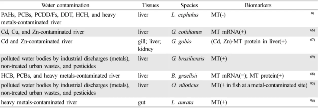
Metallothionein(MT)은 세균과 균류로부터 사람을 포 함한 척추 동물에 이르기까지 다양한 생물체에 존재하 고 있는 단백질로, 열에 안정하며 thiol기(-SH)가 풍부 하여 일부 중금속과 결합력이 큰 특성이 있다. 이 때문 에 MT는 중금속 노출시 발현하여 독성을 저감하는 작 용을 한다.65) 중금속 노출 시 생성되는 MT의 수준은 종, 개체, 기관에 따라 다르다. 중금속으로 오염된 하천 에서 포획된 물고기를 대상으로 조사한 결과 MT가 중
금속 노출을 지표하는 좋은 바이오마커임을 보여주었 다(Table 9).66-68) MT의 변화가 개체의 건강 수준과도 잘 일치하는 것으로 보고한 연구도 있었다(Table 10).
Linde-Arias 등69)은 오염원이 서로 다른 지역(인구밀집 지역, 공업지역, 농업지역)에서 포획된 어류를 대상으로 MT와 CF를 분석한 결과, 중금속 오염이 심한 공업 지 역에서 MT의 수준이 가장 높았으며, 대조지역에 비해 CF 수준이 낮은 것을 관찰하였다. 그러나 중금속 노출 을 평가하기 위해 MT와 조직수준 이상의 지표를 동시 에 분석한 사례는 3건으로 매우 제한적이어서 MT의 활용성을 판정하기 위해서는 추가적인 연구가 필요할 것으로 생각된다.
IV. 결 론
물 환경오염의 생태위해성 평가를 위한 주요 분자·
생화학적 바이오마커의 현장 적용성을 알아보기 위해
Table 9. Summary of studies which applied reproduction metallothionein of freshwater fish as biomarkers to assess the effects
of water contamination
Water contamination Tissues Species Biomarkers
PAHs, PCBs, PCDD/Fs, DDT, HCH, and heavy metals-contaminated river
liver L. cephalus MT(-) 8)
Cd, Cu, and Zn-contaminated river liver G. cotidianus MT mRNA(+) 66)
Cd and Zn-contaminated river gill; liver;
kidney
G. gobio (Cd, Zn)-MT protein in liver(+) 67)
polluted water bodies by industrial discharges (metals), non-treated urban wastes, and pesticides
liver G. brasiliensis MT(+) 69)
HCB, PCBs, and heavy metals-contaminated river liver B. graellsii MT mRNA(=); MT protein(+) 68) polluted water bodies by industrial discharges (metals),
non-treated urban wastes, and pesticides
liver O. niloticus MT(+ in fish at a metal-contaminated site)95)
heavy metals-contaminated river gut L. aurata MT(+) 96)
-, inhibition; =, no (significant) response; +, induction; The numbers in parenthesis represent references.
Table 10. Summary of studies which link biomarkers of metallothionein to the observational endpoints of higher biological levels in freshwater fish
Water contamination Tissues Species Biomarkers/higher level endpoints PAHs, PCBs, PCDD/Fs, DDT, HCH, and heavy
metals-contaminated river
liver L. cephalus MT(-); CF(=) 8)
heavy metals-contaminated river gut L. aurata gut morphological analysis(+);
histomorphological analysis of mucous goblet cells(normal); MT(+)
96)
polluted water bodies by industrial discharges (metals), non-treated urban wastes, and pesticides
liver G. brasiliensis MT(+); CF(-) 69)
polluted water bodies by industrial discharges (metals), non-treated urban wastes, and pesticides
liver O. niloticus MT(+ in fish at a metal-contaminated site); CF(- in fish at a metal- contaminated site)
95)
-, inhibition; =, no (significant) response; +, induction; The numbers in parenthesis represent references.
담수 어류를 대상으로 화학물질의 노출을 평가한 사례 를 조사하였다. 조사된 바이오마커는 대사효소, 산화적 손상 매개변수, 생식 및 내분비계교란 매개변수, 유전 독성 매개변수, MT 등이다. 대사효소로는 CYP1A1와 같은 대사효소의 단백질 수준과 효소 활성을 측정하는 방법이 주로 활용되었으며, 유기오염물질 오염을 모니 터링하기 위한 적절한 바이오마커인 것으로 나타났다.
산화적 손상 관련 바이오마커는 일부 화학물질의 노출 을 잘 지시하는 것으로 나타났다. 유전독성을 평가하기 위해 가장 빈번하게 사용하는 DNA 부가물 분석은 현 장시료에서도 일부 오염물질의 노출을 민감하게 지시 하였다. 중금속으로 오염된 하천에서 포획된 물고기를 대상으로 MT의 농도를 측정한 대부분의 연구사례들은 MT가 중금속 노출을 지표하는 좋은 바이오마커임을 보 여주었다. 이와 같이 조사된 바이오마커는 대개 현장 시료에서 유해물질의 독성기전을 지시하는데 활용성이 컸다. 그러나 분자·생화학적 바이오마커만을 가지고 개체 또는 개체군 이상에서 나타나는 영향을 추정하는 것은 어려웠다. 특히 생식 및 내분비계교란과 관련된 바이오마커를 제외한 나머지 바이오마커의 경우에는 조 직병리학적 지표 이상의 상위 단계와의 연계성이 비교 적 낮았다. 그 이유는 분자·생화학적 바이오마커는 비 교적 단기간 낮은 수준에 노출되어도 나타날 수 있지 만 쉽게 회복될 수 있기 때문에 상위단계의 영향으로 까지 진행되지 않는 경우가 많기 때문이다. 한편 일부 바이오마커는 유해물질 노출뿐만 아니라 수질변화나 질 병발생에 의해서도 반응하는 경우도 있어, 바이오마커 의 반응이 반드시 유해물질 노출의 결과라고 볼 수는 없었다. 따라서 바이오마커를 직접 환경 시료에 적용할 경우에는 이러한 제한점을 고려해야 할 것이다. 유해인 자의 물환경 생태계 영향을 정확하게 파악하기 위해서 는 분자·생화학적 바이오마커와 함께 적절한 상위단계 의 생물영향지표를 평가하는 것이 타당한 것으로 판단 된다.
감사의 글
본 연구는 환경부 Eco-STAR 수생태복원사업단 (CAER)에서 지원을 받아 수행되었다. 원고를 검토한 서울대학교 보건환경연구소 배현주 박사께 감사드린다.