pISSN 1738-6640 eISSN 2234-4020 http://dx.doi.org/10.6114/jkood.2015.28.1.023
Original Article / 원저
加味潤燥湯이 LPS로 유도된 RAW 264.7 대식세포에서의 항염 효과 연구
최종민1·김용민2·김희택1
1 세명대학교 한의과대학 안이비인후피부과학 교실
2 세명대학교 한방화장품과학과
Anti-inflammatory Effects of Gamiyunjo-tang on Lipopolysaccharide Induced Inflammatory Responses in RAW 264.7 Cells
Jong-Min Choi
1)·Yong-Min Kim
2)·Hee-Taek Kim
1)1
Dept. of Oriental Ophthalmology, Otolaryngology & Dermatology, College of Korean medicine, Semyung University
2
Dept. of Oriental Medical and Herbal Cosmetic Sciences, Semyung University
Abstract
Objectives : Allergic disease has been well known as an IgE-dependent immunologic response. Recently, interest about the late inflammatory reaction has grown up as well as early allergic reaction characterized by IgE and mast cell. The purpose of this study was to find the anti-inflammatory effect of Gamiyunjo-tang(GMYJT) in allergic reaction.
Methods : The experiment was performed using Raw 264.7 cells pretreated with GMYJT extracts. In this study, we observed the toxicity of cells by MTT analysis and measured the production of LPS-induced NO, PGE₂, IL-1β, IL-6 and TNF-α at a concentration of 50, 100, 200 and 400 ㎍/㎖.
Results : No toxicity of GMYJT (50, 100, 200, 400 ㎍/㎖) on RAW 264.7 cells was found after 24 hours incubation. LPS-induced NO production was reduced after treatment with GMYJT (100, 200, 400 ㎍/㎖)(P<0.05).
PGE₂was reduced after treatment with GMYJT (100, 200, 400 ㎍/㎖)(P<0.05). IL-1β did not decrease at any dose.
IL-6 decreased at 200, 400 ㎍/㎖(P<0.05). TNF-α production decreased only at 400 ㎍/㎖(P<0.05).
ⓒ 2015 the Society of Korean Medicine Ophthalmology & Otolaryngology & Dermatology
This is an Open Access journal distributed under the terms of the Creative Commons Attribution Non-Commercial License (http://creativecommons.org/license/by-nc/3.0/) which permits unrestricted non-commercial use, distribution, and reproduction in any medium, provided the original work is properly cited.
Conclusions : These data suggest that GMYJT has anti-inflammatory effects in late allergic reaction.
Key words : Anti-inflammation; NO; PGE
2;IL-1β; IL-6; TNF-α; Gamiyunjo-tang;
Corresponding Author : Hee-Taek Kim,
Semyung University Oriental Medicine Hospital, 66, Semyeong-ro, Jecheon-si, Chungcheongbuk-do, Korea(Tel:043-649-1817, E-mail:[email protected])
∙Recieved 2015/1/19 ∙Revised 2015/2/2 ∙Accepted 2015/2/9
Ⅰ. 서 론
알레르기 초기 면역 반응에서는 항원에 노출된 후 비만세포의 탈과립이 일어나면서 알레르기 증상을 유 발시키는 histamine, prostaglandin, leukotriene과 같은 다양한 매개물질들이 분비된다. 그리고 후기반 응은 T세포, 호염기구, 호산구로 이루어진 염증세포 들의 유입과 이로 인해 특징지어지는 복합적인 염증 반응에 의해 이루어진다1,2).
알레르기 질환의 지속적인 염증반응에 있어서는 상 피세포, 비만세포와 후기반응의 주된 역할을 하는 호 산구, 호염기구, T세포와 같은 염증세포 모두가 관여 하여 알레르기 염증반응을 유발하고 지속시킨다. 이 러한 일련의 과정에서 대식세포는 T세포에 항원을 제 공하는 기능을 하여 면역반응을 개시하는데 중요한 역할을 하고, 비특이적인 면역반응을 담당하게 된다.
그중 NO는 주로 대식세포에서 생성되는 중간물질3)로 염증반응의 정도를 가늠하는 척도가 된다4,5). 활성화 된 대식세포는 interleukin-1 beta(IL-1β), IL-6, tumor necrosis factor-α(TNF-α)와 같은 pre- inflammatory cytokine과 prostaglandin E2(PGE2) 등을 생산하고, 이를 통해 염증반응을 촉진시킴으로 써 다양한 염증 관련 질환의 발달에 기여한다6,7). 본 연구에 사용한 加味潤燥湯8)은 全身瘙痒 血燥 風 毒性症에 다용되며 피부질환의 급성기 보다는 만성기 에 사용할 수 있는 처방이다. 이에 저자는 LPS로 유 도된 RAW 264.7 대식세포에서 NO 생성과 염증 반 응에 관여하는 사이토카인인 IL-1β, IL-6, TNF-α에 대한 실험을 진행하여 유의한 결과를 얻었기에 보고
하고자 한다.
Ⅱ. 실험 방법
1. 실험 재료
1) 처방 구성 및 시료 제조
加味潤燥湯(Gamiyunjo-tang, 이하는 GMYJT)의 구성 약물을 ㈜HMAX(제천, 한국)에서 구입하여 사 용하였다(Table 1). 가미윤조탕 2첩 분량에 해당하는 112 g을 3차 증류수 2 L와 혼합하여 100℃로 4시간 동안 열수 추출 하였으며, 여과지로 여과한 추출액을 rotary evaporator를 이용하여 100 ㎖ 까지 농축하고 -80℃로 동결하였다. 농축한 동결액을 freezing dryer system (Labconco, USA)을 이용하여 7일간 동결건 조 하였다(수율 약 18%).
Herbal
Name Scientific Name Dose(g)
熟地黃 Rehmanniae Radix Preparat 6
生地黃 Rehmanniae Radix 6
白芍藥 Paeoniae Radix Alba 6
知 母 Anemarrhenae rhizoma 6
黃 芩 Scutellariae Radix 6
秦 艽 Gentianae Macrophyllae Radix 6
玄 蔘 Scrophulariae Radix 6
黑芝麻 Sesami Semen Nigrum 4
防 風 Saposhnikovia Radix 4
浮 萍 Spirodelae Herba 4
甘 草 Glycyrrhizae Radix 2
Total 56
Table 1. The Amount and Composition of GMYJT
2) 세포 배양
실험에 사용된 mouse 대식세포는 RAW 264.7 cell line (ATCC, USA)을 분양받아 사용하였다.
RAW 264.7 cells은 37℃, 5% CO2 조건에서 10%
Fetal bovine serum (FBS), penicillin (100 U/㎖) 및 streptomycin (100 ㎍/㎖) 등이 포함된 DMEM 배지 로 배양되었다.
배양세포들은 75 cm2 flask (Falcon, USA)에서 충 분히 증식된 후 배양 3일 간격으로 배양세포 표면을 PBS 용액으로 씻어준 뒤 50 ㎖ flask 당 1 ㎖의 0.25% trypsin-EDTA 용액을 넣고 실온에서 1분간 처리한 다음 trypsin을 버리고 37℃에서 5분간 보관 하여 세포를 탈착하여 계대 배양하였다. 탈착된 세포 는 10% FBS가 첨가된 DMEM 배양액 10 ㎖에 부유 시킨 다음 새로운 배양용기 (50 ㎖ culture flask)에 옮겨 1:2 split ratio로 CO2 배양기에서 배양하였다.
2. 실험방법 1) MTT assay
세포독성 유발 효과를 알아보기 위하여 3-(4,5- dimrthylthiazol-2-yl)-2,5-diphenyltetrazolium bromide (MTT) assay 방법을 사용하여 측정하였다.
96 well plate에 1×10⁵ cells/well의 cell을 100 ㎕씩 넣고 37℃, 5% CO₂가 공급되는 배양기에서 24시간 동안 배양한 후 배지를 버리고 배양세포 표면을 1×PBS 용액으로 씻어주었다. 같은 양의 배지와 PBS 에 녹인 시료를 농도별로 각 well에 처리하고 24시간 배양하였다. 배양이 끝난 후 PBS에 녹인 1 ㎍/㎖
MTT (Sigma, USA)를 100 ㎕씩 각 well에 처리하여 알루미늄 호일로 차광시킨 뒤 2시간동안 같은 조건에 서 배양하였다. 배양액을 모두 제거한 후 DMSO를 100 ㎕ 처리하고 37℃에서 2시간 방치한 다음 microplate reader를 이용하여 490 ㎚에서 흡광도를 측정하였다. 세포생존율은 다음과 같은 공식으로 계 산되었다.
Viability(%) = 100 × AT/AC
AT : absorbance of tested extract solution AC : absorbance of control
2) NO assay
96 well plate에 1×10⁵ cells/well의 cell을 100 ㎕ 씩 넣고 37℃, 5% CO₂가 공급되는 배양기에서 24 시간동안 배양하여 세포를 안정화 시켰다. 안정화 시 킨 세포에 lipopolysacharride (LPS) 1 ㎍/㎖와 가미 윤조탕 추출물을 농도별로 처리하고 24시간 동안 3 7℃, 5% CO₂가 공급되는 배양기에서 배양한 후 세 포배양 상등액 60 ㎕을 채취하여 여기에 Griess 시약 100 ㎕을 혼합하여 15분 동안 반응시킨 뒤 microplate reader를 이용하여 540 ㎚에서 흡광도를 측정하였다.
3) PGE2 생성 측정
PGE2의 측정은 commercial competitive enzyme immunoassay kit를 R&D systems (Minneapolis, USA)에서 구입하여 사용하였다. RAW 264.7 세포에 가미윤조탕과 1 ㎍/㎖의 LPS를 처리하여 24시간 배양 한 후 세포 배양 상층액을 수거하여 PGE2 측정에 사 용하였다. 배양액을 goat anti-mouse로 coating된 96 well plate에 각각의 배양액을 100 ㎕씩 loading하고 여기에 primary antibody solution 50 ㎕와 PGE2
conjugate 50 ㎕씩 첨가하여 4℃에서 overnight시켰 다. 기질용액을 200 ㎕씩 처리하여 5~20 분간 반응시 킨 후, 50 ㎕의 stop solution을 처리하고 450 ㎚에서 흡광도를 측정하였다.
4) Cytokine 생성 측정
96 well plate에 1×10⁵ cells/well의 cell을 100 ㎕ 씩 넣고 37℃, 5% CO₂가 공급되는 배양기에서 24 시간 동안 배양하여 세포를 안정화 시켰다. 안정화 시 킨 세포에 LPS 1 ㎍/㎖와 가미윤조탕 추출물을 농도 별로 처리하고 24시간 동안 배양하였다. 배양이 끝나
면 상등액을 채취하여 Bio-Plex Suspension Assay System을 이용, Quantitative Multiplexed Cytokine/
Chemokine Assay를 실시하여 IL-1β, IL-6 및 TNF- α 생성량을 측정하였다.
5) 통계 분석
실험결과는 SPSS Window program(Ver. 12.0)을 이용하였으며, 모든 측정값은 평균값 ± 표준편차 (Mean ± SD)로 나타내었고, 대조군과 각 실험군과의 평균차이는 Student's t-test로 분석하여 p-value가 0.05 미만일 때 통계학적으로 유의한 차이가 있는 것 으로 판정하였다.
Ⅲ. 실험 성적
1. 세포독성에 미치는 영향
가미윤조탕의 농도가 세포내에서 독성을 일으키는 지 확인하기 위해 MTT assay를 이용하여 측정하였 다. 정상군의 생존율을 100 ± 3.96%로 계산하였을 때 모든 농도에서 세포독성이 없음을 확인하였다 (Table 2).
Concentration (㎍/㎖) Cell Viability (% of control)
Normal 100 ± 3.96
50 94.95 ± 5.51
100 98.75 ± 4.49
200 95.56 ± 5.64
400 94.94 ± 5.51
Table 2. The Cytotoxic Effect of Gamiyunjo-tang (GMYJT) Water-extract on RAW 264.7 Macrophage Cells by MTT assay
Values are the mean ± SD of the three independent experiments.
Normal : not treated with GMYJT 50 : Treated with GMYJT (50 ㎍/㎖) 100 : Treated with GMYJT (100 ㎍/㎖) 200 : Treated with GMYJT (200 ㎍/㎖) 400 : Treated with GMYJT (400 ㎍/㎖)
2. NO 생성에 미치는 영향
가미윤조탕이 마우스 대식세포의 NO 생성에 미치 는 영향을 관찰하였다. LPS로 유도된 NO의 생성율을 100 ± 1.86%로 계산하였을 때 가미윤조탕 100 ㎍/
㎖, 200 ㎍/㎖ 및 400 ㎍/㎖ 농도로 처리한 군에서 LPS 단독 처리한 군에 비하여 통계학적으로 유의한 감소가 나타났다(Table 3).
Concentration (㎍/㎖) NO production (% of control)
Control 100 ± 1.86
LPS + 50 98.50 ± 1.28 LPS + 100 85.33 ± 0.47*
LPS + 200 82.57 ± 1.34*
LPS + 400 77.81 ± 0.16*
Table 3. The Effect of Gamiyunjo-tang (GMYJT) Water-extract on NO Production of RAW 264.7 Macrophage Cells
Values are the mean ± SD of the three independent experiments.
Control : Treated with LPS (1 ㎍/㎖)
LPS + 50 : Treated with LPS (1 ㎍/㎖) and GMYJT (50 ㎍/㎖) LPS + 100 : Treated with LPS (1 ㎍/㎖) and GMYJT (100 ㎍/㎖) LPS + 200 : Treated with LPS (1 ㎍/㎖) and GMYJT (200 ㎍/㎖) LPS + 400 : Treated with LPS (1 ㎍/㎖) and GMYJT (400 ㎍/㎖)
* p < 0.05 compared with control
Concentration (㎍/㎖) PGE
2production (% of control)
Control 100 ± 2.10
LPS + 50 98.41 ± 1.92 LPS + 100 90.29 ± 1.80*
LPS + 200 79.30 ± 1.78*
LPS + 400 74.20 ± 1.72*
Table 4. The Effect of Gamiyunjo-tang (GMYJT) Water-extract on PGE2 Production of RAW 264.7 Macrophage Cells
Values are the mean ± SD of the three independent experiments.
Control : Treated with LPS (1 ㎍/㎖)
LPS + 50 : Treated with LPS (1 ㎍/㎖) and GMYJT (50 ㎍/㎖) LPS + 100 : Treated with LPS (1 ㎍/㎖) and GMYJT (100 ㎍/㎖) LPS + 200 : Treated with LPS (1 ㎍/㎖) and GMYJT (200 ㎍/㎖) LPS + 400 : Treated with LPS (1 ㎍/㎖) and GMYJT (400 ㎍/㎖)
* p < 0.05 compared with control
3. Prostaglandin E2 생성에 미치는 영향 가미윤조탕이 마우스 대식세포의 Prostaglandin E2
생성에 미치는 영향을 관찰하였다. LPS로 유도된 PEG2의 생성율을 100 ± 1.65%로 계산하였을 때 가 미윤조탕 100 ㎍/㎖, 200 ㎍/㎖ 및 400 ㎍/㎖ 농도로 처리한 군에서 LPS 단독처리한 군에 비하여 통계학적 으로 유의한 감소가 나타났다(Table 4).
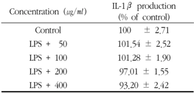
4. IL-1β 생성에 미치는 영향
가미윤조탕이 마우스 대식세포의 IL-1β 생성에 미 치는 영향을 관찰하였다. LPS로 유도된 IL-1β의 생 성율을 100 ± 2.71%로 계산하였을 때 가미윤조탕을 처리한 모든 군에서 LPS 단독처리한 군에 비하여 통 계학적으로 유의한 감소가 나타나지 않았다(Table 5).
Concentration (㎍/㎖) IL-1β production (% of control)
Control 100 ± 2.71
LPS + 50 101.54 ± 2.52 LPS + 100 101.28 ± 1.90 LPS + 200 97.01 ± 1.55 LPS + 400 93.20 ± 2.42
Table 5. The Effect of Gamiyunjo-tang (GMYJT)Water-extract on Interleukin-1β Production of RAW 264.7 Macrophage Cells
Values are the mean ± SD of the three independent experiments.
Control : Treated with LPS (1 ㎍/㎖)
LPS + 50 : Treated with LPS (1 ㎍/㎖) and GMYJT (50 ㎍/㎖) LPS + 100 : Treated with LPS (1 ㎍/㎖) and GMYJT (100 ㎍/㎖) LPS + 200 : Treated with LPS (1 ㎍/㎖) and GMYJT (200 ㎍/㎖) LPS + 400 : Treated with LPS (1 ㎍/㎖) and GMYJT (400 ㎍/㎖)
* p < 0.05 compared with control
5. IL-6 생성에 미치는 영향
가미윤조탕이 마우스 대식세포의 IL-6 생성에 미치 는 영향을 관찰하였다. LPS로 유도된 IL-6의 생성율 을 100 ± 1.23%로 계산하였을 때 가미윤조탕 200 ㎍
/㎖ 및 400 ㎍/㎖ 농도로 처리한 군에서 LPS 단독처 리한 군에 비하여 통계학적으로 유의한 감소가 나타 났다(Table 6).
Concentration (㎍/㎖) IL-6 production (% of control)
Control 100 ± 1.23
LPS + 50 99.70 ± 1.04 LPS + 100 93.09 ± 1.01 LPS + 200 85.86 ± 1.89*
LPS + 400 81.77 ± 1.83*
Table 6. The Effect of Gamiyunjo-tang (GMYJT) Water-extract on Interleukin 6 Production of RAW 264.7 Macrophage Cells
Valuse are the mean ± SD of the three independent experiments.
Control : Treated with LPS (1 ㎍/㎖)
LPS + 50 : Treated with LPS (1 ㎍/㎖) and GMYJT (50 ㎍/㎖) LPS + 100 : Treated with LPS (1 ㎍/㎖) and GMYJT (100 ㎍/㎖) LPS + 200 : Treated with LPS (1 ㎍/㎖) and GMYJT (200 ㎍/㎖) LPS + 400 : Treated with LPS (1 ㎍/㎖) and GMYJT (400 ㎍/㎖)
* p < 0.05 compared with control
Concentration (㎍/㎖) TNF-α production (%
of control)
Control 100 ± 5.91
LPS + 50 97.31 ± 4.23 LPS + 100 89.74 ± 3.03 LPS + 200 84.17 ± 3.89 LPS + 400 79.40 ± 4.73*
Table 7. The Effect of Gamiyunjotang (GMYJT) Water-extract on Tumor Necrosis Factor-α (TNF-α) Production of RAW 264.7 Macrophage Cells
Values are the mean ± SD of the three independent experiments.
Control : Treated with LPS (1 ㎍/㎖)
LPS + 50 : Treated with LPS (1 ㎍/㎖) and GMYJT (50 ㎍/㎖) LPS + 100 : Treated with LPS (1 ㎍/㎖) and GMYJT (100 ㎍/㎖) LPS + 200 : Treated with LPS (1 ㎍/㎖) and GMYJT (200 ㎍/㎖) LPS + 400 : Treated with LPS (1 ㎍/㎖) and GMYJT (400 ㎍/㎖)
* p < 0.05 compared with control
6. TNF-α 생성에 미치는 영향
가미윤조탕이 마우스 대식세포의 TNF-α 생성에 미치는 영향을 관찰하였다. LPS로 유도된 TNF-α의 생성율을 100 ± 5.91%로 계산하였을 때 가미윤조탕 400 ㎍/㎖ 처리한 군에서 LPS 단독처리한 군에 비하 여 통계학적으로 유의한 감소가 나타났다(Table 7).
Ⅳ. 고 찰
알레르기 질환은 일반적으로 정상인 사람들에게 별 로 해가 없는 외부항원에 대한 불필요한 면역반응, 즉 과민반응을 보이는 것으로 항원의 반복적인 노출에 따른 변화된 상태, 즉 이상반응이라는 광범위한 의미 에서부터 자극적이거나 해로운 작용을 일으키는 면역 반응으로 해석되고 있다9,10).
알레르기 면역반응은 항원에 대해 IgE에 의해 매개 되는 면역반응으로 초기 수분 내에 일어나는 즉각적 초기반응과 4~8시간 이후에 일어나는 후기반응으로 나눌 수 있다. 초기반응은 지속적으로 항원에 노출될 경우 B세포로부터 IgE 항체가 생성되어 비만세포 (mast cell)를 활성화시키고 탈과립으로 유리되는 다 양한 화학매개물질(histamin, tryptase, chymase, bradykinin, heparin)에 의해 혈관확장, 점막부종, 기 관지 평활근 수축, 점액 분비 증가, 콧물 생성 등이 유발되게 된다11-13).
후기반응은 조기반응 후 T세포, 호염기구, 호산구 로 이루어진 염증세포들의 유입에 의한 염증반응으로 이루어지며, 이들 염증세포에서 분비되는 매개물질에 의해 그 증상이 지속되게 된다. 즉 후기반응이 알레르 기 질환의 지속적인 염증반응과 연관이 있다. 이러한 만성적인 염증 반응으로 생성되는 결과물 중 NO는 주로 대식세포에서 생성되는 작고 불안정한 무기가스 로, 급만성 염증에 핵심적인 역할을 하며 Th1의 감 소, 사이토카인의 생성에 중요한 역할을 한다. 그리고 만성 알레르기비염과 천식 환자의 점막과 혈정, 날숨
등에서 NO의 농도가 높게 측정되었으며, 이를 이용 하여 알레르기 비염과 천식 등의 진단에 이용할 수 있다고 주장하는 연구가 있다5,14).
활성산소종인 NO는 주로 대식세포에서 L-arginine 으로부터 NO 합성경로(NO synthase pathway ; NOS)를 통해 iNOS에 의해 합성되는 작은 분자량의 자유라디칼로서 세포내 항상성 유지, 신경전달물질 운반, 항암작용 및 세포독성 등에 관여하는 신호전달 자로서, 특히 LPS나 IFN-γ, β-amyloid 등의 자극으 로 활성화된 대식세포로부터 과도하게 생성되어 세포 독성과 염증반응을 유발하는 것으로 알려져 있다15-17). 따라서 염증반응이 진행되는 동안 유의적으로 증가하 는 NO 생성을 효과적으로 억제하는 억제제 개발에 대한 연구가 최근 이루어지고 있으며 다양한 염증질 환의 유용한 치료방법으로 여겨지고 있다18-20). PGE₂는 prostaglandin endoperoxide synthase인 COX-2에 의해 합성되는 염증물질로서 NO와 마찬가 지로 다양한 염증질환의 병태생리에 기여하며21,22), 최 근 COX-2 저해제가 각종 염증질환 개선에 효능이 있 는 것이 알려지면서 치료제 개발 연구가 활발히 진행 되고 있다.
Bacterial endotoxin인 LPS는 염증을 유발하는 주 요 inflammagen이며, 종양괴사인자(tumor necrosis factor, TNF)와 관련하여 강력한 염증반응을 유도하 는 물질로서 TNF-ɑ, IL-1β를 포함한 다양한 사이토 카인 분비를 통해 신체 각 부위의 면역반응을 담당하 게 된다23). 특히 TNF-ɑ, IL-1β는 LPS에 의해 활성 화된 대식세포로부터 다량 분비되어 각종 염증 질환 에서 염증유발에 핵심적인 역할을 한다. 따라서 최근 에는 대식세포에서 LPS 인식기전의 이해 및 염증 유 발 관련 유전자의 발굴, 나아가 이들의 조절을 통한 퇴행성 뇌질환 치료에 대한 연구가 활발히 이루어지 고 있다24). LPS는 또한 대식세포를 활성화시킴으로써 NO, TNF-α, monocyte chemoattractant protein-1 (MCP-1), macrophage inflammatory peptide-1α (MIP-1α), IL-1β, IL-6, IL-8, IL-18, macrophage
colony-stimulating factor(M-CSF) 등의 염증반응물 질을 분비하도록 자극함으로써 이를 통해 조직 내 염 증반응을 촉진시킴으로써 다양한 염증 관련 질환의 발달에 기여하게 된다25).
TNF-ɑ와 IL-1β는 주로 단핵구나 대식세포에서 생 성되는데, 특히 기관지천식 환자의 경우 TNF-ɑ가 알 레르기성 염증 반응과 관련이 있고 실제 환자의 혈청 과 기관지세척액에서 TNF-ɑ의 농도가 증가되어 있으 며 mRNA와 단백질 발현이 증명되어 TNF-ɑ는 기관 지 천식이나 만성 폐질환 등의 병인 기전에 중요한 역할을 하는 사이토카인으로 인식되고 있다26,27). 加味潤燥湯은 韓方臨床40年8)에 수록되어 全身瘙痒 血燥 風毒性症을 치료하는 처방으로 熟地黃, 生地黃, 白芍藥, 知母, 黃芩, 秦艽, 玄蔘, 胡麻子, 防風, 浮萍 草, 甘草로 구성되어 있다. 임상에서는 陰氣를 보충해 주고 祛風淸熱, 潤燥除濕하는 효능으로 피부질환의 급성기 보다는 만성기에 사용되고 있지만 이에 대한 연구로는 이28)의 아토피성 피부염 환자에 대한 임상 연구 외에는 처방의 기전에 대한 연구는 없었다.
이에 본 연구에서는 가미윤조탕이 알레르기 후기반 응에 관련한 염증 억제 효과를 확인하기 위하여 가미 윤조탕이 LPS로 유도된 RAW 264.7 대식세포에서 NO생성, PGE₂생성, 사이토카인인 IL-1β, IL-6, TNF-α 등의 생성에 미치는 영향을 관찰하여 항염증 에 유의한 결과를 얻었다.
실험에 사용된 가미윤조탕 추출물(이하 GMYGT water-extract)의 세포독성을 평가하기 위해 시행한 MTT assay 결과 모든 처리 농도에서 세포독성은 보 이지 않았다(Table 2).
GMYGT water-extract의 항염작용을 알아보기 위 해 100, 200 및 400 ㎍/㎖ 농도에서 시행한 NO assay에서 모든 군에서 LPS 단독처리한 군에 비하여 통계학적으로 유의한 감소가 나타났으며(Table 3), Prostaglandin E2 생성에 미치는 영향을 실험한 결과 에서도 100, 200 및 400 ㎍/㎖ 농도로 처리한 군에서 LPS 단독처리한 군에 비하여 통계학적으로 유의한 감
소가 나타났다(Table 4). IL-1β 생성에 미치는 영향 에 대한 실험에서는 모든 농도에서 LPS 단독처리한 군에 비하여 통계학적으로 유의한 감소가 나타나지 않았다(Table 5). IL-6 생성에 미치는 영향에 대한 실 험에서는 200 및 400 ㎍/㎖ 농도 처리한 군에서 LPS 단독처리한 군에 비하여 통계학적으로 유의한 감소가 나타났으며(Table 6), TNF-α 생성에 미치는 영향에 대한 실험에서는 400 ㎍/㎖ 처리한 군에서 LPS 단독 처리한 군에 비하여 통계학적으로 유의한 감소가 나 타났다(Table 7).
본 연구를 통해 GMYGT water-extract가 만성 염 증반응에 중요한 역할을 하는 염증물질인 NO, PGE2, IL-6, TNF-ɑ 생성의 억제 효과를 확인할 수 있었지 만 향후 이에 대한 기전 연구 등 후속 연구가 이루어 져야 할 것으로 생각된다.
Ⅴ. 결 론
알레르기 후기 염증반응에 있어 가미윤조탕의 염증 억제 기전을 확인하기 위하여 LPS를 처리하여 염증을 유도한 RAW 264.7 대식세포에 가미윤조탕을 처리하 여 NO생성 정도, Prostaglandin E2, IL-1β, IL-6, TNF-α의 생성에 미치는 영향을 관찰하여 다음과 같 은 결과를 얻었다.
1. 가미윤조탕 추출물은 모든 농도에서 세포독성이 없었다.
2. 가미윤조탕 추출물은 100, 200 및 400 ㎍/㎖ 농도 에서 농도 비례하여 NO의 생성을 억제하였다.
3. 가미윤조탕 추출물은 100, 200 및 400 ㎍/㎖ 농도 에서 농도 비례하여 PGE₂생성을 감소시켰다.
4. 가미윤조탕 추출물은 실험한 모든 농도에서 IL-1 β 생성을 감소시키지 못했다.
5. 가미윤조탕 추출물은 200 및 400 ㎍/㎖ 농도에서 농도 비례하여 IL-6 생성을 감소시켰다.
6. 가미윤조탕 추출물은 400 ㎍/㎖ 농도에서 TNF-α 생성을 감소시켰다.
감사의 글
이 논문은 2013학년도 세명대학교 교내학술연구비 지원에 의해 수행된 연구임
References
1. Bradding P, Feather IH, Wilson S, Bardin PG, Heusser CH, Holgate ST et al.
Immunolocalization of cytokines in the nasal mucosa of normal and perennial rhinitic subjects. The mast cell as a source of IL-4, IL-5, and IL-6 in human allergic mucosal flammation. J Immunol. 1993;151:3853-65.
2. Pawankar R, Ra C. Heterogeneity of mast cells and T cells in the nasal mucosa. The Journal of allergy and clinical immunology.
1996;98:S248-62.
3. Kim HK, Shin WS, Park JH. Inhibitory Effect of Galgeunhaegi-Tang on Compound48/80 Stimulated Allergic Reaction. Korean J.
Oriental Physiology & Pathology. 2009;23 (2):381-8
4. Kim DN, Kim JY, Han EH, Oh KN, Kim SH, Jin MR, Jeong HG, Kim DH.
Gamipaidok-san Possesses Antiallergic and Anti-inflammatory Activities. Korean J.
Oriental Physiology & Pathology. 2005;19 (6):1659-65.
5. Frieri M. Nitric oxide in allergic rhinitis and asthma. Allergy Asthma Proc. 1998;19(6):
349-51.
6. Guha M, Mackman N. LPS induction of gene expression in human monocytes. Cellular Signaling. 2001;13:85-94.
7. Raabe T, Bukrinsky M, Currie RA Relative contribution of transcription and translation ti the induction of tumor necrosis factor -alpha by lipopolysaccharide. The journal of biological chemistry. 1998;273:974-80.
8. Park BG. Herbal Clinical 40 Years.
Daekwang Munhwasa. 1990. 445.
9. Park YC, Lim JD, Park YK, Yoon MS, Lee SD. Review : Clinical application and efficacy of herbal medicines by modulating cytokines in atopic dermatitis-induced animal model.
Kor J Herbology. 2012;27(4):33-44.
10. The Korean Academy of Asthma, Allergy and Clinical Immunology. Asthma and Allergic Diseases. Seoul:Ryo Moon Gak. 2012;431-3.
11. Ahn KM. Role of mast cell in allergic inflammation and innate immunity. Korean J Pediatrics. 2004;47(11):1137-41.
12. Kim SH, Kim SA, Park MK, Kim SH, Park YD, NaHJ, Kim HM, Shin MK, Ahn KS.
Paeonol inhibits anaphylactic reaction by regulation histamine and TNF-α.
International Immunopharmacology. 2004;4:
279-87.
13. Kawakami T, Galli SJ. Regulation of mast cell and basophil function and survival by IgE.
Nature Reviews Immunology. 2002;2:773-86.
14. Struben VM. Wieringa MH, Feenstra L, de Jongste JC. Nasal nitric oxise and nasal allergy. Allergy. 2006;61(6):665-70.
15. Bosca L, Zeini M, Traves PG, Hortelano S.
Nitric oxide and cell viability in inflammatory cells: a role for NO in macrophage function
and fate. Toxicology. 2005;208(2):249-58.
16. Moncada S. Nitric oxide: discovery and impact on clinical medicine. Journal of the Royal Society of Medicine. 1999;92(4):164-9.
17. Nathan C. Nitric oxide as a secretory product of mammalian cells. The FASEB journal.
1992;6(12)3051-64.
18. Park KM, Kim YS, Jeong TC, Joe CO, Shin HJ, Lee YH, Nam KY, Park JD. Nitric oxide is involved in the immunomodulating activities of acidic polysaccharide from Panax ginseng. Planta medica. 2001;67(2):122-6.
19. Gao H, Wang F, Lien EJ, Trousdale MD.
Immunostimulating polysaccharides from Panax notoginseng. Pharmaceutical research.
1996;13(8):1196-200.
20. Ng TB. Pharmacological activity of sanchi ginseng (Panax notoginseng). The Journal of pharmacy and pharmacology. 2006;58:1007-19.
21. Turini ME, DuBois RN. Cyclooxygenase-2: a therapeutic target. Annual review of medicine. 2002;53:35-57.
22. Rocca B, FitzGerald GA. Cyclooxygenases and prostaglandins: shaping up the immune response. International immunopharmacology.
2002;2(5):603-30.
23. Guha M, Mackman N. LPS induction of gene expression in human monocytes. Cellular Signaling. 2001;13:85-94.
24. Hanada T, Yoshimura A. Regulation of cytokine signaling and inflammation. Cytokine
& growth factor reviews. 2002;3(4-5):413-21.
25. Raabe T, Bukrinsky M, Currie RA. Relative contribution of transcription and translation to the induction of tumor necrosis factor- alpha by lipopolysaccharide. The Journal of
biological chemistry. 1998;273:974-80.
26. WR Lee, BY Pyun. Serum Level of TNF-alpha and Soluble TNF Receptor I in Infants with RDS and Their Significance as a Prospective Indicator for Development of Infantile Asthma. The Journal of Korean Academy of Pediatric Allergy and Respiratory Disease. 1999;9(3):280-9.
27. Lampinen M, Carlson M, Håkansson LD, Venge P. Cytokine-regulated accumulation of eosinophils in inflammatory disease. Allergy.
2004;59(8):793-805.
28. Lee GB. Study on the Effect of Gamiyunjotang to the Atopic Dermatitis. College of Oriental Medicine Graduate School of WonKwang University. 2003.