Worldwide experiences of endoscopic submucosal dissection: Not just Eastern acrobatics
Kwang Bum Cho, Won Joong Jeon, Jae J Kim
Kwang Bum Cho, Division of Gastroenterology, Department of Internal Medicine, Keimyung University School of Medi- cine, Daegu 700-712, South Korea
Won Joong Jeon, Department of Internal Medicine, Cheju Halla General Hospital, Cheju 690-766, South Korea
Jae J Kim, Department of Medicine, Samsung Medical Center, Sungkyunkwan University School of Medicine, Seoul 135-710, South Korea
Author contributions: Cho KB drafted the manuscript; Jeon WJ gathered the data; Kim JJ reviewed and edited the manuscript.
Correspondence to: Jae J Kim, MD, PhD, Department of Medicine, Samsung Medical Center, Sungkyunkwan Univer- sity School of Medicine, 50, Irwon-dong, Gangnam-gu, Seoul 135-710, South Korea. [email protected]
Telephone: +82-2-34103404 Fax: +82-2-34106983 Received: June 26, 2010 Revised: September 2, 2010 Accepted: September 9, 2010
Published online: June 7, 2011
Abstract
The high incidence of gastric cancer has led to the ini- tiation of cancer screening programs. As a result, the number of early gastric cancer cases has increased and consequentially, the cancer mortality rate has decreased.
Moreover, the development of minimally invasive endo- scopic treatment has been introduced for these early lesions. Endoscopic submucosal dissection (ESD) is now recognized as one of the preferred treatment modalities for premalignant gastrointestinal epithelial lesions and early gastric cancer without lymph node metastasis. We review the results of ESD including experiences in Japan and Korea, as well as western countries.
© 2011 Baishideng. All rights reserved.
Key words: Experiences; Endoscopic submucosal dis- section
Peer reviewer: Nageshwar D Reddy, Professor, Asian Institute
of Gastroenterology, 6-3-652, Somajiguda, Hyderabad-500 082, India
Cho KB, Jeon WJ, Kim JJ. Worldwide experiences of endoscop- ic submucosal dissection: Not just Eastern acrobatics. World J Gastroenterol 2011; 17(21): 2611-2617 Available from: URL:
http://www.wjgnet.com/1007-9327/full/v17/i21/2611.htm DOI:
http://dx.doi.org/10.3748/wjg.v17.i21.2611
INTRODUCTION
The high incidence of gastric cancer in Japan and Ko- rea has led to the initiation of national cancer screening programs. As a result, the number of early gastric cancer (EGC) cases has increased dramatically and accounts for up to 50% of all gastric cancer diagnoses[1,2]. In addition, as a result of these programs, the mortality rate from gas- tric cancer has decreased[3]. Moreover, the development of minimally invasive endoscopic treatment has been intro- duced for these early lesions.
Initially, EGC was treated using endoscopic mucosal resection (EMR) techniques that included an injection that lifts the lesions, which are then cut. EMR can be used with a cap (EMR-C) and with ligation (EMR-L). These techniques are limited by the size of the lesions; only le- sions of a relatively small size (< 2 cm) can be resected en bloc, due to the restricted size of the snare, cap and ligation devices. If EMR is attempted for lesions > 2 cm, the risk of piecemeal resection might increase, which makes it difficult to determine whether the lateral resec- tion margins are free of disease. Previous studies of EMR have reported an approximately 75% en bloc resection rate.
However, there is a high risk of local recurrence (2%-35%) with this procedure, especially when EMR cannot achieve en bloc resection[4-7].
To avoid the problems associated with EMR, endo- scopic submucosal dissection (ESD) has been introduced
© 2011 Baishideng. All rights reserved.
doi:10.3748/wjg.v17.i21.2611
TOPIC HIGHLIGHT Hoon Jai Chun, MD, PhD, AGAF, Professor, Series Editor
for safe en bloc resection. ESD can be used for larger le- sions and those with ulceration, regardless of their loca-
tion[7-9]. Although ESD techniques require advanced skill
and might have a higher complication rate, including bleeding and perforation, they increase the rate of en bloc resection and complete histological analysis, and might ultimately reduce local disease recurrence rate[7,10].
ESD is now recognized as one of the preferred treat- ment modalities for premalignant gastrointestinal epithe- lial lesions and EGC without lymph node metastasis. The ESD procedure starts by making several marking dots around the lesion; a lifting solution is injected into the submucosal layer, followed by endoscopic circumferen- tial incision of the lesion with various knives; dissection starts at the lateral edges, and proceeds through the lifted submucosal layer until the lesion is resected in one piece.
According to a PubMed search, a total of 517 articles on ESD have been published up to January 2010. Most of the studies have been published from Asia, mainly Japan;
however, recently there have been an increasing number of papers from Korea and China.
In this chapter, ESD for EGC is reviewed, including experiences from Japan and Korea, as well as western countries.
BACKGROUND OF ENDOSCOPIC RESECTION
EGC is defined by tumor invasion confined to the muco- sa or submucosa, regardless of lymph node metastasis[11]. According to the outcomes after gastrectomy, 5- and 10-year survival rates for patients that have a diagnosis of mucosal EGC have been reported to be 96% and 92%, respectively[12]. The long-term outcomes after EMR for EGC < 2 cm has demonstrated excellent results with disease-specific 5- and 10-year survival rates of 99% and 99%, respectively; this was even with patients that had major organ complications, who were not good candi- dates for surgery[13]. Clinical experience suggests that com- plete resection of the cancer is possible, and cure can be achieved as long as the potential for metastatic spread is accurately excluded.
The accepted indications for EMR and ESD are le- sions diagnosed as well-differentiated adenocarcinoma by histology, which are elevated and < 2 cm in diameter, and small (≤ 10 mm), depressed, well-differentiated tumors without ulcer formation[14]. However, these indications are rather strict, which leads to many patients being subjected to unnecessary surgery. Actually, endoscopic resection has also been used for larger lesions without lymph node metastasis by many Japanese investigators. Lymphovascu- lar involvement, ulcer formation, and tumor size > 3 cm are independent risk factors for lymph node metastasis in EGC that is limited to the mucosa[15]. A large study on post-gastrectomy outcomes with lymph node dissection has shown that the overall risk for lymph node metastasis among patients with EGC that involved the mucosa was 2.7%. The risk increased to 18.6% when the cancer invad- ed the submucosa. The absence of submucosal lympho-
vascular involvement in moderately or well-differentiated adenocarcinoma was found to correlate with a nominal risk for lymph node metastasis. In lesions < 3 cm, the risk of lymph node metastasis is very low regardless of the presence of ulceration. In lesions without ulceration, the risk is unaffected by the size of the tumor[16]. Therefore, according to the Treatment Guidelines for Gastric Cancer in Japan, the expanded criteria for ESD are as follows: (1) a differentiated mucosal cancer without ulceration, no lymphatic-vascular invasion, regardless of size; (2) a dif- ferentiated mucosal cancer with ulceration, no lymphatic- vascular invasion, tumor < 3 cm; (3) an undifferentiated type of mucosal cancer without ulceration and tumor
< 2 cm in diameter, and absence of lymphatic-vascular invasion; and (4) when a differentiated adenocarcinoma, which has not invaded deeper than submucosal level 1 (<
500 μm) and lymphovascular invasion is absent, additional lymph node dissection is not necessary[14].
However, expanding the indications for endoscopic resection remains controversial because the long-term outcomes of these indications have not been fully docu- mented. Several publications have reported lymph node metastasis in EGC that meet the extended criteria[17-19]. Jee et al[19] have reported lymph node status in a total of 181 patients who met expanded indications for ESD and had undergone surgical resection. They reported lymph node metastasis in 2.3% of 129 patients with mucosal cancer.
This included one ulcerated differentiated cancer < 3 cm in diameter, and two undifferentiated cancers < 2 cm in diameter without ulceration. Also, in a study of lymph node metastasis in 4% of 52 patients with submucosal cancer, those were two differentiated tumors.
Therefore, considering the indications for ESD, sur- gery is preferentially recommended for undifferentiated mucosal cancer, although several recent studies have shown that the rate of lymph node metastasis is negligible in small and undifferentiated mucosal cancer[20-24].
WORlDWIDE ESD RESUlTS
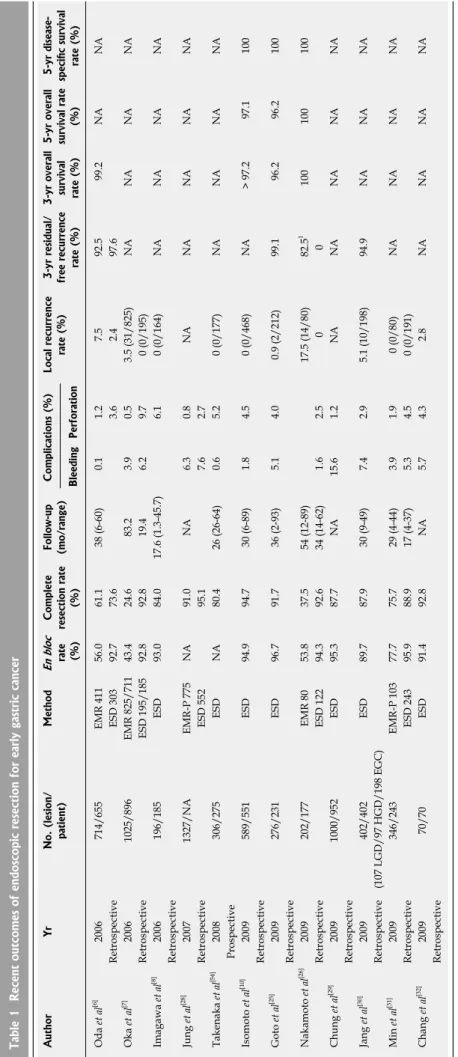
The results of recent studies have suggested that the technique of ESD achieves a high rate of en bloc resec- tion (92%-97%) and complete resection (73.6%-94.7%) with various rates of complications including bleeding (0.1%-15.6%) and perforation (1.2%-9.7%). Furthermore, they have revealed excellent long-term outcomes (5-year overall and disease-specific survival rates of 97.1% and 100%, respectively) (Table 1).
Japan
Oda et al[6] have performed a multicenter retrospective study to determine the nationwide results of endoscopic resection for EGC. Seven hundred and fourteen EGCs (EMR, 411; ESD, 303) that met the expanded criteria, ex- cept for the undifferentiated type of mucosal cancer, from 655 consecutive patients and 11 Japanese institutions were evaluated. Technically, 71.6% of the lesions were resected in one piece. The rate of en bloc resection by ESD (92.7%) was significantly higher than by EMR (56.0%). The rate of
Table 1 Recent outcomes of endoscopic resection for early gastric cancer AuthorYrNo. (lesion/ patient)MethodEn bloc rate (%) Complete resection rate (%) Follow-up (mo/range)Complications (%)Local recurrence rate (%)3-yr residual/ free recurrence rate (%) 3-yr overall survival rate (%) 5-yr overall survival rate (%)
5-yr disease- specific survival rate (%)BleedingPerforation Oda et al[6] 2006714/655EMR 41156.061.138 (6-60)0.11.27.592.5 99.2NANA RetrospectiveESD 30392.773.63.62.497.6 Oka et al[7]20061025/896EMR 825/71143.424.683.23.90.53.5 (31/825)NANANANA RetrospectiveESD 195/18592.892.819.46.29.70 (0/195) Imagawa et al[8] 2006196/185ESD93.084.017.6 (1.3-45.7)6.10 (0/164)NANANANA Retrospective Jung et al[28]20071327/NAEMR-P 775NA91.0NA6.30.8NANANANANA RetrospectiveESD 55295.17.62.7 Takenaka et al[54] 2008306/275ESDNA80.426 (26-64)0.65.20 (0/177)NANANANA Prospective Isomoto et al[10]2009589/551ESD94.994.730 (6-89)1.84.50 (0/468)NA > 97.2 97.1100 Retrospective Goto et al[25] 2009276/231ESD96.791.736 (2-93)5.14.00.9 (2/212)99.1 96.2 96.2100 Retrospective Nakamoto et al[26]2009202/177EMR 8053.837.554 (12-89)17.5 (14/80) 82.51100100100 RetrospectiveESD 12294.392.634 (14-62)1.62.500 Chung et al[29] 20091000/952ESD95.387.7NA15.61.2NANANANANA Retrospective Jang et al[30]2009402/402ESD89.787.930 (9-49)7.42.95.1 (10/198)94.9NANANA Retrospective (107 LGD/97 HGD/198 EGC) Min et al[31] 2009346/243EMR-P 10377.775.729 (4-44)3.91.90 (0/80)NANANANA RetrospectiveESD 24395.988.917 (4-37)5.34.50 (0/191) Chang et al[32]200970/70ESD91.492.8NA5.74.32.8NANANANA Retrospective 1Overall 5-year recurrence free rate. LGD: Low-grade dysplasia; HGD: High-grade dysplasia; NA: Not applicable; EGC: Early gastric cancer; ESD: Endoscopic submucosal dissection; EMR: Endoscopic mucosal resection; EMR-P: precutting followed by snare resection.
curative resection by ESD (73.6%) was significantly higher than by EMR (61.1%). Bleeding was found in 0.1% of cases. The frequency of perforation with ESD and EMR was 3.6% and 1.2%, respectively. All complications were managed endoscopically, and there was no procedure- related mortality. The median follow-up period was 3.2 years. The 3-year cumulative residual-free/recurrence-free rate in the ESD group (97.6%) was significantly higher than that in the EMR group (92.5%).
Oka et al[7] have reported on a comparative study between EMR and ESD of 1020 EGCs that met the ex- panded criteria. Eight hundred and twenty-five EMRs and 195 ESDs were performed. In cases without ulceration, the en bloc and curative resection rates were significantly higher with ESD (both 92.8%) than with EMR (43.4%
and 24.6%), regardless of tumor size. The average opera- tion time was significantly longer for ESD than for EMR (84.4 min vs 12.6 min), regardless of tumor size. In addi- tion, the frequency of intraoperative bleeding was signifi- cantly higher with ESD (22.6%) than with EMR (7.6%).
The frequency of delayed bleeding did not differ. No patient experienced recurrence after ESD.
Imagawa et al[8] have analyzed 196 EGCs that met the expanded criteria and were treated by ESD, in relation to lesion size, location and the presence or absence of ulcer- ation. The rate of en bloc resection was 93%, the curative resection rate was 84%, with a perforation rate of 6.1%, and a mean procedure time of 68 min. The rate of curative en bloc resection differed significantly depending on the lo- cation of the lesion (upper vs middle vs lower, 74% vs 77%
vs 91%), as well as on the size of the lesion (> 20 mm vs ≤ 20 mm, 59% vs 89%). There were also significant differenc- es in the mean procedure time in relation to the location of the lesion (upper vs middle vs lower, 105 min vs 81 min vs 45 min) and the size of the lesion (> 20 mm vs ≤ 20 mm, 124 min vs 55 min), as well as the presence of ulceration (positive vs negative, 97 min vs 65 min). They showed that the difficulty of ESD depends on the location and size of the lesion, as well as on the presence of ulceration.
Isomoto et al[10] have published the first long-term follow-up results of 589 EGCs treated by ESD, which met the expanded criteria. En bloc resection was achieved in 94.9%. Curative resection was achieved in 94.7%. En bloc resection was the only significant factor associated with curative ESD. Patients with a non-curative resection devel- oped local recurrence more frequently. The 5-year overall and disease-specific survival rates were 97.1% and 100%, respectively.
Goto et al[25] have carried out a retrospective investiga- tion of ESD to determine long-term outcomes. Two hun- dred and seventy-six node-negative EGCs that met the expanded criteria, except for the undifferentiated type of mucosal cancer, were enrolled. The en bloc and complete resection rates were 96.7% and 91.7%, respectively. Dur- ing a median follow-up of 3 years, there were two local recurrences (0.9%). The 5-year overall and disease-specific survival rates were 96.2% and 100%, respectively.
Nakamoto et al[26] have performed a comparative study
of EMR and ESD for 202 EGCs. The overall en bloc and complete resection rates were lower in patients undergoing EMR compared to ESD (en bloc: 53.8% vs 94.3%, complete:
37.5% vs 92.6%). The overall 5-year recurrence-free rate was lower in the EMR group than in the ESD group (82.5%
vs 100%). However, with regard to tumor size, EMR was comparable to ESD for the small lesions (< 5 mm).
Korea
The number of publications on ESD in Korea has been increasing. The results of ESD including the en bloc resec- tion rate, complete resection rate, and long-term follow-up survival rates are similar to those from Japan. The Health Insurance Review and Assessment Service of Korea have reported that 74 institutions, mainly tertiary hospitals, have performed ESD in 2008.
Kim et al[27] have published the first multicenter ret- rospective study of endoscopic resection in Korea. They collected 514 EGCs in 506 patients during January 2000 to December 2002 by use of the on-line database registry system. The most commonly used technique was circum- ferential precutting followed by snare resection (EMR-P, 52.3%). The second most common procedure was the in- jection and cut technique (24.3%). ESD was used only in 6.6% of cases at that time. Complete resection was con- firmed in 77.6% of the lesions, and the mean tumor size was 1.76 cm. However, ESD is now in the mainstream for endoscopic resection of early gastric lesions in Korea.
Jung et al[28] have reported an 11-year experience of endoscopic resection performed by a single endoscopist.
Seven hundred and seventy-five EMR procedures were performed after precutting (EMR-P) during the first 9 years and 552 ESDs over the following 2 years. The median spec- imen sizes were 33 mm for EMR-P and 45 mm for ESD.
The complete resection rates were 91% and 95.1% and the respective complication rates were 7.1% and 10.7%.
The Korean ESD study group has published a retro- spective six university hospital experience with ESD for 1000 gastric neoplasms[29]. The rate of en bloc resection and complete resection was 95.3% and 87.7%, respectively.
The rate of delayed bleeding and perforation was 15.6%
and 1.2%, respectively. The rate of en bloc resection differed significantly based on the location of the lesions and pres- ence of a scar. Procedure times were increased in cases in the upper stomach that had a large lesion (> 40 mm), with the presence of an ulcer, and the presence of a scar.
Jang et al[30] have reported the results of ESD for 402 gastrointestinal neoplasms at a single hospital. En bloc resection and complete resection were achieved in 89.7%
and 87.9%, respectively, and the local recurrence rate was 5.1%. The 3-year cancer-free survival rate was 94.9%.
Min et al[31] have published a comparative study of ESD and EMR after circumferential precutting (EMR-P) of 346 EGCs. En bloc resection and complete resection were achieved in 77.7% and 75.7% of the EMR-P group, respectively, and 95.9% and 88.9% of the ESD group. For EGCs > 20 mm, ESD demonstrated a significantly higher en bloc resection and complete resection rate compared to
EMR-P. In cases with completely resected differentiated cancer, neither group showed local recurrence during a median 29 and 17 mo follow-up, respectively.
Taiwan
Chang et al[32] have published a retrospective multicenter review of ESD of 70 EGCs in Taiwan. The en bloc resec- tion rate was 91.4%. The bleeding and perforation rates were 5.7% and 4.3%, respectively. Emergency surgery was performed in the patients with perforations. The local recurrence rate was 2.8%. Another small study has been published in Taiwan[33]; however, the results have indicated that the procedure requires more experience.
Western countries
As a result of the low incidence of EGC in the west, rela- tively few institutions use ESD and there have been only a few clinical studies on the outcomes[34-42]. However, the reported outcomes of western studies have not been sub- stantially different from those in eastern countries. Car- doso et al[35] have published an initial experience with ESD for 15 EGCs < 30 mm with no ulceration or scaring from Brazil. The mean procedure time was 140 min. The en bloc and complete resection rate was 80% and perforations occurred in 20% of cases. Catalano et al[36] have published their experience with EMR and ESD on 48 gastric lesions in Italy. After an initial experience with 36 EMRs, the pro- cedure was changed to ESD. Out of 36 EMR procedures, en bloc and complete resection were achieved in 72% and 56%, respectively. However, among the 12 ESD cases, the rates both increased to 92%. Bleeding and perforation occurred in one case each. Dinis-Ribeiro et al[37] have pub- lished the results of ESD for 19 gastric lesions in Europe.
ESD was performed under general anesthesia with a 79%
en bloc resection rate. Probst et al[38] have published ESD results for 71 epithelial or submucosal tumors, and have demonstrated a learning curve that resulted in a decrease in the procedure duration and increased rate of complete en bloc resection over time (65.7% to 72.2%). Although there have been a limited number of studies outside Asia, the number of endoscopy centers that are performing ESD is slowly increasing worldwide[43].
WhAT IS UNIqUE ABOUT ESD IN EASTERN ASIA?
In Eastern Asia, endoscopic resection methods have been widely accepted as the standard treatment for gastric tu- mors, and many trained endoscopists are familiar with EMR techniques. Compared to other organs including the esophagus or colon, tumors of the stomach are rela- tively easy to remove by endoscopic resection. The basic techniques of EMR overlap with those of ESD. This al- lows for a stepwise approach to a variety of lesions. For example, one might start with the frequently encountered easier lesions in the distal portion of the stomach, move to lesions in the proximal stomach, and then lesions in the esophagus or colon as the final step[9,44]. However, in the
west, because of the lower frequency of gastric cancer, endoscopists have relatively less experience with EMR techniques. In addition, the high incidence of Barrett’s esophagus, the treatment for which is technically de- manding, makes it difficult for beginners to gain extensive experience with ESD[45]. Training programs specifically aimed at advancing experience with ESD are useful[46,47]. Although data on the learning curve for ESD are limited, Choi et al[48] have shown that, for an experienced endos- copist, approximately 40 cases of gastric EMR with a circumferential mucosal incision in a low risk location are necessary for satisfactory training.
Kakushima et al[49] have reported a retrospective study on the learning curve for 383 ESD procedures by two principal operators and 11 (< 30 cases) endoscopists with less experience. For the two main operators, there was no significant difference between 25 consecutive patients with regard to the en bloc resection and complication rates. The size of the lesions increased as the number of patients in- creased, whereas the average procedure time decreased sig- nificantly. For the endoscopists with less experience, there was a similar treatment outcome and complication rate, mainly due to the easier location of the tumors in their cas- es. A constant rate of both treatment outcomes and com- plications was achieved over a 5-year period of experience with ESD. A decrease in the procedure time was found to be a marker for operator proficiency with this technique.
Yamamoto et al[44] have reported on the learning curve for three resident endoscopists that had already learned the basic procedures. They performed ESD under supervi- sion for 30 consecutive lesions each. They obtained a good overall complete resection rate of 93%, with an acceptable complication rate of 4.4% with appropriate supervision;
however, there was difficulty in achieving a sufficient rate of finishing up alone for submucosal dissection.
ESD was first performed in Korea in 1999. The Ko- rean Society of Gastrointestinal Endoscopy (KSGE) or- ganized the ESD research group to investigate and expand the ESD procedure nationwide in 2003. Prior to 2006, only 22 hospitals had the facilities to perform ESD. The KSGE developed ESD hands-on courses and traveled nationwide to introduce the ESD procedure and the devices with ani- mal models on eight occasions[50]. As a result, the number of registered ESD facilities increased to 77 according to data from National Health Insurance Review and Assess- ment Service in 2008. Furthermore, an international ESD live demonstration has been held every year since 2006.
Such support from the KSGE, including the ESD research group, a joint symposium with the Korean Pathology So- ciety, ESD live demonstrations, multicenter studies, and training models for teaching ESD have made it possible to standardize ESD guidelines and techniques in Korea.
ESD EAST TO WEST
Minimally invasive approaches provide a substantially bet- ter quality of life compared to conventional open surgery.
There is currently not enough long-term follow-up out- come data on ESD compared to open surgery; however,
if careful patient selection is maintained, excellent onco- logical outcomes with ESD are attainable by experienced endoscopists. Although ESD procedures have not been performed in western countries with as much experi- ence as in eastern countries, improved techniques can be achieved with additional experience. In spite of the low incidence of gastric lesions in the west, there is a relatively high incidence of colon lesions, including large sessile and flat polyps. The novice might start with a modified EMR technique including the adoption of some of the methods used for ESD, such as circumferential marginal incision, which has been used to teach ESD techniques during the past decade in Korea. Furthermore, training programs can be developed to teach ESD[47], and close collaboration be- tween western and Asian centers and attending live dem- onstrations can help to gain such experience. On the other hand, modern medical science and technology continue to develop at a rapid pace. The recent development of ac- cessories and traction devices might help in the acquisition of the skill and experience needed to make ESD easier to learn and apply[51-53].
CONClUSION
ESD is an effective and safe therapeutic modality for man- agement of early gastrointestinal tract neoplasms, although it has a relatively longer operation time and high risk of complications. In spite of the late start for ESD in the west, the results have been being similar to those from Japan. In the same manner, although there is a problem to overcome the flat learning curve in view of the low number of detect- ed cases in western countries, close collaboration between western and Asian centers is required for improvement of the ESD technique and its clinical application.
REFERENCES
1 Nakamura K, Ueyama T, Yao T, Xuan ZX, Ambe K, Adachi Y, Yakeishi Y, Matsukuma A, Enjoji M. Pathology and prog- nosis of gastric carcinoma. Findings in 10,000 patients who underwent primary gastrectomy. Cancer 1992; 70: 1030-1037 2 Park IS, Lee YC, Kim WH, Noh SH, Lee KS, Kim H. Clini- copathologic characteristics of early gastric cancer in Korea.
Yonsei Med J 2000; 41: 607-614
3 Tsubono Y, Hisamichi S. Screening for gastric cancer in Ja- pan. Gastric Cancer 2000; 3: 9-18
4 Kojima T, Parra-Blanco A, Takahashi H, Fujita R. Outcome of endoscopic mucosal resection for early gastric cancer: re- view of the Japanese literature. Gastrointest Endosc 1998; 48:
550-554; discussion 550-554
5 Soetikno R, Kaltenbach T, Yeh R, Gotoda T. Endoscopic mucosal resection for early cancers of the upper gastrointes- tinal tract. J Clin Oncol 2005; 23: 4490-4498
6 Oda I, Saito D, Tada M, Iishi H, Tanabe S, Oyama T, Doi T, Otani Y, Fujisaki J, Ajioka Y, Hamada T, Inoue H, Gotoda T, Yoshida S. A multicenter retrospective study of endoscopic resection for early gastric cancer. Gastric Cancer 2006; 9:
262-270
7 Oka S, Tanaka S, Kaneko I, Mouri R, Hirata M, Kawamura T, Yoshihara M, Chayama K. Advantage of endoscopic sub- mucosal dissection compared with EMR for early gastric cancer. Gastrointest Endosc 2006; 64: 877-883
8 Imagawa A, Okada H, Kawahara Y, Takenaka R, Kato J,
Kawamoto H, Fujiki S, Takata R, Yoshino T, Shiratori Y.
Endoscopic submucosal dissection for early gastric cancer:
results and degrees of technical difficulty as well as success.
Endoscopy 2006; 38: 987-990
9 Gotoda T, Yamamoto H, Soetikno RM. Endoscopic submu- cosal dissection of early gastric cancer. J Gastroenterol 2006;
41: 929-942
10 Isomoto H, Shikuwa S, Yamaguchi N, Fukuda E, Ikeda K, Nishiyama H, Ohnita K, Mizuta Y, Shiozawa J, Kohno S.
Endoscopic submucosal dissection for early gastric cancer: a large-scale feasibility study. Gut 2009; 58: 331-336
11 Japanese Classification of Gastric Carcinoma - 2nd English Edition. Gastric Cancer 1998; 1: 10-24
12 Itoh H, Oohata Y, Nakamura K, Nagata T, Mibu R, Na- kayama F. Complete ten-year postgastrectomy follow-up of early gastric cancer. Am J Surg 1989; 158: 14-16
13 Uedo N, Iishi H, Tatsuta M, Ishihara R, Higashino K, Takeu- chi Y, Imanaka K, Yamada T, Yamamoto S, Yamamoto S, Tsukuma H, Ishiguro S. Longterm outcomes after endo- scopic mucosal resection for early gastric cancer. Gastric Cancer 2006; 9: 88-92
14 Shimada Y. JGCA (The Japan Gastric Cancer Association).
Gastric cancer treatment guidelines. Jpn J Clin Oncol 2004;
34: 58
15 Yamao T, Shirao K, Ono H, Kondo H, Saito D, Yamaguchi H, Sasako M, Sano T, Ochiai A, Yoshida S. Risk factors for lymph node metastasis from intramucosal gastric carci- noma. Cancer 1996; 77: 602-606
16 Gotoda T, Yanagisawa A, Sasako M, Ono H, Nakanishi Y, Shimoda T, Kato Y. Incidence of lymph node metastasis from early gastric cancer: estimation with a large number of cases at two large centers. Gastric Cancer 2000; 3: 219-225 17 Ishikawa S, Togashi A, Inoue M, Honda S, Nozawa F, Toya-
ma E, Miyanari N, Tabira Y, Baba H. Indications for EMR/
ESD in cases of early gastric cancer: relationship between histological type, depth of wall invasion, and lymph node metastasis. Gastric Cancer 2007; 10: 35-38
18 Nagano H, Ohyama S, Fukunaga T, Hiki N, Seto Y, Yama- guchi T, Kato Y, Yamaguchi A. Two rare cases of node-pos- itive differentiated gastric cancer despite their infiltration to sm1, their small size, and lack of lymphatic invasion into the submucosal layer. Gastric Cancer 2008; 11: 53-57; discussion 57-58
19 Jee YS, Hwang SH, Rao J, Park DJ, Kim HH, Lee HJ, Yang HK, Lee KU. Safety of extended endoscopic mucosal resec- tion and endoscopic submucosal dissection following the Japanese Gastric Cancer Association treatment guidelines.
Br J Surg 2009; 96: 1157-1161
20 Abe N, Watanabe T, Sugiyama M, Yanagida O, Masaki T, Mori T, Atomi Y. Endoscopic treatment or surgery for undif- ferentiated early gastric cancer? Am J Surg 2004; 188: 181-184 21 Ha TK, An JY, Youn HK, Noh JH, Sohn TS, Kim S. Indica-
tion for endoscopic mucosal resection in early signet ring cell gastric cancer. Ann Surg Oncol 2008; 15: 508-513
22 Park YD, Chung YJ, Chung HY, Yu W, Bae HI, Jeon SW, Cho CM, Tak WY, Kweon YO. Factors related to lymph node metastasis and the feasibility of endoscopic mucosal resection for treating poorly differentiated adenocarcinoma of the stomach. Endoscopy 2008; 40: 7-10
23 Ye BD, Kim SG, Lee JY, Kim JS, Yang HK, Kim WH, Jung HC, Lee KU, Song IS. Predictive factors for lymph node me- tastasis and endoscopic treatment strategies for undifferenti- ated early gastric cancer. J Gastroenterol Hepatol 2008; 23: 46-50 24 Kang HY, Kim SG, Kim JS, Jung HC, Song IS. Clinical out- comes of endoscopic submucosal dissection for undifferen- tiated early gastric cancer. Surg Endosc 2010; 24: 509-516 25 Goto O, Fujishiro M, Kodashima S, Ono S, Omata M. Out-
comes of endoscopic submucosal dissection for early gastric cancer with special reference to validation for curability cri- teria. Endoscopy 2009; 41: 118-122
26 Nakamoto S, Sakai Y, Kasanuki J, Kondo F, Ooka Y, Kato K,
Arai M, Suzuki T, Matsumura T, Bekku D, Ito K, Tanaka T, Yokosuka O. Indications for the use of endoscopic mucosal resection for early gastric cancer in Japan: a comparative study with endoscopic submucosal dissection. Endoscopy 2009; 41: 746-750
27 Kim JJ, Lee JH, Jung HY, Lee GH, Cho JY, Ryu CB, Chun HJ, Park JJ, Lee WS, Kim HS, Chung MG, Moon JS, Choi SR, Song GA, Jeong HY, Jee SR, Seol SY, Yoon YB. EMR for early gastric cancer in Korea: a multicenter retrospective study. Gastrointest Endosc 2007; 66: 693-700
28 Jung HY, Choi KD, Song HJ, Lee GH, Kim JH. Risk man- agement in endoscopic submucosal dissection using needle knife in Korea. Dig Endosc 2007; 19 Suppl 1: S5-S8
29 Chung IK, Lee JH, Lee SH, Kim SJ, Cho JY, Cho WY, Hwangbo Y, Keum BR, Park JJ, Chun HJ, Kim HJ, Kim JJ, Ji SR, Seol SY. Therapeutic outcomes in 1000 cases of endo- scopic submucosal dissection for early gastric neoplasms:
Korean ESD Study Group multicenter study. Gastrointest Endosc 2009; 69: 1228-1235
30 Jang JS, Choi SR, Qureshi W, Kim MC, Kim SJ, Jeung JS, Han SY, Noh MH, Lee JH, Lee SW, Baek YH, Kim SH, Choi PJ. Long-term outcomes of endoscopic submucosal dissec- tion in gastric neoplastic lesions at a single institution in South Korea. Scand J Gastroenterol 2009; 44: 1315-1322 31 Min BH, Lee JH, Kim JJ, Shim SG, Chang DK, Kim YH,
Rhee PL, Kim KM, Park CK, Rhee JC. Clinical outcomes of endoscopic submucosal dissection (ESD) for treating early gastric cancer: comparison with endoscopic mucosal resec- tion after circumferential precutting (EMR-P). Dig Liver Dis 2009; 41: 201-209
32 Chang CC, Lee IL, Chen PJ, Wang HP, Hou MC, Lee CT, Chen YY, Cho YP, Lin JT. Endoscopic submucosal dissec- tion for gastric epithelial tumors: a multicenter study in Tai- wan. J Formos Med Assoc 2009; 108: 38-44
33 Lee IL, Wu CS, Tung SY, Lin PY, Shen CH, Wei KL, Chang TS. Endoscopic submucosal dissection for early gastric can- cers: experience from a new endoscopic center in Taiwan. J Clin Gastroenterol 2008; 42: 42-47
34 Rösch T, Sarbia M, Schumacher B, Deinert K, Frimberger E, Toermer T, Stolte M, Neuhaus H. Attempted endoscopic en bloc resection of mucosal and submucosal tumors using in- sulated-tip knives: a pilot series. Endoscopy 2004; 36: 788-801 35 Cardoso DM, Campoli PM, Yokoi C, Ejima FH, Barreto PA, de Brito AM, Mota ED, de Fraga Júnior AC, da Mota OM.
Initial experience in Brazil with endoscopic submucosal dis- section for early gastric cancer using insulation-tipped knife:
a safety and feasibility study. Gastric Cancer 2008; 11: 226-232 36 Catalano F, Trecca A, Rodella L, Lombardo F, Tomezzoli A,
Battista S, Silano M, Gaj F, de Manzoni G. The modern treat- ment of early gastric cancer: our experience in an Italian cohort. Surg Endosc 2009; 23: 1581-1586
37 Dinis-Ribeiro M, Pimentel-Nunes P, Afonso M, Costa N, Lopes C, Moreira-Dias L. A European case series of endo- scopic submucosal dissection for gastric superficial lesions.
Gastrointest Endosc 2009; 69: 350-355
38 Probst A, Golger D, Arnholdt H, Messmann H. Endoscopic submucosal dissection of early cancers, flat adenomas, and submucosal tumors in the gastrointestinal tract. Clin Gastro- enterol Hepatol 2009; 7: 149-155
39 Cipolletta L, Rotondano G, Bianco MA, Garofano ML, Meucci C, Prisco A, Cipolletta F, Piscopo R. Self-assembled hydro-jet system for submucosal elevation before endoscop- ic resection of nonpolypoid colorectal lesions (with video).
Gastrointest Endosc 2009; 70: 1018-1022
40 Hurlstone DP, Atkinson R, Sanders DS, Thomson M, Cross SS, Brown S. Achieving R0 resection in the colorectum us- ing endoscopic submucosal dissection. Br J Surg 2007; 94:
1536-1542
41 Hurlstone DP, Fu KI, Brown SR, Thomson M, Atkinson R, Tiffin N, Cross SS. EMR using dextrose solution versus so- dium hyaluronate for colorectal Paris type I and 0-II lesions:
a randomized endoscopist-blinded study. Endoscopy 2008;
40: 110-114
42 Smith LA, Baraza W, Tiffin N, Cross SS, Hurlstone DP. En- doscopic resection of adenoma-like mass in chronic ulcer- ative colitis using a combined endoscopic mucosal resection and cap assisted submucosal dissection technique. Inflamm Bowel Dis 2008; 14: 1380-1386
43 Neuhaus H. Endoscopic submucosal dissection in the upper gastrointestinal tract: present and future view of Europe.
Dig Endosc 2009; 21 Suppl 1: S4-S6
44 Yamamoto S, Uedo N, Ishihara R, Kajimoto N, Ogiyama H, Fukushima Y, Yamamoto S, Takeuchi Y, Higashino K, Iishi H, Tatsuta M. Endoscopic submucosal dissection for early gastric cancer performed by supervised residents: assessment of feasibility and learning curve. Endoscopy 2009; 41: 923-928 45 Bergman JJ. How to justify endoscopic submucosal dissec-
tion in the Western world. Endoscopy 2009; 41: 988-990 46 Neuhaus H, Costamagna G, Devière J, Fockens P, Ponchon T,
Rösch T. Endoscopic submucosal dissection (ESD) of early neoplastic gastric lesions using a new double-channel endo- scope (the “R-scope”). Endoscopy 2006; 38: 1016-1023 47 Vázquez-Sequeiros E, de Miquel DB, Olcina JR, Martín JA,
García M, Lucas DJ, Garrido E, González C, Blanco AP, Arnau MR, Buenadicha A, Vicente VM, de Argila CM, Milicua JM.
Training model for teaching endoscopic submucosal dissec- tion of gastric tumors. Rev Esp Enferm Dig 2009; 101: 546-552 48 Choi IJ, Kim CG, Chang HJ, Kim SG, Kook MC, Bae JM. The
learning curve for EMR with circumferential mucosal inci- sion in treating intramucosal gastric neoplasm. Gastrointest Endosc 2005; 62: 860-865
49 Kakushima N, Fujishiro M, Kodashima S, Muraki Y, Tatei- shi A, Omata M. A learning curve for endoscopic submu- cosal dissection of gastric epithelial neoplasms. Endoscopy 2006; 38: 991-995
50 Cho JY, Cho WY. Toward the global standardization of en- doscopic submucosal dissection proposal for 10 years from now - present and future view of Korea. Dig Endosc 2009; 21 Suppl 1: S2-S3
51 Gotoda T, Oda I, Tamakawa K, Ueda H, Kobayashi T, Kak- izoe T. Prospective clinical trial of magnetic-anchor-guided endoscopic submucosal dissection for large early gastric cancer (with videos). Gastrointest Endosc 2009; 69: 10-15 52 Jeon WJ, You IY, Chae HB, Park SM, Youn SJ. A new tech-
nique for gastric endoscopic submucosal dissection: peroral traction-assisted endoscopic submucosal dissection. Gastro- intest Endosc 2009; 69: 29-33
53 Sakurazawa N, Kato S, Miyashita M, Kiyama T, Fujita I, Yamashita N, Saitou Y, Tajiri T, Uchida E. An innovative technique for endoscopic submucosal dissection of early gastric cancer using a new spring device. Endoscopy 2009; 41:
929-933
54 Takenaka R, Kawahara Y, Okada H, Hori K, Inoue M, Kawa- no S, Tanioka D, Tsuzuki T, Yagi S, Kato J, Uemura M, Ohara N, Yoshino T, Imagawa A, Fujiki S, Takata R, Yamamoto K.
Risk factors associated with local recurrence of early gastric cancers after endoscopic submucosal dissection. Gastrointest Endosc 2008; 68: 887-894
S- Editor Sun H L- Editor Kerr C E- Editor Ma WH