CASE REPORT
Subacute Thyroiditis Presenting as a Focal Lesion on [ 18 F] Fluorodeoxyglucose Whole-Body
Positron-Emission Tomography/CT
S.H. Yeo S.K. Lee I. Hwang E.J. Ahn
SUMMARY:A 52-year-old man presented with subacute thyroiditis and showed an intensely hyper- metabolic thyroid lesion on18F-FDG PET/CT. The patient also had coexisting chronic lymphocytic thyroiditis. The lesion of subacute thyroiditis revealed an SUVmaxof 9.1 g/mL and was ill-defined, heterogeneous, markedly hypoechoic, and hypovascular on US. Biopsy demonstrated histologic findings of subacute thyroiditis, which should be included in the differential diagnosis of an intensely hypermetabolic thyroid lesion on18F-FDG PET/CT.
ABBREVIATIONS:18F-FDG PET/CT ⫽ [18F] fluorodeoxyglucose whole-body positron-emission to- mography and CT; FDG⫽ fluorodeoxyglucose; US ⫽ sonography; PD ⫽ power Doppler; PTC ⫽ papillary thyroid carcinoma; SUVmax⫽ maximum standardized uptake value; US-FNAB ⫽ sonogra- phy-guided fine-needle aspiration biopsy
S ubacute thyroiditis is an uncommon inflammatory dis- ease of the thyroid gland that occurs most often in middle- aged women. It is usually preceded by an upper respiratory tract infection and is presumed to be caused by a viral infec- tion. A postviral inflammatory response leads to giant cell in- filtration into the thyroid follicles, ultimately resulting in fol- licular swelling, which in turn results in stretching of the thyroid capsule with subsequent pain and tenderness on pal- pation, symptoms that are characteristic clinical features of this illness.1It is a self-limited illness, which often resolves spontaneously. Thyroid US is useful for the initial diagnosis and follow-up of patients with subacute thyroiditis.
2,3
Thyroid lesions identified by combined
18F-FDG PET/CT can create a dilemma for both patients and physicians; the probability of malignancy in thyroid focal uptake on
18F-FDG PET/CT ranges from 14% to 63%.
4,5Diffuse FDG uptake can be an indicator of chronic lymphocytic thyroiditis,
6which can also present as focal FDG uptake, mimicking thyroid malig- nancy.
7Cases of subacute thyroiditis presenting as diffuse FDG uptake have also been reported
8,9; however, focal FDG uptake in the case of subacute thyroiditis has not appeared in the English literature, to our knowledge.
We report a case of subacute thyroiditis presenting as an intensely hypermetabolic thyroid lesion on
18F-FDG PET/CT, along with findings at US and US-FNAB that mimicked a ma- lignancy of the thyroid gland, in a patient with coexisting chronic lymphocytic thyroiditis.
Case Report
A 52-year-old man was referred to our department for further evalu- ation of an incidentally identified hypermetabolic mass in the right thyroid lobe on18F-FDG PET/CT. Physical examination was unre- markable except for a tender palpable mass at the right anterior neck.
Approximately 17 days prior, he was admitted to our medical center for 2 days for general aches and mild febrile sensation. Findings of his thyroid function test and serum radioimmunoassay were normal ex- cept for elevated antithyroglobulin antibodies (153.7 IU/mL [normal,
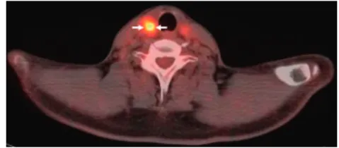
⬍70 IU/mL]). An 18F-FDG PET/CT was performed for cancer screening, which showed an intensely hypermetabolic mass with an SUVmaxof 9.1 g/mL in the right thyroid lobe (Fig 1), which had not been present on the previous18F-FDG PET/CT performed 20 months earlier. Background thyroid parenchyma showed mild diffuse FDG uptake in both thyroid lobes, which suggested chronic lymphocytic thyroiditis (Fig 1).
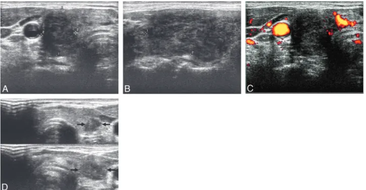
Thyroid US, performed 1 day after the18F-FDG PET/CT exami- nation, demonstrated an approximately 21⫻ 19 ⫻ 37 mm mass in the right thyroid lobe (Fig 2A⫺C), the location of which coincided with the hypermetabolic focus on18F-FDG PET/CT. The mass was ill- defined, markedly hypoechoic, and heterogeneous, with scattered hy- perechoic areas on gray-scale US (Fig 2A, -B), and was hypovascular compared with the background parenchyma on PD US (Fig 2C). Ten- derness was noted just on compression of the mass. These sono- graphic findings suggested subacute thyroiditis. In addition, 2 small focal ill-defined markedly hypoechoic nodular lesions were present in the left thyroid lobe (Fig 2D). They could not be distinguished from background thyroid parenchyma that showed mild diffuse FDG up- take on18F-FDG PET/CT. We performed US-FNAB of the right thy- roid mass because the patient and referring physician were anxious about the newly developed intensely hypermetabolic focus on18F- FDG PET/CT and because we could not definitively exclude infiltra- tive malignancies such as PTC or primary thyroid lymphoma.
US-FNAB was also performed on the 2 small nodular lesions of the left thyroid lobe to exclude PTC. The results of US-FNAB of the
Received September 24, 2009; accepted after revision December 2.
From the Departments of Radiology (S.H.Y., S.K.L., E.J.A.) and Pathology (I.H.), Dongsan Medical Center, Keimyung University School of Medicine, Daegu, South Korea.
Please address correspondence to Sang Kwon Lee, MD, Department of Radiology, Dongsan Medical Center, Keimyung University School of Medicine, 194 Dongsan-dong, Jung-gu, Daegu 700 –712, South Korea; e-mail: [email protected]
DOI 10.3174/ajnr.A2017
Fig 1.18F-FDG PET/CT image demonstrates an intensely hypermetabolic mass (SUVmax⫽ 9.1 g/mL) in the right thyroid lobe (arrows). Background thyroid parenchyma shows mild diffuse FDG uptake.
E58 Yeo 兩 AJNR 32 兩 Apr 2011 兩 www.ajnr.org
right and left thyroid lesions were consistent with subacute thyroiditis (Fig 3A) and chronic lymphocytic thyroiditis (Fig 3B), respectively.
Discussion
In subacute thyroiditis, the clinical and laboratory data are usually sufficient for making the diagnosis, and biopsy is rarely indicated. Our case presented a clinical dilemma because of unexpected focal, newly developed, intense FDG uptake of the lesion of subacute thyroiditis on
18F-FDG PET/CT, which raised clinical concern about thyroid malignancy.
Focal FDG uptake in the thyroid gland is likely caused by malignancy.
10However, focal FDG uptake may also be attrib- uted to several benign conditions, including nodular hyper- plasia, Hu¨rthle cell and follicular neoplasms, and even chronic lymphocytic thyroiditis.
7,10Subacute thyroiditis can involve 1 or both lobes of the thy- roid gland diffusely or focally.
11Several reports concerning US
features of subacute thyroiditis have been published in the English literature.
2,3In a study of 27 patients with subacute thyroiditis,
2unilateral thyroid involvement was demonstrated in 23 and bilateral involvement, in 4 patients. All the lesions were ill-defined and hypoechoic. Nine patients had a hy- poechoic nodular lesion with an irregular or microlobulated margin, which mimicked thyroid carcinoma. No hypervascu- larity was noted in any of the lesions on color or PD US.
Zacharia et al
3reported the utility of color Doppler US in a case of subacute thyroiditis that mimicked thyroid carcinoma clinically. They reported that color Doppler US showed no hypervascularity at the acute stage and slightly increased vas- cularity at the recovery stage.
Two cases of subacute thyroiditis with diffuse hypermetab- olism on
18F-FDG PET, involving a single lobe and both lobes of the thyroid gland, respectively, have been reported.
8,9How- ever, cases of subacute thyroiditis with focal hypermetabolism
Fig 2. A, Transverse gray-scale US reveals a markedly hypoechoic mass with a taller-than-wide shape in the right thyroid lobe (delineated by electronic calipers). B, A longitudinal gray-scale US shows an ill-demarcated heterogeneous mass with scattered hyperechoic areas (delineated by electronic calipers). C, A transverse PD US demonstrates hypovascularity of the mass compared with the background thyroid parenchyma. D, Split transverse gray-scale USs show 2 small focal ill-defined markedly hypoechoic nodular lesions in the left thyroid lobe (arrows).
Fig 3. A, Photographs of the cytologic examination of the specimen obtained by US-FNAB of the right thyroid mass show several multinucleated giant cells (arrows) with the background of smaller inflammatory cells, consistent with subacute thyroiditis (Papanicolaou, original magnification⫻200). B, Photomicrograph of the cytologic examination of the specimen obtained by US-FNAB of 1 of the 2 left thyroid nodules reveals a lymphoid tangle (arrow) in the background of small lymphoid cells, suggestive of chronic lymphocytic thyroiditis (Papanicolaou, original magnification⫻200).
HEAD&NECKCASEREPORT
AJNR Am J Neuroradiol 32:E58 –E60 兩 Apr 2011 兩 www.ajnr.org E59
have not appeared in the English literature. Our case repre- sents the first of subacute thyroiditis that showed focal hyper- metabolism on
18F-FDG PET/CT, mimicking a thyroid malig- nancy. In view of clinical and US features, subacute thyroiditis was suggested as a probable diagnosis. However, because the patient and referring physician were anxious about the newly developed intensely hypermetabolic focus on
18F-FDG PET/CT and because we could not definitively exclude infil- trative malignancies such as PTC and primary thyroid lym- phoma, we proceeded to US-FNAB. The results of cytologic examination were consistent with subacute thyroiditis. In ad- dition, 2 small nodular lesions of the left thyroid lobe proved to be chronic lymphocytic thyroiditis by US-FNAB.
Thus, this case represents coexistence of subacute thyroid- itis and chronic lymphocytic thyroiditis. Interestingly, the le- sion of subacute thyroiditis showed intense hypermetabolism, while the lesions of focal chronic lymphocytic thyroiditis were not significantly hypermetabolic compared with the back- ground parenchyma. We hypothesized that the intense hyper- metabolism of the lesion of subacute thyroiditis might be caused by active inflammation, while the lesions of focal chronic lymphocytic thyroiditis might be relatively quiescent;
thus they did not show hypermetabolism on
18F-FDG PET/CT.
In conclusion, subacute thyroiditis should be included in the differential diagnosis of focal FDG uptake on
18F-FDG PET/CT. Gray-scale and PD US are useful for the character-
ization of this lesion, and US-FNAB can provide a definitive diagnosis of subacute thyroiditis that presents as an intensely hypermetabolic thyroid incidentaloma on
18F-FDG PET/CT.
References
1. Volpe´ R. Subacute thyroiditis. In: Burrow GN, Oppenheimer JH, Volpe´ R, eds.
Thyroid Function and Disease. Philadelphia: Saunders; 1989:179 – 87 2. Park SY, Kim EK, Kim MJ, et al. Ultrasonographic characteristics of subacute
granulomatous thyroiditis. Korean J Radiol 2006;7:229 –34
3. Zacharia TT, Perumpallichira JJ, Sindhwani V, et al. Gray-scale and color Doppler sonographic findings in a case of subacute granulomatous thyroid- itis mimicking thyroid carcinoma. J Clin Ultrasound 2002;30:442– 44 4. King DL, Stack BC Jr, Spring PM, et al. Incidence of thyroid carcinoma in
fluorodeoxyglucose positron emission tomography-positive thyroid inciden- talomas. Otolaryngol Head Neck Surg 2007;137:400 – 04
5. Van den Bruel A, Maes A, De Potter T, et al. Clinical relevance of thyroid fluorodeoxyglucose-whole body positron emission tomography incidenta- loma. J Clin Endocrinol Metab 2002;87:1517–20
6. Yasuda S, Shohtsu A, Ide M, et al. Chronic thyroiditis: diffuse uptake of FDG at PET. Radiology 1998;207:775–78
7. Schmid DT, Kneifel S, Stoeckli SJ, et al. Increased 18F-FDG uptake mimicking thyroid cancer in a patient with Hashimoto’s thyroiditis. Eur Radiol 2003;13:
2119 –21
8. Song YS, Jang SJ, Chung JK, et al. F-18 fluorodeoxyglucose (FDG) positron emission tomography (PET) and Tc-99m pertechnate scan findings of a pa- tient with unilateral subacute thyroiditis. Clin Nucl Med 2009;34:456 –58 9. Meller J, Sahlmann CO, Scheel AK. 18F-FDG PET and PET/CT in fever of
unknown origin. J Nucl Med 2007;48:35– 45
10. Kim TY, Kim WB, Ryu JS, et al. 18F-fluorodeoxyglucose uptake in thyroid from positron emission tomogram (PET) for evaluation in cancer patients:
high prevalence of malignancy in thyroid PET incidentaloma. Laryngoscope 2005;115:1074 –78
11. Greene JN. Subacute thyroiditis. Am J Med 1971;51:97–108
E60 Yeo 兩 AJNR 32 兩 Apr 2011 兩 www.ajnr.org