대한임상신경생리학회지 10(1):66~69,2007 ISSN 1229-6414
Copyright 2008 by The Korean Society for Clinical Neurophysiology 66
유전자분석으로 진단한 얼굴어깨위팔근육디스트로피 1예
한양대학교 의과대학 신경과학교실, 성균관의대 삼성서울병원 진단검사의학과1
이석호․기창석1․이승철․박진석․고성호․이규용
A Case of Facioscapulohumeral Muscular Dystrophy Confirmed by Genetic Analysis
Seok-Ho Lee, M.D., Chang-Seok Ki, M.D., Ph.D.1, Seung-Chul Lee, M.D., Jin-seok Park, M.D., Seong-Ho Koh, M.D., Ph.D., Kyu-Yong Lee, M.D., Ph.D.
Department of Neurology, College of Medicine, Hanyang University, Guri, Korea
1
Department of Laboratory Medicine, Sungkyunkwan University School of Medicine, Samsung Medical Center, Seoul, Korea
Received 6 April 2008; received in revised form 3 June 2008; accepted 4 June 2008.
Facioscapulohumeral muscular dystrophy (FSHD), the third most common inherited muscular dystrophy, is an auto- somal dominant disease characterized by progressive weakness and wasting of the facial, shoulder-girdle, upper arm, foot extensor, and pelvic girdle muscles. FSHD is caused by contraction of the polymorphic D4Z4 repeat in the subtelomere of chromosome 4q. However, there has been no report of genetically confirmed FSHD in Korea. We report a patient with FSHD who was found to have a deletion of D4Z4 repeat on chromosome 4q35.
Key Words: Facioscapulohumeral muscular dystrophy, D4Z4 repeat
Address for correspondence;
Kyu-Yong Lee, M.D. Ph.D.
Department of Neurology, Hanyang University Guri Hospital
249-1 Gyomun-dong, Guri-si, Gyeonggi-do, 471-701, Korea Tel: +82-31-560-2260 Fax: +82-31-560-2261
E-mail: [email protected]
얼굴어깨위팔근육디스트로피(facioscapulohumeral muscular dystrophy, FSHD)는 1884년 Landouzy와 Dejerine이 처음으로 기술한 질환으로 대개 30세 이전에 발병하고 초기에 안면근육과 어깨주위근육(shoulder gir- dle muscles)을 침범한다. 상염색체 우성의 유전형태를 가지고 있으나 10~30%에서는 산발형으로 발생한다.
1국내에서는 임상증상과 유전형태를 바탕으로 얼굴어깨
위팔근육디스트로피를 진단한 보고가 몇 차례 있었으나, 아직까지 얼굴어깨위팔근육디스트로피를 유전자분석으로 진단한 보고는 없었다.
2-4저자들은 가족력이 전혀 없는 안면근육과 어깨주위근육 의 위약을 보인 환자에서 유전자분석을 통하여 얼굴어깨 위팔근육디스트로피로 확진한 1예를 경험하였기에 이를 보고한다.
증 례
42세 남자가 중학교 때부터 발생하여 느리게 진행하는
보행장애와 사지의 위약감을 주소로 내원하였다. 환자는
중학교 시절부터 우측 어깨의 근위약이 서서히 진행하였
고, 5년 전부터 일을 하고 나서 심해지는 하지의 위약과 통
유전자분석으로 진단한 얼굴어깨위팔근육디스트로피 1예
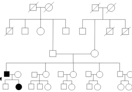
Korean J Clin Neurophysiol / Volume 10 / June, 2008 67 Figure 1. Pedigree of facioscapulohu- meral muscular dystrophy family. The dark filled symbols represent affected male ( ■ ) and female ( ● ).
Figure 2. Hematoxylin and Eosin(H&E) stain of muscle biopsy (×200). The muscle biopsy also shows nonspecific myopathic changes, often with inflammatory infiltrates.
증이 있었으며, 1~2년 전부터 계단 올라가기가 힘들었다.
2남 3녀 중 장남으로 아들과 딸, 두 명의 자녀를 두고 있었 다. 아버지와 어머니 형제들 중에서 근위약을 호소하는 사 람은 없었고 자녀들도 특별한 증상이 없었다(Fig. 1). 입원 당시 혈압은 120/80 mmHg이었고 다른 생체 징후는 정상 이었으며, 이학적검사도 정상이었다. 신경학적검사에서 의 식은 명료하였고, 언어기능은 정상이었으며, MMSE (mini- mental state examination)검사는 30점/30점이었다. 양 측 동공반사와 안구운동은 정상이었고 안저검사도 정상이 었다. 안면의 감각은 정상이었으나. 얼굴의 표정이 없었으 며, 휘파람불기나 풍선불기를 하지 못하였다. 발음곤란, 삼킴곤란은 없었고, 목젖과 혀의 편위는 보이지 않았다.
경부근위축이 심했으며, 양측에 익상견갑(winging sca- pula)이 있었고, 돌출된 하복부와 요추전만증(lumbar lordosis)이 있었다. 양상지의 근육은 상완이두근(biceps brachii)과 어깨세모근(deltoid), 극상근육(supraspina- tus)의 위축이 있었고, 양하지의 근육은 외측광근(vastus lateralis)과 전경골근(tibialis anterior)의 위축이 있었 다. 양측 상하지의 근력은 MRC grade IV 정도로 굽힘운 동시 근력약화가 더 심하였다. 사지의 감각과 건반사는 정 상이었고 바빈스키징후는 없었다. 소뇌기능은 정상이었고, 보행 시 근육병증보행(waddling gait)을 보이고 있었다.
입원 당일 검사한 혈청 젖산탈수효소, 혈청 크레아틴키 나아제, 갑상선호르몬은 정상이었다. 심전도검사와 청력 검사는 모두 정상이었으나, 폐기능검사에서 최대 환기량 (maximal voluntary ventilation, MVV)이 85.4 L/min 으로 경한 제한성 환기장애(mild restrictive pattern)가 있 었다. 신경전도속도검사에서 우측 정중신경(median ner- ve), 자신경(ulnar nerve)의 운동신경전도속도와 감각신
경전도속도는 정상이었고, 종아리신경(peroneal nerve)과 뒤정강신경(posterior tibial nerve)의 운동신경전속도와 장딴지신경(sural nerve)의 감각신경전도속도도 정상이었 다. 근전도검사는 우측 상완이두근, 어깨세모근, 외측광근 및 전경골근에서 저진폭의 다상성전위(small amplitude, polyphasic MUP pattern)를 보여 근육병증임을 확인하 였다.
좌측 어깨세모근에서 근생검(muscle biopsy)을 하였다.
광학현미경에서 산재되어 변성되고 괴사된 근섬유들(sca-
ttered degenerating and necrotic myofibers)과 소수의
재생되는 근섬유들(a few regenerating myofibers)이 보
이고, 혈관주위와 근내막에 염증세포침윤(perivascular
and endomysial inflammatory cell infiltration)이 있어
이석호
․
기창석․
이승철․
박진석․
고성호․
이규용Korean J Clin Neurophysiol / Volume 10 / June, 2008 68
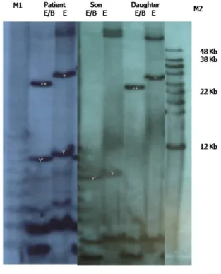
Figure 3. Southern blot analysis result. Convential electrophoresis and Southern blot analysis of EcoRI (E) and EcoRI/BlnI (E/B) digested DNA from the patient and daughter revealed a 28-kb sized EcoRI fragment (*), which is reduced to 25-kb fragment by double indigestion (lanes labeled E/B, **). M1, 1 kb size marker; M2, high molecular weight size marker.
비특이적 근육병증 소견에 합당하였다(Fig. 2).
환자는 임상 소견에서 얼굴어깨위팔근육디스트로피가 의심되어 외래 통원치료 중, 2007년 10월에 특별한 증상 이 없는 16살의 아들과 14살의 딸과 함께 유전자검사를 하 였다. 환자와 딸은 유전자검사(D4Z4 analysis)에서 4q염 색체에서
EcoRI fragment의 크기가 28 Kb, D4Z4 repeat 가 7 units으로 밝혀져 얼굴어깨위팔근육디스트로피로 확 진되었으며, 아들은 정상이었다(Fig. 3).
고 찰
얼굴어깨위팔근육디스트로피는 현재 임상적, 병리학적, 그리고 유전학적으로 확립된 유전성 디스트로피로 다양한 인종에서 발생한다.
5얼굴어깨위팔근육디스트로피는 Du- chenne 근디스트로피와 근육긴장디스트로피 다음으로 흔 한 디스트로피로 유병률(prevalence)은 10만 명 당 0.2~
6.7명으로 다양하게 보고가 되고 있으며, 일본에서는 10만 명당 0.3~0.4명의 유병률을 보이고 있다.
6우리나라에서 도 몇몇 얼굴어깨위팔근육디스트로피의 보고가 있었다.
그러나 이전 보고는 모두 임상 소견, 근육병리 소견 그리 고 유전 형태에 의해 진단하였던 가족형 얼굴어깨위팔근
육디스트로피의 증례들로서
2-4유전자검사로 진단된 경우 는 본 증례가 처음이라고 생각한다.
얼굴어깨위팔근육디스트로피는 4q염색체의 subtelo- mere에 위치한
EcoRI fragments에 포함된 다형의(poly- morphic) D4Z4 repeat의 수축(contraction)에 의하여 발 생한다.
7정상인에서
EcoRI fragments는 40 Kb에서 300 Kb까지 다양한 크기를 가지고 있으나, 얼굴어깨위팔근육 디스트로피 환자에서는
EcoRI fragments의 크기가 10~
35 Kb로 작아져 있다.
7임상증상은
EcoRI fragments의 크기에 의하여 결정되는데, 크기가 작아질수록 더 일찍 발 병하고 임상증상이 심해지며, 간질이나 정신 지연 등의 증 상이 나타난다.
1본 증례는
EcoRI fragments의 크기가 28 Kb으로 작아져 있어 다른 얼굴어깨위팔근육디스트로피 환자에 비하여 얼굴과 어깨주위 근육의 근위축 이외에 다 른 심한 임상증상이 나타나지는 않았다.
얼굴어깨위팔근육디스트로피의 유전자의 이상 부위는
비교적 정확히 밝혀져 있지만 그에 반하여 병태생리학적
으로 병을 설명할 수 있는 유전자나 단백질 이상에 대해서
는 아직 모른다.
1,7최근 ANT1 단백질의 과다 생산이 사립
체의 기능에 이상을 초래하여 병이 발생한다는 가설이 있
지만 정확하게 입증되지 않았다.
8,9유전자분석으로 진단한 얼굴어깨위팔근육디스트로피 1예