<Review>
Identification of the Gene Responsible for Chicken Muscular Dystrophy
Hirokazu Matsumoto, Shinji Sasazaki and Hideyuki Mannen† Graduate School of Agricultural Science, Kobe University, Kobe 657-8501, Japan
ABSTRACT By a series of positional cloning, we successfully narrowed down the AM candidate region to approximately 1.2 Mbp on GGA2q including 7 functional genes. Subsequently, we identified WWP1 gene as the most likely AM candidate by sequence comparison. The amino acid sequence around the candidate mutation was highly conserved among tetrapods, sug- gesting that WWP1 is the causative gene of chicken muscular dystrophy. Transfection of mutated WWP1 gene into C2C12
myoblasts disrupted muscle differentiation process. The abnormal muscle differentiation is a characteristic of chicken muscular dystrophy, so we could demonstrate a part of phenotype of the disease. Furthermore, western blotting revealed that accu- mulation of caveolin-3 protein is limited in damaged muscle of muscular dystrophic chicken, suggesting caveolin-3 may be associated with the pathological change of the disease. We could conclude that WWP1 gene is the responsible one for chicken muscular dystrophy from these results, but the mechanism leading the onset should be clarified in the future. The information will contribute to the study of chicken muscular dystrophy and the corresponding human dystrophies.
(Key words : chicken, muscular dystrophy, WWP1)
†To whom correspondence should be addressed : [email protected]
BACKGROUND
Muscular dystrophy is defined as the group of inherited di- seases with progressive weakness and degeneration of skele- tal muscle, and there exist plural genetical backgrounds (Par- tridge, 1991). Main symptoms of the muscular dystrophy in- clude progressive muscle wasting, limited range of movement, walking difficulty, respiratory difficulty and so on. Some types of muscular dystrophies can also affect the heart, causing car- diomyopathy or arrhythmias (Nonaka, 1987a). The prognosis for the patients varies widely. Some patients of muscular dys- trophies die in infancy, while others are affected only mode- rately and live into adulthood. In addition, the affected mus- cles vary according to the type of the disease. Many types of muscular dystrophies tend to occur in early childhood, though some forms can affect adults.
The most famous and earliest identified protein responsible for muscular dystrophies is dystrophin. Dystrophin is one of muscle proteins to compose of the linkage between sarcolemma and basal lamina. Mutations in dystrophin gene lead loss or lack of dystrophin protein from sarcolemma, which results in two types of muscular dystrophies, Duchenne and Becker
muscular dystrophy (Nonaka, 1987a). Duchenne muscular dys- trophy (dystrophin-deficient) is among the most severe forms of muscular dystrophies; hence the function of dystrophin and its related proteins has been intensively studied for these years. However, numbers of muscular dystrophies and related diseases need elucidating the responsible genes or the mecha- nisms (Lisi and Cohn, 2007).
Animal models are very useful tools to examine a patho- physiologic role of certain molecule, to elucidate a mechanism of a disease and to promote a therapeutic research. From this perspective, various animal models for muscular dystrophies have been established and utilized. Representative models in Japan are mdx mouse (dystrophin-deficient), grmd dog (dys- trophin-deficient), dy mouse (merosin-deficient), muscular dys- trophic hamster (δ-sarcoglycan-deficient) and muscular dystro- phic chicken (Nonaka, 1987b). These models are ones with abnormalities of dystrophin or other related proteins, except for muscular dystrophic chicken. Thanks to these animal mo- dels, the studies about muscular dystrophies lacking dystro- phin and/or its related proteins have been intensively pro- gressed, while those have not about other types of muscular dystrophies.
Matsumoto et al. : Identification of the Gene Responsible for Chicken Muscular Dystrophy 146
Chicken muscular dystrophy with abnormal muscle (AM) has been known since 1956 (Asmundson and Julian, 1956), and several strains for this disease have been established. One of them is New Hampshire 413-strain (NH-413). This strain was introduced from University of California, Davis to Japan in 1976 (Kondo et al., 1982). The phenotype of AM dystro- phic chickens is relatively mild and they are able to breed, which makes it easy to obtain fertilized eggs with AM gene and AM/AM homozygous chicks. Therefore, this animal model is regarded as the suitable for a diachronic study to examine when and how muscular dystrophies start to affect (Nonaka, 1987b). The disease in NH-413 is transmitted codominantly by a single gene, and the phenotype is modified by other back- cross genes (Asmundson and Julian, 1956; Asmundson et al., 1966; Kikuchi et al., 1981; Wagner and Peterson, 1970). The responsible gene has not been identified yet, and dystrophin and/or other known causative proteins seem not to be respon- sible gene for this disease (Saito et al., 2005).
Elucidating the responsible gene and the mechanism leading the onset of chicken muscular dystrophy would be useful to understand muscular dystrophies and related diseases whose causes are unknown. We attempted to identify the responsible gene of the disease by genetical and biochemical technique.
In this article, we will give an outline of our findings.
GENETICAL ANALYSIS FOR CHICKEN MUSCULAR DYSTROPHY
Previously, the AM locus was mapped to chicken chro- mosome 2q (GGA2q) using a linkage map constructed with the Kobe University resource family (Lee et al., 2002). Chi- cken consensus map by Schmid et al. (2000) revealed that this region was syntenic to the human chromosome 8q11-24.3, indi- cating the genes on this region were candidates for the disease, although approximately 600 functional genes exist on the hu- man chromosome 8q11-24.3. Subsequently, the candidate region was narrowed down to 3.6 Mbp range on GGA2q by linkage analysis with 361 backcross chickens (Yoshizawa et al., 2004).
This region included 34 functional genes, but there did not exist any known causative genes in this region. Therefore, fur- ther analyses were required to identify the responsible gene.
In order to narrow down the AM candidate region further
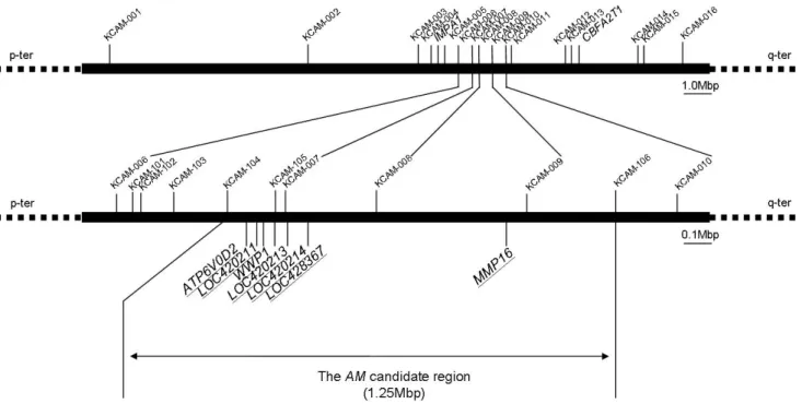
by positional cloning, we newly established F2 resource family for chicken muscular dystrophy. One F2 chicken, generated by F1 parents, has the information equivalent to that of 2 backcross individuals theoretically, since the recombination events occurred both in a F1 male and a female can be analyzed with a F2 chicken. Highly polymorphic microsatellites were focused on as DNA markers. By haplotype analysis with 487 F2 chickens and 22 DNA markers, AM candidate region was reduced to approximately 1.2 Mbp on GGA2q (Fig. 1). This region included seven functional genes, ATP6V0D2, LOC420211, WWP1, LOC420213, LOC420214, LOC428367 and MMP16, indicating them as the most likely AM candidates (Matsumoto et al., 2007). None of the candidates have been determined to be responsible genes for other muscular dystrophies so far.
Subsequently, to identify the responsible gene and muta- tion, we carried out sequence comparison and expression ana- lysis in normal and dystrophic chickens. The sequence com- parison of seven candidate genes revealed three synonymous mutations and one missense mutation specific to the pheno- type of muscular dystrophy. Two of the synonymous muta- tions were detected in LOC420214 (C660T and C1009A), and the other in LOC428367 (C954T). The WWP1 missense muta- tion (G1321A) caused amino acid substitution from arginine to glutamine, leading to the molecular alteration from a basic side chain to an uncharged polar side chain (Fig. 2). We focused further studies on this missense mutation since it was predicted to influence the function of the WWP1 protein.
The single nucleotide polymorphism (SNP) in WWP1 gene was genotyped in 111 normal birds from 16 strains with gene- tically varied backgrounds. None of the normal birds exhi- bited this type of substitution. The region of WWP1 including the SNP specific to chicken muscular dystrophy was highly conserved among normal birds. Additional homology research among tetrapods was conducted to estimate amino acid conser- vation in this region. The sequences of chicken, human, chim- panzee, monkey, mouse, rat, dog and cattle were available in the NCBI database. To obtain further information from other tetrapod species, we sequenced partial WWP1 gene of pigeon, snake, alligator, lizard, turtle and frog. The amino acid sequence around the region was highly conserved among these tetra- pods (Fig. 3), suggesting that the region was important for the function of WWP1 protein. These data suggested that
Fig. 1. The abnormal muscle (AM) candidate region and locations of genetic markers used in this study. The region between IMPA1 and CBFA2T1 is the AM candidate region by Yoshizawa et al. (2004). The upper figure shows the region of 16 informative gene markers on first step. The lower figure shows reduced AM candidate region by 11 informative gene markers on the second step. The genes underlined are the AM candidate genes.
Fig. 2. The domain structure of chicken WWP1 and the site of missense mutation. Chicken WWP1 protein is composed of 922 amino acids. Here are shown WWP1 functional domains: C2 domain, three WW domains and HECT domain. C in HECT domain indicates an active cysteine residue. The arrow indicates the site of missense muta- tion detected in this study.
WWP1 is the causative gene of chicken muscular dystrophy (Matsumoto et al., 2008).
The expression analysis of seven candidate genes were performed with the pectoral muscle derived from both normal and dystrophic chicken. Pectoral muscle is the one selectively affected in chicken muscular dystrophy (Barnard et al., 1982).
Therefore, we expected that the expression of the AM respon- sible gene might differ between normal and dystrophic chi- cken. The expression of WWP1, LOC420213 and LOC420214 could be detected by Northern blotting. To ATP6V0D2, MMP16
and LOC420211, RT-PCR analysis was applied because the expression of these genes was not detected by Northern blo- tting. The expression of LOC428367 was not confirmed in either genotype, which meant that this gene was not expressed or expressed at extremely low level in the pectoral muscle.
The expression of human LOC428367 (CNGB3) is limited only in retina (Kohl et al., 2000), suggesting this gene is not involved in muscular dystrophic phenotype.
There was no difference in size of mRNA between affected and normal birds in any of these genes. LOC420213 was highly expressed in affected individuals. Two bands were de- tected in WWP1 and LOC420214. WWP1 was expressed sligh- tly higher in normal than in affected chickens, while a slightly higher level of expression was exhibited for LOC420214 from affected chickens. Higher expression of ATP6V0D2 and MMP16 in dystrophic chickens was observed than in normal birds. The pectoral muscles from both genotypes expressed LOC420211 to identical level. Since no extreme alteration of expression level and abnormal splicing were observed, the onset of chicken muscular dystrophy might not be attributed to aberrant expression of these genes (Matsumoto et al., 2008).
Matsumoto et al. : Identification of the Gene Responsible for Chicken Muscular Dystrophy 148
Chicken N1) Chicken A Human1) Chimpanzee1) Monkey1) Mouse1) Rat1) Dog1) Cattle1) Pigeon Snake Alligator Lizard Turtle Frog
*
RNQLQGAMQQFNQRYLYSASMLSAENDPLGPLPPGWERRVDSNDRVYFVNHNTKTTQWED ...Q...
...A...Y...K...T...
...A...Y...K...T...
...A...Y...K...T...
...A...Y...K...T...
...A...Y...K...T...
...A...Y...K...T...
AD...A...Y...K...T...
...
...
...T...
...
...
...T...T...
Fig. 3. Homology study of WWP1 among tetrapods. Chicken N and A show amino acid sequences of normal and dystrophic chickens, respectively. R441Q WWP1 mutation is specific to chicken muscular dystrophy, and the amino acid sequence on this region is highly conserved among tetrapods. Dots indicate the same amino acids with above sequences. Asterisk indicates the residue that the substitution was detected in dystrophic chicken. (1) Amino acid sequence published in NCBI.
The WW domain containing E3 ubiquitin protein ligase 1 (WWP1) is classified into a ubiquitin ligase (E3) which plays an important role in ubiquitin-proteasome pathway (UPP) to degrade unneeded or damaged proteins (Scheffner and Staub, 2007). In chicken, the WWP1 expression has not been studied so far. Therefore, the mRNA expression of WWP1 in various skeletal muscles and other tissues of normal and dystrophic chickens was analyzed to know the differences in the general expression pattern between these chickens. The mRNA ex- pression of WWP1 gene was detected by Northern blotting in various muscles and other tissues of normal and muscular dystrophic chickens (Fig. 4). Two bands were detected in all tissues examined, and revealed almost equally expression level in any muscles and tissues observed. Flasza et al. (2002) reported that there are plural splice variants of human WWP1 gene, and our results indicated that chicken WWP1 gene also has plural splice variants. The downward alteration of WWP1 expression was observed commonly in almost all dystrophic muscles examined, but even in unaffected muscles the expression was lowered. Not only affected muscles but unaffected ones exhibited the same pattern (Fig. 5). Moreover, the expression was altered in liver and kidney, unaffected tissues. The result suggested that WWP1 expression level lowered in dystrophic phenotype is not directly related to the cause of disease in chicken muscular dystrophy, whereas mutated WWP1 does
Fig. 4. Expression of chicken WWP1 in various tissues. A WWP1 cDNA probe was used to detect WWP1 mRNA transcripts by Northern blotting using blots containing 2 μg of mRNAs from chicken muscles or various other tissues.
M. pectoralis superficialis (PS), M. tensor fascia lata (TFL), M. biceps femoris (BF), M. triceps surae (TS), M. pero- neus longus (PL), heart (H), brain (B), liver (L), kidney (K) and embryo (E) were analyzed. A doublet band is detected at variable levels in all tissues. “A” indicates mRNAs from dystrophic chickens. “N” indicates mRNAs from normal chickens. The numbers below the GAPDH bands represent the relative ratios of WWP1/GAPDH.
not function properly to cause the onset of chicken muscular dystrophy (Matsumoto et al., 2009).
FUNCTIONAL ANALYSIS TOWARD THE MOST LIKELY CANDIDATE GENE OF
CHICKEN MUSCULAR DYSTROPHY Though oncogenic character of WWP1 is intensively stu- died (Chen and Matesic, 2007), the role of WWP1 in muscle
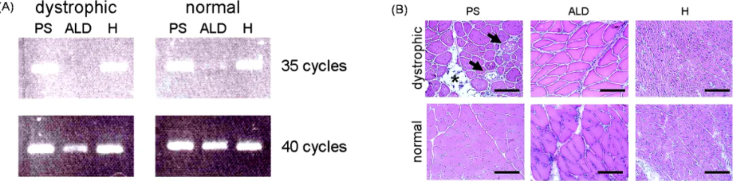
Fig. 5. RT-PCR detection of WWP1 gene and histological analysis for three representative muscle types. M. pectoralis superficialis (PS), M. anterior latissimus dorsi (ALD) and heart (H) expressed WWP1 less in muscular dystrophic chicken, but only dystro- phic PS was severely harmed. A) Expression of WWP1 in PS, ALD and H was analyzed by RT-PCR method. PCR was performed for 35 or 40 cycles. B) The PS, ALD and H of affected and normal chickens were analyzed with HE staining.
Vacuoles (arrows) and fatty infiltration (asterisk) are observed in PS of dystrophic chickens. It is also remarkable that, in dystrophic PS, many muscle fibers have many nuclei in cytoplasm and vary widely in size. These pathological features are not observed in ALD and H of dystrophic chicken. Scale bar = 120 μm.
remains elucidated. The switching of adult phenotype in fast muscle is inhibited in chicken muscular dystrophy, resulting in the continued expression of slow twitch myosin heavy chain (MyHC) isoform in adult fast muscles (Bandman, 1985;
Bandman and Bennett, 1988; Kaprielian et al., 1991; Tidy- man et al., 1997). This inhibition is proposed to contribute to the onset of chicken muscular dystrophy. To investigate the effects of the overexpression and the expression of mutated WWP1 gene on the MyHC genes expressions, we transfected the wild and mutated types of WWP1 gene into C2C12 cells which are myoblasts derived from mice skeletal muscle. The expression of muscle-differentiation markers, Myogenin (Myog), myogenic differentiation 1 (MyoD) and MyHCs (MyHC Ia and MyHC IIb), was analyzed by real-time PCR. Myog and MyoD were analyzed as the markers for early stages (Langlands et al., 1997) and MyHCs for later stages of muscle differen- tiation (Silberstein et al., 1986). MyHCs are divided into two classes, type I composed of fast twitch fibers and type II of slow twitch fibers (Larsson and Salviati, 1989).
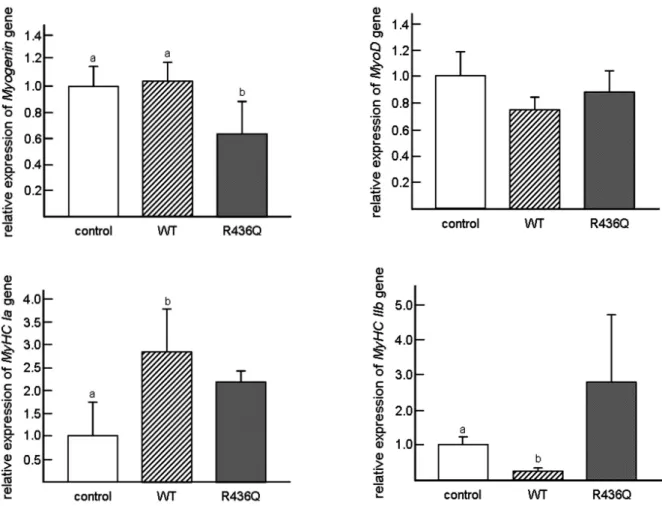
Transfecting mouse WWP1 gene into C2C12 mouse myoblasts resulted in the disturbed expressions of MyHC genes, though clear difference was not observed in the myotube morphology and the proliferation rate of myoblasts. The expression level of MyHC Ia gene was higher in wild type WWP1-introduced cells, but MyHC IIb gene expression was lower (Fig. 6), indicating that WWP1 promotes to transform C2C12 cells into fast twitch characteristics. However, R436Q-transfected cells
persisted in the high expression of both fast MyHC Ia and slow MyHC IIb isoforms, suggesting retainment in slow and fast twitch isoforms characteristic (Matsumoto et al., 2010a).
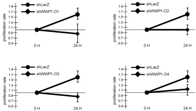
Furthermore, the WWP1 suppression by RNAi technique reduced the proliferation rate of C2C12 myoblasts, while clear difference was not observed in the proliferation rate by the transfection of the mutated WWP1 gene into the cells (Fig. 7). These data indicated that the mutation responsible for chicken muscular dystrophy does not eliminate the enzymatic activity and pro- vides some new function for the gene (Matsumoto et al., 2010b). Taken together, the R436Q mutation in WWP1 gene seems to inhibit the normal fiber type differentiation. The results suggest that WWP1 plays an important role in myo- blasts’ differentiating process.
The WWP1 is classified into a ubiquitin ligase to degrade unneeded or damaged proteins (Scheffner and Staub, 2007), suggesting that muscular dystrophic phenotype might be trig- gered by aberrant regulation of some WWP1’s substrates. From this perspective, we focused on caveolin-3 as a candidate protein interacting with WWP1, since an electron microscopic analysis revealed increase of caveolae in dystrophic chickens (Costello and Shafiq, 1979), and caveolin-3 is a main com- ponent of caveolae in muscle cells (Song et al., 1996). Gene- encoding caveolin-3 is responsible for limb-girdle muscular dystrophy type 1C (LGMD-1C) and other muscle diseases (Dowling et al., 2008). Caveolin-3 deficiency induces a mus- cular dystrophic phenotype (Galbiati et al., 2000a; Hagiwara
Matsumoto et al. : Identification of the Gene Responsible for Chicken Muscular Dystrophy 150
Fig. 6. Muscle-differentiation markers in WWP1-transfected (WT and R436Q) and empty vector-transfected (control) C2C12 cells. After transfection of wild-type (WT) and mutated (R436Q) WWP1 into C2C12 cells, expression levels of muscle-differentiation markers (Myog, MyoD, MyHC Ia and MyHC IIb) in differentiating day 6 cells were analyzed by real-time PCR. The expression of Myog in R436Q group was significantly lower than other groups, while no significant difference was seen in the expression level of MyoD gene. However, the expression of fast MyHC Ia in WWP1-transfected cells was significantly higher than in control cells, while that of slow MyHC IIb was significantly lowered in WWP1-transfected cells compared to control cells. The R436Q-transfected cells retained the high expression of both fast MyHC Ia and slow MyHC IIb isoforms compared to control cells. Y-axis indicates relative expression level of each gene to the GAPDH gene expression. Bars indicate standard deviations.
Different letters indicate significantly differences (p<0.05) among column graphs.
et al., 2000), while its overexpression is also harmful to muscle cells (Galbiati et al., 2000b).
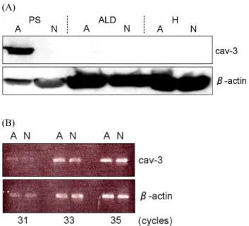
Western blotting analysis revealed the higher expression of caveolin-3 protein in only affected muscle in dystrophic chi ckens (Fig. 8(A)), suggesting a close correlation between higher caveolin-3 expression and dystrophic phenotype. On the other hand, semi-quantitative RT-PCR analysis revealed that affected muscle did not present such higher caveolin-3 expression at the mRNA level (Fig. 8(B)). The amount of caveolin-3 pro- tein might be controlled in a posttranslational fashion such as ubiquitination. Furthermore, the expression of other caveolae-
related proteins showed similar tendency as that of caveolin- 3. These results suggest that the accumulation of caveolin-3 protein may be associated with the causative process of chicken muscular dystrophy (Matsumoto et al., 2010c). However, more detailed analysis is required in order to elucidate the relationship between WWP1 and caveolin-3.
Abnormality in WWP1 protein might lead to aberrant regu- lation of its substrates. Therefore, identification of WWP1’s substrates in muscle will greatly contribute to elucidation of the mechanism leading to the onset of chicken muscular dys- trophy. Recently, some WWP1’s substrates have been iden-
Fig. 7. The effects of four WWP1-shRNA sequences on C2C12 cells. Four WWP1-shRNA sequences (shWWP1-O1-4) were prepared and their effects on the proliferation rate of C2C12 cells were observed. All cells with shWWP1-O1-4 vectors showed significant downward tendencies twenty-four hours after transfection (24 H) (shWWP1-O1: 89 ± 35 %, shWWP1-O2: 101 ± 22 %, shWWP1-O3: 83 ± 16 % and shWWP1-O4: 110 ± 22 %). Y-axis indicates relative proliferation rate based on the cell numbers of immediately after transfection (0 H). Proliferation rates were calculated as relative levels toward the cell numbers of immediately after transfection. Three individual samples were prepared for each calculation.
tified, but any of them are oncogenic proteins and might not be involved in the causative event of chicken muscular dys- trophy (Chen and Matesic, 2007). To identify novel substrates, study with WWP1-specific antibody is inevitable. Unfortuna- tely, any commercially available WWP1-specific antibody was not suitable for chicken muscle sample. WWP1-specific anti- body for chicken muscle sample must be developed for fur- ther study to understand the mechanism of chicken muscular dystrophy.
RESENT FINDINGS OF THE MOST LIKELY CANDIDATE GENE OF CHICKEN
MUSCULAR DYSTROPHY
Aberrant glycosylation of α-dystroglycan in muscular dystro- phic chicken was reported (Saito et al., 2005). α-dystroglycan is one of dystrophin-related proteins and shortage of its gly- cosylation causes muscular dystrophic phenotype (Dalkilic and Kunkel, 2003). In addition, certain mutations in glycosyltrans-
ferases result in muscular dystrophic phenotypes including Fukuyama type congenital muscular dystrophy, Walker-War- burg syndrome and Muscle-Eye-Brain disease (Nonaka, 1987a).
Therefore, abnormality of α-dystroglycan’s glycosylation was also expected to trigger the onset of chicken muscular dys- trophy. However, there was not glycosyltransferases and their related proteins in the AM candidate region narrowed down by haplotype analysis and the most likely AM candidate gene identified by sequence comparison, WWP1, has not been re- ported to associate with glycosyltransferases so far. Some F-box type ubiquitin ligases, such as Fbx2 and Fbs2, are known as the ones that recognize sugar chain (Yoshida et al., 2002, 2003), while WWP1 is classified into HECT type ubiquitin ligase (Scheffner and Staub, 2007). HECT type ubiquitin ligases responsible for sugar chain regulation have not been reported so far. The possibility that WWP1 might control glycosylation of α-dystroglycan still remains, but short sugar chain of α-dystroglycan in muscular dystrophic chicken may be the secondary result of the pathological change and new
Matsumoto et al. : Identification of the Gene Responsible for Chicken Muscular Dystrophy 152
(A)
(B)
Fig. 8. Expression of caveolin-3 at protein and mRNA level.
(A) Expression of caveolin-3 in M. pectoralis super- ficialis (PS), M. anterior latissimus dorsi (ALD) and heart (H) was analyzed by Western blotting. PS expressed higher amount of caveolin-3 protein in muscular dystrophic chickens (A), while the expression in ALD and H was undetectable as in normal chickens (N). (B) The caveolin-3 expression in PS was assessed at mRNA level. No drastic difference was observed between D and N.
explanation for this disease may be required.
Many HECT type ubiquitin ligases ubiquitinate membrane proteins (Chen and Matesic, 2007). Therefore, WWP1 may also catalyze some membrane protein in muscle. In fact, WWP1 can bind a membrane protein, β-dystroglycan which is one of dystrophin-related proteins, in vitro (Pirozzi et al., 1997). β-dystroglycan is normally expressed in chicken mus- cular dystrophy (Saito et al., 2005); hence, this interaction between WWP1 and β-dystroglycan is unlikely to relate with the pathological change of chicken muscular dystrophy. Iden- tification of WWP1’s new substrates, which may accumulate in muscle and harm muscle physiology, will be needed in order to elucidate the mechanism for mutated WWP1 to cause the muscular dystrophic phenotype.
ACKNOWLEDGEMENTS
This work was supported by many people. Especially we would like to express our sincere gratitude to Prof. T. Ki-
kuchi of National Institute of Neuroscience, Prof. N. Ichihara of Azabu University and Dr. A. Fujiwara of Nippon Institute for Biological Science.
LITERATURE CITED
Asmundson VS, Julian LM 1956 Inherited muscle abnormality in the domestic fowl. J Hered 47:248-252.
Asmundson VS, Kratzer FH, Julian LM 1966 Inherited myo- pathy in the chicken. Ann NY Acad Sci 138:49-60.
Bandman E 1985 Continued expression of neonatal myosin heavy chain in adult dystrophic skeletal muscle. Science 227:780-782.
Bandman E, Bennett T 1988 Diversity of fast myosin heavy chain expression during development of gastrocnemius, bicep brachii, and posterior latissimus dorsi muscles in normal and dystrophic chickens. Dev Biol 130:220-231.
Barnard EA, Lyles JM, Pizzey JA 1982 Fiber types in chicken skeletal muscles and their changes in muscular dystrophy.
J Physiol 331:333-354.
Chen C, Matesic LE 2007 The Nedd4-like family of E3 ubi- quitin ligases and cancer. Cancer Metastasis Rev 3-4:587- 604.
Costello BR, Shafiq SA 1979 Freeze-fracture study of muscle plasmalemma in normal and dystrophic chickens. Muscle Nerve 2:191-201.
Dalkilic I, Kunkel LM 2003 Muscular dystrophies: genes to pathogenesis. Curr Opin Genet Dev 13:231-238.
Dowling JJ, Gibbs EM, Feldman EL 2008 Membrane traffic and muscle: Lessons from human disease. Traffic 9:1035- 1043.
Flasza M, Gorman P, Roylance R, Canfield AE, Baron M 2002 Alternative splicing determines the domain structure of WWP1, a Nedd4 family protein. Biochem Biophys Res Commun 290:431-437.
Galbiati F, Volonte D, Minetti C, Bregman DB, Lisanti MP 2000a Limb-girdle muscular dystrophy (LGMD-1C) mu- tants of caveolin-3 undergo ubiquitination and proteasomal degradation. J Biol Chem 275:37702-37711.
Galbiati F, Volonte D, Chu JB, Li M, Fine SW, Fu M, Ber- mudez J, Pedemonte M, Weidenheim KM, Pestell RG, Minetti C, Lisanti MP 2000b Transgenic overexpression
of caveolin-3 in skeletal muscle fibers induces a Duchenne- like muscular dystrophy phenotype. Proc Natl Acad Sci USA 97:9689-9694.
Hagiwara Y, Sasaoka T, Araishi K, Imamura M, Yorifuji H, Nonaka I, Ozawa E, Kikuchi T 2000 Caveolin-3 deficiency causes muscle degeneration in mice. Hum Mol Genet 9:
3047-3054.
Kaprielian Z, Bandman E, Fambrough DM 1991 Expression of Ca2+-ATPase isoforms in denervated, regenerating, and dystrophic chicken skeletal muscle. Dev Biol 144:199- 211.
Kikuchi T, Ishiura S, Nonaka I, Ebashi S 1981 Genetic he- terozygous carriers in hereditary muscular dystrophy of chickens. Tohoku J Agric Res 32:14-26.
Kohl S, Baumann B, Broghammer M, Jagle H, Sieving P, Kellner U, Spegal R, Anastasi M, Zrenner E, Sharpe LT, Wissinger B 2000 Mutations in the CNGB3 gene enco- ding the beta-subunit of the cone photoreceptor cGMP-gated channel are responsible for achromatopsia (ACHM3) linked to chromosome 8q21. Hum Mol Genet 9:2107-2116.
Kondo K, Kikuchi T, Mizutani M 1982 Breeding of the chicken as an animal model for muscular dystrophy. Page 19-24 In: Muscular Dystrophy, Tokyo University Press, Tokyo.
Langlands K, Yin X, Anand G, Prochownik EV 1997 Diffe- rential interactions of Id proteins with basic-helix-loop- helix transcription factors. J Biol Chem 32:19785-19793.
Larsson L, Salviati G 1989 Effects of age on calcium tran- sport activity of sarcoplasmic reticulum in fast- and slow- twitch rat muscle fibres. J Physiol 419:253-264.
Lee EJ, Yoshizawa K, Mannen H, Kikuchi H, Kikuchi T, Mizutani M, Tsuji S 2002 Localization of the muscular dystrophy AM locus using a chicken linkage map cons- tructed with the Kobe University resource family. Anim Genet 33:42-48.
Lisi MT, Cohn RD 2007 Congenital muscular dystrophies:
new aspects of an expanding group of disorders. Biochim Biophys Acta 1772:159-172.
Matsumoto H, Maruse H, Yoshizawa K, Sasazaki S, Fujiwara A, Kikuchi T, Ichihara N, Mukai F, Mannen H 2007 Pin- pointing the candidate region for muscular dystrophy in chickens with an abnormal muscle gene. Anim Sci J 78:
476-483.
Matsumoto H, Maruse H, Inaba Y, Yoshizawa K, Sasazaki S, Fujiwara A, Nishibori M, Nakamura A, Takeda S, Ichihara N, Kikuchi T, Mukai F, Mannen H 2008 The ubiquitin ligase gene (WWP1) is responsible for the chicken mu- scular dystrophy. FEBS Lett 582:2212-2218.
Matsumoto H, Maruse H, Sasazaki S, Fujiwara A, Takeda S, Ichihara N, Kikuchi T, Mukai F, Mannen H 2009 Ex- pression pattern of WWP1 in muscular dystrophic and normal chickens. J Poult Sci 46:95-99.
Matsumoto H, Inba Y, Sasazaki S, Fujiwara A, Ichihara N, Kikuchi T, Mannen H 2010a Mutated WWP1 induces an aberrant expression of myosin heavy chain gene in C2C12
skeletal muscle cells. J Poult Sci 47:115-119.
Matsumoto H, Takahama M, Kajiyama R, Sasazaki S, Oyama K, Mannen H 2010b Suppression of WWP1 gene via RNAi induced the reduction of proliferation rate of C2C12 myo- blasts. J Poult Sci 47:288-293.
Matsumoto H, Sasazaki S, Fujiwara A, Ichihara N, Kikuchi T, Mannen H 2010c Accumulation of caveolin-3 protein is limited in damaged muscle in chicken muscular dys- trophy. Comp Biochem Physiol A Mol Integr Physiol 157:68-72.
Nonaka I 1987a Muscular dystrophy. pp 42-75 In: Muscular Dystrophy, Japan Medical Journal, Tokyo.
Nonaka I 1987b Animal models for muscular dystrophy. pp 178-185 In: Muscular Dystrophy, Japan Medical Journal, Tokyo.
Partridge T 1991 Animal models of muscular dystrophy:
What can they teach us? Neuropathol Appl Neurobiol 17:
353-363.
Pirozzi G, McConnell SJ, Uveges AJ, Carter JM, Sparksi AB, Kayi BK, Fowlkes DM 1997 Identification of novel human WW domain-containing proteins by cloning of li- gand targets. J Biol Chem 272:14611-14616.
Saito F, Blank M, Schroder J, Manya H, Shimizu T, Camp- bell KP, Endo T, Mizutani M, Kroger S, Matsumura K 2005 Aberrant glycosylation of α-dystroglycan causes de- fective binding of laminin in the muscle of chicken mus- cular dystrophy. FEBS Lett 579:2359-2363.
Scheffner M, Staub O 2007 HECT E3s and human disease.
BMC Biochem 8 Suppl 1:S6.
Matsumoto et al. : Identification of the Gene Responsible for Chicken Muscular Dystrophy 154
Schmid M, Nanda I, Guttenbach M, Steinlein C, Hoehn M, Schartl M, Haaf T, Weigend S, Fries R, Buerstedde JM, Wimmers K, Burt DW, Smith J, A'Hara S, Law A, Gri- ffin DK, Bumstead N, Kaufman J, Thomson PA, Burke T, Groenen MA, Crooijmans RP, Vignal A, Fillon V, Mo- risson M, Pitel F, Tixier-Boichard M, Ladjali-Mohammedi K, Hillel J, Maki-Tanila A, Cheng HH, Delany ME, Burnside J, Mizuno S 2000 First report on chicken genes and chromosomes. Cytogenet Cell Genet 90:169-218.
Silberstein L, Webster SG, Travis M, Blau HM 1986 Deve- lopmental progression of myosin gene expression in cul- tured muscle cells. Cell 46:1075-1081.
Song KS, Scherer PE, Tang Z, Okamoto T, Li S, Chafel M, Chu C, Kohtz DS, Lisanti MP 1996 Expression of caveolin-3 in skeletal, cardiac, and smooth muscle cells. Caveolin-3 is a component of the sarcolemma and co-fractionates with dystrophin and dystrophin-associated glycoproteins. J Biol Chem 271:15160-15165.
Tidyman WE, Moore LA, Bandman E 1997 Expression of fast myosin heavy chain transcripts in developing and dys- trophic chicken skeletal muscle. Dev Dyn 208:491-504.
Wagner WD, Peterson RA 1970 Muscular dystrophy synd- rome in the Cornish chicken. Am J Vet Res 31:331-338.
Yoshizawa K, Inaba K, Mannen H, Kikuchi T, Mizutani M, Tsuji S 2004 Fine mapping of the muscular dystrophy (AM) gene on chicken chromosome 2q. Anim Genet 35:
397-400.
Yoshida Y, Chiba T, Tokunaga F, Kawasaki H, Iwai K, Suzuki T, Ito Y, Matsuoka K, Yoshida M, Tanaka K, Tai T 2002 E3 ubiquitin ligase that recognizes sugar chains.
Nature 418:438-442.
Yoshida Y, Tokunaga F, Chiba T, Iwai K, Tanaka K, Tai T 2003 Fbs2 is a new member of the E3 ubiquitin ligase fa- mily that recognizes sugar chains. J Biol Chem 278:
43877-43884.
(접수: 2011. 2. 23, 수정: 2011. 4. 27, 채택: 2011. 4. 27)