Received:October 19, 2020, Revised:November 22, 2020, Accepted:November 25, 2020 Corresponding to:Kichul Shin http://orcid.org/0000-0002-6749-7598
Division of Rheumatology, Seoul Metropolitan Government-Seoul National University Boramae Medical Center, 20 Boramae-ro 5-gil, Dongjak-gu, Seoul 07061, Korea. E-mail:[email protected]
Copyright ⓒ 2021 by The Korean College of Rheumatology. All rights reserved.
This is an Open Access article, which permits unrestricted non-commerical use, distribution, and reproduction in any medium, provided the original work is properly cited.
A Patient With Late-onset Limb-girdle Muscular Dystrophy Type 2B Mimicking Dermatomyositis: A Case Report and Review
Min Jung Kim, M.D.1, Yoon-Jeong Oh, M.D.2, Yoon Ho Hong, M.D., Ph.D.3, Sung-Hye Park, M.D., Ph.D.4, Ji Seon Oh, M.D., Ph.D.5, Min Jung Kim, M.S.6, Jong-Hee Chae, M.D., Ph.D.6, Kichul Shin, M.D., Ph.D.1
1Division of Rheumatology, Seoul Metropolitan Government-Seoul National University Boramae Medical Center, Seoul, 2Division of Rheumatology, Department of Internal Medicine, Kangwon National University School of Medicine, Chuncheon, 3Department of Neurology, Seoul Metropolitan Government-Seoul National University Boramae Medical Center, 4Department of Pathology, Seoul National University College of Medicine, 5Clinical Research Center, Asan Medical Center, University of Ulsan College of Medicine, 6Department of Pediatrics, Seoul National University Children’s Hospital, Seoul National University College of Medicine, Seoul, Korea
Limb-Girdle Muscular Dystrophy 2B (LGMD2B) presents with proximal and/or distal muscle weakness and markedly high crea- tine kinase level. It is caused by the loss of dysferlin due to mutations in the DYSF gene. Due to its similar clinical features as inflammatory myopathy, it is often difficult to distinguish between the two. We present a case of a 48-year-old male who devel- oped progressive proximal muscle weakness, papulosquamous lesions on the knuckles, elevated levels of muscle enzymes, and electromyogram abnormalities. Based on the clinical presentation, the initial impression was dermatomyositis, yet it was refractory to immunosuppressive therapy. Subsequently, dysferlin immunostaining and genetic analysis led to the final diag- nosis of LGMD2B. This case shows that LGMD2B can present with extramuscular symptoms mimicking inflammatory myo- pathy in later stages of life. Dysferlin immunostaining and/or genetic analysis of the DYSF gene are essential for its diagnosis.
(J Rheum Dis 2021;28:101-106)
Key Words. Muscular dystrophies, Limb-girdle, Dermatomyositis
INTRODUCTION
Inflammatory myopathy is a chronic inflammatory mus- cle disease caused by immune-mediated muscle injury and consists of dermatomyositis (DM), polymyositis (PM), inclusion body myositis and immune-mediated ne- crotizing myopathy (IMNM). Patients with DM or PM present with progressive proximal muscle weakness over weeks to months and show good responses to im- munosuppressive therapy [1]. DM is associated with dis- tinct skin manifestations such as Gottron’s papules or a heliotrope eruption, which are useful in distinguishing between inflammatory myopathies. Additionally, other muscle diseases such as muscular dystrophy, metabolic,
endocrine or drug-induced myopathies may mimic in- flammatory myopathy, but none of these disorders are as- sociated with the skin lesions characteristic of DM [2].
Limb-girdle muscular dystrophy 2B (LGMD2B), a sub- type of dysferlinopathy, is characterized by predom- inantly proximal muscle weakness and markedly high creatine kinase (CK) levels [3]. LGMD2B, caused by the dysferlin gene (DYSF) mutation, usually occurs in child- hood or early adolescence [4]. It may present as similar as inflammatory myopathy in terms of clinical, laboratory, and electromyography (EMG) features. Histologically, in- flammatory cell infiltrations in myofibers could be pres- ent in LGMD2B [5]. Therefore, a late-onset presentation of LGMD2B can at times be mistaken for inflammatory
Figure 1. Changes in serum creatine kinase levels and daily doses of glucocorticoid during the follow-up period.
myopathy.
Herein we report a case of a 48-year-old male with pro- gressive proximal muscle weakness, papulosquamous le- sions in the knuckles, elevated CK levels, and myopathic changes on the EMG. The initial impression of his diag- nosis was DM, yet the final diagnosis turned out to be LGMD2B with psoriasis.
CASE REPORT
A 48-year-old male presented with proximal muscle weakness in his lower extremities followed by skin rashes on the hands. Muscle weakness had slowly progressed over the past eight months. He had difficulty in sitting up and could only move around using a wheelchair or a walker. He denied myalgia, arthralgia, or dysphagia. He was diagnosed with type 2 diabetes mellitus, hyper- tension, and dyslipidemia during the past three years. He had a second degree burn over 70% of his face ten years ago. He was a 15-pack-year smoker with no occupation and drank alcohol once a week. He was taking aspirin, atorvastatin, perindopril and nateglinide. His family his- tory of medical illness was unremarkable.
On physical examination, his vital signs were stable, and the body mass index was 19.8 kg/m2 (height 165 cm, body weight 54 kg). Proximal muscle strength in both lower extremities was grade 3 out of 5 based on the Medical Research Council scale. The strength in his distal lower extremities and the proximal and distal upper ex- tremities was grade 4+. Calf muscle atrophy was not observed. Erythematous papulosquamous skin lesions with scales were present on the dorsal surface of his meta- carpophalangeal joints, fingers, and elbows. Physical find- ings in other systems and organs were unremarkable. At his first visit, CK level was 4,512 IU/L (normal 45∼163), aldolase was 18.2 U/L (0∼7.6), and lactate dehydrogen- ase was 432 IU/L (100∼225). Aspartate transaminase and alanine transaminase levels increased to 75 IU/L (1∼
40) and 77 IU/L (1∼40), respectively. His complete blood count was as follows: white blood cell count 6,220 /μL, hemoglobin 14.7 g/dL, and platelets 267× 103 /μL.
The erythrocyte sedimentation rate and C-reactive pro- tein levels were 8 mm/hr (0∼9) and 1.57 mg/dL (0∼
0.5), respectively. Anti-nuclear antibody, rheumatoid fac- tor, and anti-Jo-1 antibody were not detected. EMG re- vealed small-amplitude, short-duration and polyphasic motor-unit potentials without increased spontaneous ac- tivity in the deltoid, biceps brachii, and pronator teres,
and high amplitude and long duration motor-unit poten- tials in the vastus lateralis, tibialis anterior, and gastro- cnemius muscles. Nerve conduction studies were normal. Results of the electrocardiogram and chest radio- graph were unremarkable. There was no evidence of ma- lignancy on further evaluation. For economic reasons, muscle imaging or biopsy was not initially performed.
With the suspicion of DM (symmetric proximal muscle weakness, scaly patchy redness over knuckles, elevated muscle enzyme levels, and EMG abnormalities), 20 mg/day of prednisolone and 300 mg/day of hydroxy- chloroquine were given. However, muscle weakness did not respond to the initial combination of medications within 6 months. Erythematous papules and plaques with silvery scales on his elbows, hands, and legs were confirmed as psoriasis by a Dermatologist; topical oint- ment and 12.5 mg/week of methotrexate were prescribed.
Despite the immunosuppressive treatment, muscle weakness continued to worsen and CK levels fluctuated throughout the following 12 months (Figure 1). Eventually, a muscle biopsy of the vastus lateralis was performed.
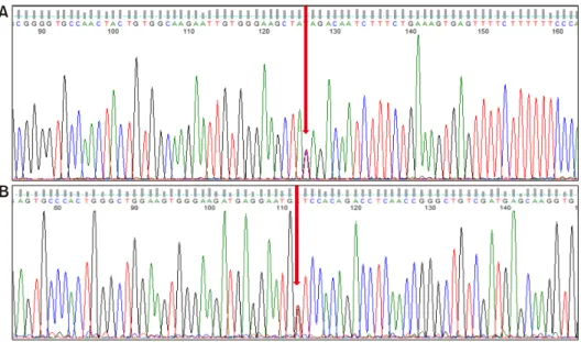
The specimen showed muscle cell degeneration, as well as regeneration and atrophy. Fatty replacement of muscle tissue was severe. There were no muscle cell necrosis or inflammatory cell infiltrates in endomysial and peri- vascular regions. Neural cell adhesion molecule (CD56) and dystrophin were present, yet dysferlin was absent on immunostaining (Figure 2). Electron microscopy did not show any tubuloreticular inclusion (Figure 3). Furthermore, a molecular analysis with Next Generation Sequencing disclosed heterozygous mutations in the DYSF gene;
c.2548C>T (p.Gln850*) and c.3051G>T (p.Trp1017Cys)
Figure 2. H&E stain (A) and im- munohistochemistry (B∼D) of the muscle biopsy. (A) Myofibers show moderate size variation with degenerating cells with en- domysial fibrofatty change (H&E,
×200). (B) Many degenerated myofibers show variation of size and positivity for CD56 (CD56 immunohistochemistry,
×100). Dystrophin (C) was ro- bustly positive (Dystrophin 1 immunohistochemistry, ×200), yet dysferlin (D) was totally ab- sent in the sarcolemmal mem- brane (Dysferlin immunohisto- chemistry, ×200).
Figure 3. Electron microscopy of the muscle. Myofibers show size variation with mild atrophic change. Lay-down of colla- gen are present within the endomysium (Uranyl acetate and lead citrate, ×6,000).
[6]. Sanger sequencing confirmed that the mutations seg- regated with the disease phenotype (Figure 4). A final di- agnosis of LGMD2B was made, and all immuno- suppressive agents were tapered off. The patient re- mained stable on physiotherapy.
The study was approved by the Institutional Review Board of Seoul Metropolitan Government-Seoul National University Boramae Medical Center (IRB No. 16-2017- 61). Informed consent was obtained from the patient.
DISCUSSION
Our case presents a 48-year-old male with progressive proximal muscle weakness in the lower limbs, distinctive skin rashes, high CK levels, and a myopathic pattern in the EMG. He was initially diagnosed with DM, but showed poor treatment response to immunosuppressive agents. After dysferlin immunostaining of the muscle tis- sue and genetic analysis, a final diagnosis of LGMD2B was made.
Dysferlin, a protein encoded by the DYSF gene located on chromosome 2p13, plays a role in vesicle docking and fusion with the plasma membrane to repair sarcolemma disruption. Thus, mutations in the DYSF gene are respon- sible for a defective repair mechanism in the sarcolemma leading to muscular dystrophy: a LGMD2B affecting the proximal lower limbs at the onset, and a Miyoshi myo- pathy (MM) affecting posterior distal compartment of the legs [7]. Both LGMD2B and MM have similar features, however, muscle weakness with pelvic girdle distribution distinguishes between LGMD2B and MM. Interestingly, the type of DYSF gene mutation does not correlate with phenotypic features of dysferlinopathy and shows clinical heterogeneity even with the same mutation [8]. Patients with dysferlinopathy, especially LGMD2B, have sym- metric proximal weakness, high CK levels and small poly- phasic motor-unit potentials in the EMG, which may be confused with inflammatory myopathy. Nguyen et al. [4]
Figure 4. Sanger sequencing of the mutations in allele c.2548 C>T (p.Gln850*) (nonsense mutation) (A) and c.3051G>T (p.Trp1017Cys) (missense mu- tation) (B) (arrows).
reported that approximately 25% of patients with dysfer- linopathy are initially misdiagnosed with inflammatory myopathy. Nevertheless, the two entities display several different clinical features. First, patients with dysferlin- opathy commonly demonstrate inability to stand or walk on toes due to distal muscle involvement and atrophy of the gastrocnemius [9]. Second, the disease onset of dys- ferlinopathy usually occurs during early adulthood (typically 15 to 35 years), and patients may have a pre-clinical period with relatively high CK levels ranging from 1,000 to 40,000 IU/L [3,10]. Third, treatment re- sponse to glucocorticoid and/or other immunosuppressive agents is far from adequate. The mainstay management of dysferlinopathy is exercise to maintain muscle strength [11]. Table 1 compares the characteristics of dysferlinop- athy with those of inflammatory myopathies.
The precise distinction between dysferlinopathy and in- flammatory myopathy is based on muscle biopsy and dys- ferlin immunostaining, which shows a significant reduc- tion or absence of dysferlin protein in the sarcolemma in dysferlinopathy [12]. Interestingly, inflammatory cell in- filtrates in muscles are often present in dysferlinopathy [13]. Inflammatory cell types in dysferlinopathy shows high CD4+/CD8+ T cell ratios predominantly in the per- ivascular region similar to DM. But unlike inflammatory myopathy, major histocompatibility complex class I ex- pression is low and sarcolemmal membrane attack com- plex is deposited on non-necrotic fibers [5]. In our case, IMNM should also be considered since the patient was on statin therapy, but no muscle cell necrosis or macrophage infiltration was found in the biopsy specimen [14].
Our patient demonstrated several unusual features of dysferlinopathy that made the diagnosis difficult. First, age of onset was later in life compared with that presented in previous studies. Park et al. [15] reported that the age of onset varied from 9 to 36 years in Korean patients with dysferlinopathy. A Japanese study showed a wider range of age; 14 to 58 years [16]. Late-onset LGMD2B is asso- ciated with the homozygous mutation of the c.2997G>T (p.Trp999Cys) allele in Japanese [16]. Two of 23 Korean patients with dysferlinopathy showed a heterozygous mutation in the c.2997G>T allele; one was a 41-year-old male with Miyoshi myopathy and the other was a 27-year-old female with LGMD2B [6]. The mutations in our patient and its association with a late-onset pheno- type need to be further investigated. Second, psoriasis sporadically appeared in multiple regions in our patient along with muscle weakness. There is no report on the as- sociation between dysferlinopathy with psoriasis to date.
It is sometimes challenging to distinguish between psor- iasis and Gottron’s papules seen in DM as the two often develop at similar anatomical sites.
SUMMARY
In summary, due to the clinical heterogeneity of dysfer- linopathy and its overlapping features with inflammatory myopathy, it could sometimes be difficult to discern the two entities. Dysferlinopathy should be considered in pa- tients with myopathy who have markedly high CK levels, calf muscle atrophy, and poor response to immuno- suppressive therapy. Muscle dysferlin immunostaining
Table 1. Differential diagnoses of dysferlinopathy CharacteristicDysferlinopathy DermatomyositisPolymyositisInclusion body myositisImmune-mediated necrotizing myopathyLGMD2BMiyoshi myopathy Age of onset17∼25 yearsChildren and adultsAdults>50 yearsPatients treated with statins (increased risk with advanced age >80 years) Sex preferenceNot foundFemale>maleFemale>maleMale>femaleFemale>male Onset & courseUsually slowly progressive; Occasionally subacute presentation (25%)Subacute progressive weaknessAcute or subacute progressive weaknessInsidious onset, slowly progressiveAcute or subacute progressive weakness Muscle weaknessPredominantly pelvic and shoulder girdle muscle weakness, distal involvement may present Most marked only in distal and posterior parts of the legs, initial muscle atrophy of gastrocnemius and soleus Proximal>distalProximal>distalDistal and proximal; usually asymmetric finger flexor and proximal leg weakness
Proximal>distal DysphagiaNoYesYesYesYes Dermatologic involvementNoYesNoNoRare CKElevated (more than 10 to 70-fold, often >100-fold)Normal or elevated (up to 50-fold)Elevated (often >10-fold, up to 50-fold) Normal or elevated (<10-fold)Elevated (often >10-fold) EMGMyopathicMyopathic MyopathicMyopathicMyopathic MRIFat suppressed T2 and STIR weighted sequences may demonstrate hyperintensities (muscle edema), difficult to differentiate from inflammatory myopathies. Dysferlinopathy shows diffuse muscle edema and fatty degeneration, while inflammatory myopathies show predominant patchy muscle edema and mild fatty degeneration PathologyInflammatory features in 40% or more cases, similar to those of DM, Dystrophic muscle pattern and variable morphologic findings related to duration of symptoms; Absence or partial deficiency of dysferlin by immunoblot. Plasma membrane microlesions and subsarcolemmal vesical accumulation on electron microscopy
Perifascicular, perimysial or perivascular inflammatory cell infiltrate; CD4+ T cell and B cell dominant, perifascicular atrophy, MAC deposition within necrotic fibers Endomysial, perimysial, perivascular inflammatory cell infiltrate, non-necrotic fibers invaded by predominantly CD8+ T cell, CD8/MHC-1 complex, MAC deposition within necrotic fibers Endomysial inflammatory cell infiltrate; rimmed vacuoles and eosinophilic inclusions
Muscle-cell necrosis and regeneration, macrophage infiltrates in endomysial and perivascular regions, relative absence of lymphocyte infiltration, MHC-1 complex and MAC deposition Response to immunosuppressive therapy
PoorGoodGoodGenerally poorGood LGMD 2B: Limb-Girdle Muscular Dystrophy type 2B, CK: creatine kinase, EMG: electromyography, MAC: membrane attack complex, MRI: magnetic resonance imaging, STIR: short-tau inversion recovery, MHC: major histocompatibility complex, DM: dermatomyositis.
plus genetic analysis would be essential for its diagnosis.
CONFLICT OF INTEREST
No potential conflict of interest relevant to this article was reported.
AUTHOR CONTRIBUTIONS
J.S.O. and K.S. contributed to the study conception and design. Material preparation, data collection and analysis were performed by M.J.K.1, Y.O., Y.H.H., S.P., J.S.O., M.J.K.6 and J.C. The first draft of the manuscript was writ- ten by M.J.K.1 and all authors commented on previous versions of the manuscript. All authors read and ap- proved the final manuscript.
REFERENCES
1. Dalakas MC. Inflammatory muscle diseases. N Engl J Med 2015;372:1734-47.
2. Bohan A, Peter JB. Polymyositis and dermatomyositis (first of two parts). N Engl J Med 1975;292:344-7.
3. Wicklund MP, Kissel JT. The limb-girdle muscular dystrophies. Neurol Clin 2014;32:729-49.
4. Nguyen K, Bassez G, Bernard R, Krahn M, Labelle V, Figarella-Branger D, et al. Dysferlin mutations in LGMD2B, Miyoshi myopathy, and atypical dysferlinopathies. Hum Mutat 2005;26:165.
5. Choi JH, Park YE, Kim SI, Kim JI, Lee CH, Park KH, et al.
Differential immunohistological features of inflammatory myopathies and dysferlinopathy. J Korean Med Sci 2009;
24:1015-23.
6. Park YE, Kim HS, Lee CH, Nam TS, Choi YC, Kim DS. Two common mutations (p.Gln832X and c.663+1G>C) ac- count for about a third of the DYSF mutations in Korean pa- tients with dysferlinopathy. Neuromuscul Disord 2012;22:
505-10.
7. Bansal D, Miyake K, Vogel SS, Groh S, Chen CC, Williamson R, et al. Defective membrane repair in dysferlin-deficient muscular dystrophy. Nature 2003;423:168-72.
8. Petersen JA, Kuntzer T, Fischer D, von der Hagen M, Huebner A, Kana V, et al. Dysferlinopathy in Switzerland:
clinical phenotypes and potential founder effects. BMC Neurol 2015;15:182.
9. Tarnopolsky MA, Hatcher E, Shupak R. Genetic myopathies initially diagnosed and treated as inflammatory myopathy.
Can J Neurol Sci 2016;43:381-4.
10. Klinge L, Aboumousa A, Eagle M, Hudson J, Sarkozy A, Vita G, et al. New aspects on patients affected by dysferlin defi- cient muscular dystrophy. J Neurol Neurosurg Psychiatry 2010;81:946-53.
11. Biondi O, Villemeur M, Marchand A, Chretien F, Bourg N, Gherardi RK, et al. Dual effects of exercise in dysferlinopathy.
Am J Pathol 2013;182:2298-309.
12. Fanin M, Angelini C. Progress and challenges in diagnosis of dysferlinopathy. Muscle Nerve 2016;54:821-35.
13. Vinit J, Samson M Jr, Gaultier JB, Laquerriere A, Ollagnon E, Petiot P, et al. Dysferlin deficiency treated like refractory polymyositis. Clin Rheumatol 2010;29:103-6.
14. Mammen AL. Statin-associated autoimmune myopathy. N Engl J Med 2016;374:664-9.
15. Park HJ, Hong JM, Suh GI, Shin HY, Kim SM, Sunwoo IN, et al. Heterogeneous characteristics of Korean patients with dysferlinopathy. J Korean Med Sci 2012;27:423-9.
16. Takahashi T, Aoki M, Suzuki N, Tateyama M, Yaginuma C, Sato H, et al. Clinical features and a mutation with late onset of limb girdle muscular dystrophy 2B. J Neurol Neurosurg Psychiatry 2013;84:433-40.