Insights
this and thousands of other papers at http://www.la-press.com.
Contrast-Enhanced Magnetic Resonance Imaging in Pediatric Patients:
Review and Recommendations for Current Practice
Ravi Bhargava
1, gabriele hahn
2, Wolfgang hirsch
3, Myung-Joon Kim
4, hans-Joachim Mentzel
5,
Øystein e. olsen
6, eira stokland
7, Fabio triulzi
8and elida Vazquez
91Division of Pediatric Radiology, Department of Radiology and Diagnostic Imaging, Stollery Children’s Hospital, University of Alberta, Edmonton, Alberta, Canada. 2Institut und Poliklinik für Radiologische Diagnostik,
Universitätsklinikum Carl Gustav Carus, Dresden, Germany. 3Department of Paediatric Radiology, University of Leipzig, Germany. 4Department of Diagnostic Radiology, Yonsei University College of Medicine, Seoul, South Korea. 5Department of Pediatric Radiology, Jena University Hospital, Jena, Germany. 6Radiology Department, Great Ormond Street Hospital for Children NHS Trust, London, UK. 7Department of Paediatric Radiology,
Sahlgrenska University Hospital, Gothenburg, Sweden. 8Department of Radiology and Neuroradiology, Ospedale Vittore Buzzi Pediatric Hospital, Milan, Italy. 9Radiology Department, Hospital Materno-Infantil Vall d’Hebron, Barcelona, Spain.
ABSTRACT: Magnetic resonance imaging (MRI), frequently with contrast enhancement, is the preferred imaging modality for many
indications in children. Practice varies widely between centers, reflecting the rapid pace of change and the need for further research. Guide-line changes, for example on contrast-medium choice, require continued practice reappraisal. This article reviews recent developments in pediatric contrast-enhanced MRI and offers recommendations on current best practice. Nine leading pediatric radiologists from interna-tionally recognized radiology centers convened at a consensus meeting in Bordeaux, France, to discuss applications of contrast-enhanced MRI across a range of indications in children. Review of the literature indicated that few published data provide guidance on best practice in pediatric MRI. Discussion among the experts concluded that MRI is preferred over ionizing-radiation modalities for many indications, with advantages in safety and efficacy. Awareness of age-specific adaptations in MRI technique can optimize image quality. Gadolinium-based contrast media are recommended for enhancing imaging quality. The choice of most appropriate contrast medium should be Gadolinium-based on criteria of safety, tolerability, and efficacy, characterized in age-specific clinical trials and personal experience.
KEYWORDS: magnetic resonance imaging, contrast-enhanced, pediatrics, gadolinium, gadobutrol, expert consensus
CITATION: Bhargava et al. contrast-enhanced Magnetic Resonance imaging in Pediatric Patients: Review and Recommendations for current Practice. Magnetic Resonance Insights 2013:6 95–111 doi:10.4137/MRi.s12561.
TYPE: Review
FUNDING: the preparation of the review article and the consensus meeting on which it is based were funded by an unrestricted educational grant from Bayer healthcare.
editorial assistance was funded by Bayer healthcare.
COMPETING INTERESTS: Authors disclose no potential conflicts of interest.
COPYRIGHT: © the authors, publisher and licensee Libertas academica Limited. this is an open-access article distributed under the terms of the creative commons
cc-BY-nc 3.0 License.
CORRESPONDENCE: [email protected]
Introduction
Magnetic resonance imaging (MRI) is an important modal-ity for diagnosing and monitoring a wide range of child-hood diseases. Gadolinium-based contrast media enhance the efficacy of MRI for many applications. Until recently,
evidence to direct best practice in pediatric MRI was based largely on adult studies, but pediatric-specific data are now increasingly available. However, a number of open issues remain, indicated by the large variations in practice between centers.
This review article reports current perceptions on the practice of MRI in children, based on discussions and consen-sus statements developed at an international expert meeting, attended by nine pediatric radiologists from internationally recognized radiology centers in Canada, Germany, Italy, South Korea, Spain, Sweden, and the UK, combined with follow-up collegiate revisions during manuscript develop-ment. The review is not intended to be comprehensive, but focuses on areas of topical interest while noting areas for fur-ther investigation. Reflecting recent clinical trial activity, the role of gadolinium-based contrast media in pediatric MRI receives particular attention.
The recommendations expressed in this review are inte-nded solely as general guidance on best practice in pediatric MRI. Clinical decision-making must be based on the require-ments of each patient, guided by the latest sources of informa-tion available, including local guidelines and newly published trial data.
Advantages of MRI in pediatric radiology. Imaging modalities available in children may be classified as invasive (eg, intra-arterial digital subtraction angiography and endoscopy) and noninvasive (eg, ultrasound, X-ray, computed tomography [CT], nuclear medicine, positron emission tomography, and MRI). In practice, all these techniques are routinely employed, since no single modality can fully replace another. The preference for particular methods depends on the local availability of each modality and on the clinical scenario, taking into account the degree of invasiveness and potential associated morbidities, including those from exposure to ionizing radiation.
CT has developed rapidly as an imaging modality. This is explained by the increasing availability of multidetec-tor CT scanners and the ability of the technique to provide rapid, high-quality image acquisition. However, the radiation associated with CT represents a major concern, particularly for children, who are more sensitive to the effects of ioniz-ing radiation than adults.1,2 The risk of cancer due to radia-tion exposure is two to three times higher in children than in adults.1,2 While specific protocols have been developed for CT with scanning parameters specifically designed for children, the best way to reduce the radiation dose to pediatric patients is to avoid unnecessary CT exams.3 Thus, alternative imaging modalities without ionizing radiation exposure, commonly ultrasound and MRI, are preferred for diagnosis in pediatric clinical practice.
Ultrasound is easy to perform and provides real-time imaging of dynamic processes at relatively low cost. In addi-tion, there is no need for sedation. However, ultrasound does not always suffice to confirm or exclude pathology, charac-terize lesions, or display exact anatomic limits to plan patient management.
MRI has the capacity to provide high-resolution images of tissue anatomy in multiple planes, combined with quan-titative functional imaging. A particular advantage of MRI
is its ability to differentiate soft tissues. The main drawbacks of MRI relevant to pediatric imaging are the potential need for sedation or anesthesia and the limited availability of MR equipment tailored to pediatric use outside specialized cen-ters. The long sequence time utilized in conventional MRI has the potential drawback of making timed scanning difficult, for example when both an arterial and a portal venous phase scan of the liver is required. This drawback has been mini-mized with newer, shorter acquisition sequences designed for contrast-enhanced MRI, such as VIBE or FLASH.
Consensus statement. MRI offers the major safety advantage of a lack of ionizing radiation, combined with efficacy benefits of excellent three-dimensional anatomic representation, tissue characterization, and quantitative/functional capabilities.
Applications of MRI in pediatric radiology. MRI is an established technique for the detection, evaluation, staging, and follow-up of a range of disease processes.4 MRI provides data on anatomy and physiologic processes (flow, diffusion, and perfusion) with high sensitivity and specificity.
The extensive experience of MRI in adult patients is often—but not always—directly transferable to the pediat-ric population. Pediatpediat-ric MRI presents challenges that relate primarily to: (a) anatomic differences in structures, including developmental changes, (b) different physiologic parameters, (c) characteristic diseases of this age-group, and (d) behaviors typical of this age-group that limit adequate performance of an MRI study.
Specific applications of MRI in children include anatomic imaging of the central nervous system (CNS), chest, abdo-men, pelvis, and musculoskeletal tissue for disorders includ-ing congenital malformations, tumors, infections, metabolic disorders, and inflammatory diseases (Table 1).4 Additional, quantitative information for the characterization of disorders can be provided by techniques including diffusion-weighted
MRI, MR spectroscopy (MRS), and perfusion MRI.5–8
Diffusion-weighted MRI has particular applications to detect early cerebral ischemia and infarction, to differentiate intra-cranial cysts from solid masses, to diagnose encephalopathy or encephalitis, and to identify congenital anomalies; recent applications based on technological developments extend beyond the CNS to include tissue characterization (eg distin-guishing benign from malignant tissue), organ function (such as for liver and kidneys), and monitoring response to therapy in extra-neurological tumors.9,10 MRS combines information from MRI with nuclear magnetic resonance to provide infor-mation on tissue metabolites that can help differentiate abnor-malities such as certain types of tumors. MRS has been used to evaluate neurodegenerative diseases, including early detec-tion and monitoring of response to therapy for demyelinating diseases (where N-acetyl aspartate [NAA] and choline levels may be increased), as well as in epilepsy and trauma (where NAA levels may be decreased); a widespread role for MRS is not yet established.11–13 Perfusion MRI, such as by arterial spin labeling (ASL), assesses relative cerebral blood flow and
Table 1. applications of pediatric MRi by body region.
BODY REGION APPLICATIONS OF MRI ADVANTAGES OF MRI MR PROTOCOL Brain and spine – tumors (eg, ependymoma,
medulloblas-toma, cerebellar low-grade astrocytoma) – congenital malformations – Demyelinating diseases – neurodegenerative disease – Inflammatory diseases – epilepsy extensive experience of anatomic and functional characterization of cns pathologies
Brain: axial t2-weighted, coronal FLaiR, and coronal and sagittal t1-weighted images
spine: sagittal, fast spin-echo t1- and t2-weighted sequences gadolinium enhancement used in suspected inflammation, tumors/ metastases, white matter disorders, neurocutaneous disorders
chest – Pulmonary diseases of alveolar infiltration or exudation patterns (eg, segmental pneumonia or bronchopneumonia, pulmonary edema)
– tumors
– Interstitial pulmonary diseases, fibrotic processes
– Lung malformations
superior visualization of interstitial processes, inflammatory disease
t2-weighted (turbo spin echo) t1-weighted spin echo or 3D- gradient echo sequence after applying gadolinium contrast (if suspicion of abscess, assess-ment of fibrosis activity)
combination of cardiac and ventila-tor gating often required (use fast imaging technique)
cardiovascular
system – congenital malformations (eg, shunts, fistulae, regurgitant valves) Provision of 3D ana-tomic and hemodynamic information, beyond echocardiography and catheterization
Breath-held, ecg-gated, bal-anced steady-state free precession (b-ssFP) cine image
gadolinium enhancement used in b-ssFP and MRa
abdomen – acute abdomen
– Unexplained abdominal pain – appendicitis
– Inflammatory bowel disease (Crohn’s disease, ulcerative colitis)
– Motility disorders
– congenital gi malformations, eg, biliary atresia, cloacal malformations
– gi tumors – Pancreatitis – ovarian pathology – trauma anatomic depiction of complete abdominal organ systems
coronal t2-weighted or stiR images in combination with axial t2-weighted and/or fat suppressed (stiR) t2-weighted images enterography (oral
con-trast distention of the bowel combined with intravenous gadolinium) provides increased sensitivity for bowel wall abnormalities
Further sequences obtained according to underlying pathology intravenous hyoscine or glucagon to reduce peristalsis
gadolinium enhancement used in suspected inflammatory bowel disease
Musculoskeletal
system – skeletal, congenital, and developmental disorders (eg, hip dysplasia, Meyer’s dysplasia)
– Rheumatic diseases (eg, juvenile spondyloarthropathies)
– trauma (bone fracture, tendon, and muscle) – Bone tumors (benign, malignant)
– soft tissue masses (eg, vascular malforma-tions, cysts, fibromatous tumors, neurofibro-mas, soft tissue malignancies)
Versatile depiction of bone marrow, cartilage, joints, and soft tissues to identify and localize pathology
t1-weighted, t2-weighted, and pro-ton density sequences (at least one combined with fat saturation), short tau inversion recovery sequence t1-weighted images performed with gadolinium contrast
genitourinary tract
(urography) – congenital anatomic abnormalities– Vesicoureteric reflux – hydronephrosis
– obstructive uropathy
evolving technique for generating high-quality anatomic scans (kidneys, ureters, and bladder) and renal function assess-ments (eg, split renal function and drainage)
T2-weighted imaging (static-fluid MR urography)
t1-weighted fat suppressed post-contrast imaging (excretory urography)
infections cns
– Bacterial intracranial infection (eg, epidural and subdural empyema, meningitis, pyogenic abscess)
– spinal infection (eg, spondylodiscitis, epidural abscess)
– Viral meningoencephalitis (eg, herpes simplex virus)
– hiV non-cns
– Musculoskeletal (eg, osteomyelitis) – gastrointestinal (eg, cholangitis) – Vascular (eg, vasculitis)
Sensitive and specific imaging, providing early diagnosis
t1- (pre- and post-gadolinium) and t2-weighted images
gadolinium enhancement provides additional information for differential diagnosis
Table 1. (Continued).
BODY REGION APPLICATIONS OF MRI ADVANTAGES OF MRI MR PROTOCOL Metabolic disorders
and malformations –stroke (arterial, venous, hemorrhagic)–hypoxic–ischemic brain injury – hereditary metabolic diseases (eg,
peroxi-somal disorders, lysoperoxi-somal storage disor-ders, disorders of amino acid and organic acid metabolism)
–Brain malformations –Vascular malformations
Depiction of small/subtle
pathology axial t2-weighted turbo spin echo, an axial FLaiR, t2*-weighted gradient-echo sequences, diffusion-weighted imaging and sagittal t1-weighted acquisition
gadolinium-enhanced t1-weighted images for inflammatory diseases or tumors
Whole body –tumors
– Multifocal lesions (eg, metastases, storage disorders, soft tissue disorders, multifocal osteomyelitis)
–Fever of unknown origin –non-accidental trauma
3D-anatomic visualiza-tion for determining loca-tion and extent of lesions; functional/quantitative capabilities
stiR or fat suppressed t2 spin echo, diffusion-weighted imag-ing, fat suppressed t1 spin echo, 3D-spoiled gradient echo sequences in arterial or portove-nous phase following gadolinium contrast, fat suppressed t1 se (post-gadolinium)
volume, and can be used to better characterize tumors and detect areas of ischemia during stroke.14 Increasingly, func-tional and quantitative techniques are being incorporated into standard MRI protocols.15
The majority of MRI procedures in children are for CNS disorders, most frequently congenital malformations, inflam-matory diseases, epilepsy, stroke, or brain tumors; the recent availability of age-specific MRI templates for neuroimaging during pediatric development provides a reference resource for normal structural changes over time.16–18 Also common are abdominal MRI to identify tumors and infections, and mus-culoskeletal MRI to diagnose arthritis, osteomyelitis, other bone and soft tissue infections, and tumors. MRI of the cardio-vascular system is being more widely used, both alone and in combination with echocardiography, as it provides exceptional visualization of three-dimensional anatomy and reliable mea-sures of function.19 Additional emerging applications for pedi-atric MRI include urography, enterography (see Fig. 7), and cine airway imaging.20 Whole body MRI, while technically demanding in children, can aid detection of disease through the entire body, with particular applications for locating multifocal lesions (eg, metastases, storage disorders, and multifocal osteo-myelitis) and determining the extent of soft tissue disorders.21,22 MRI has therefore become the modality of choice, in place of CT, in children because of the variety and types of tis-sue contrast it provides, combined with its non- invasiveness. Use of MRI is recommended in most clinical scenarios, par-ticularly in follow-up to avoid repeated radiation exposure. Nonetheless, there are specific exceptions where other imag-ing modalities are preferred, such as the followimag-ing examples:
• Lung pathology: conventional X-ray and CT are preferred
• Pathology of small bones (eg, temporal bone) and cortical bone lesions: CT is preferred in the emergency setting. MRI may misdiagnose lesions, but is useful for imaging complications as in acute mastoiditis23
• Congenital heart disease in the newborn: CT offers greater speed in diagnosis
• Multitrauma: CT offers greater speed and, usually, no requirement for sedation.
The selection of MRI over ionizing-radiation modalities is based on the availability of high-quality and high-field MR scanners, coils, and software and reflects the expertise and experience of the operator. Despite variability between centers in the current first-choice indications for imaging techniques, MRI will likely become the modality of choice for most indi-cations in future.
Consensus statement. MRI is the modality of choice for diagnosing a broad spectrum of clinical disorders and for evaluating abnormalities detected at ultrasound or X-ray. Alternative imaging modalities currently have advantages in specific situations. In future, MRI is likely to become the first-choice modality across most indications.
Practical issues in pediatric MRI.
Preparing the child. Aspects of the MRI procedure, such as the enclosed space and the loud noise from the scanner, can cause anxiety in children, especially those of younger age. An adult family member or guardian should be encouraged to stay with the child during the scan. Child life specialists are a resource available at many hospitals, offering expertise to assist pediatric patients and their parents/guardians to cope with the procedure and to provide educational information, as required.24
Sedation or anesthesia is effective for reducing anxiety and movement in approximately 90% of cases. Sedatives/ hypnotics at the lowest possible dose are preferred.25 Widely used agents for sedation include:
• Propofol: administered by infusion at 2–5 mg/kg/h for sedation, with advantages of short induction (2 min) and recovery (8 min) times and a low incidence of complications.26,27
• Dexmedetomidine: administered as a loading dose (2–3 µg/kg, over 10 min) and maintenance infusion (1–2 µg/kg) for sedation. Dexmedetomidine is unsuitable in patients with cardiac compromise; however, less air-way support may be required for dexmedetomidine than for propofol.27,28
• Pentobarbital: oral or rectal dosing at 3–6 mg/kg, with a time to onset of 15–60 min and duration of effect 60–120 min.27 Pentobarbital may be associated with cardiovascular and respiratory depression.
• Chloral hydrate is not recommended at many centers, based on high incidences of nausea and vomiting, long recovery time, postoperative agitation, and high failure rates for MRI.27
General anesthesia (GA) may be chosen in selected chil-dren (eg, with congenital heart defects or airway abnormali-ties) and particularly in patients requiring long-duration scans (eg, with staging investigations, in cases of malignancies) or with a history of failed sedation.29 In small children, the pre-dictable safety of GA may be preferred over deep sedation; sedation also has a lower success rate.27,30
Sedation and GA carry risks of complications that necessitate continuous monitoring.25 Adverse events (AEs) of sedation, including respiratory depression and hypoxemia, may occur in up to 20% of children. Conversely, inadequate sedation, potentially leading to failure of the MRI procedure, is reported in 13% of children.31 GA can impact adversely on data acquisition, such as brain chemistry assessments in MRS.32 Sedation and GA are also costly, may be impracti-cal, and require a recovery period. For these reasons, sedation and GA are generally avoided where possible and alternative approaches are employed. The choice of the agent and tech-nique used for sedation or GA reflects the experience of the practitioner, potential constraints imposed by the patient and procedure, the availability of appropriate monitoring equip-ment (including electrocardiography, pulse oximetry, blood pressure, and body temperature assessments), and the institu-tional policies in place.27 All members of the anesthetic team should be familiar with MRI-specific safety issues and the requirements of the diagnostic procedure before induction.33
Familiarizing the child and parent or guardian with MRI can facilitate the MR procedure. Verbal explanation supported by explanatory literature or cartoons (for very young children) represents good practice. Novel approaches to familiarizing young patients include interactive online pro-grams and recordings of MRI scanner noise that can be played at home.34 Exposing the patient to “mock MRI” using a scan-ner “shell” has been reported effective.35,36 Audio and video entertainment can be integrated into the scanner to distract the patient during the procedure.
Another approach that is especially suitable for infants is to time the scan to coincide with normal sleep patterns or following breastfeeding, or to encourage the child to remain
awake until the scan, aiming for natural sleep during the examination.33 A feed-and-sleep technique with use of swad-dling to reduce movement (“feed and swaddle” protocol) can successfully avoid the need for sedation in neonates and infants.37–40 Preparing the child before transfer to the scanner (eg, removing intravenous therapy equipment and monitors) can help lower anxiety. The anxiety and pain of procedure-related injections can be reduced by using anesthetic cream at the venipuncture site and, in inpatients, performing intrave-nous access on the ward.
Many centers offer their own recommendations on prac-tical methods for preparing the child, including information on food intake before the scan, what to bring to the appoint-ment, the duration of the test, and how the scan results will be communicated. Adult family members or guardians can be encouraged to become familiar with these recommendations.
Consensus statement. Staff and environment should help the patient and parent or guardian feel secure and remain calm during the MR examination. Patients can be familiarized with the procedure before the scan. Younger children may be encouraged into natural sleep during the examination. In selected cases, sedation or GA may be used, according to institutional preference.
Performing the pediatric MR examination.
Scanning times. As described elsewhere, a short scan time is a desirable objective in pediatric MRI. Hardware-based strategies to minimize scan times include high field-strength magnets and multi-channel phased-array coils for enhanced image quality. Software-based strategies include fast imaging sequences (mentioned in Table 1), parallel image processing, compressed sensing, and respiratory triggering or combined respiratory-cardiac triggering methods.41,42 MR applications that utilize parallel imaging with potential to reduce scan times in wider practice include contrast-enhanced dynamic imaging, volumetric (3D) T2-weighted imaging, and single shot imaging (SSFSE, HASTE).42 Continuing advances in hardware and software are predicted to reduce scan times further in the future.
Equipment. Knowledge of the field of view of the imaging coils available in the department dictates coil choice. The size of the imaging coil should be approximately 1.5 times the size of the body region imaged. Institutions that scan children frequently may consider obtaining a selection of dedicated coils with fields of view that fit the range of anatomy to be scanned.43 Use of an array of multichannel coils permits parallel imaging, which can substantially reduce the duration of pediatric MRI, particularly of the abdomen and cardiovascular system.
The progressive introduction of 3 Tesla (T) imaging offers improved spatial resolution and signal-to-noise ratio (SNR) compared with 1.5 T.44 3 T imaging may be particu-larly beneficial for children because of their smaller body size, although specific coils are required (detailed in21). Low-field MRI (0.2–0.5 T) cannot be recommended in children.
Consensus statement. To achieve optimal resolution, coils should be selected according to the body region. At minimum, a 1.5 T system should be used, but a 3 T system will provide superior imaging if specific coils are available.
MRI protocol. The procedures used and their sequence in the protocol have a substantial impact on the efficacy of the MR examination. Selection of the optimal protocol for individual patients is complex, especially in children. Continued changes in technology and the relative rarity of some disorders largely preclude an evidence-based approach to protocol choice. At individual centers, factors influencing protocol choice include the equipment available, staff experience, and guidelines in place.
Fluid-attenuated inversion-recovery (FLAIR) imaging is an important component of MR examination of the brain in adults, but FLAIR sequences are not routinely recommended for patients under 1 year old, because pathology may be masked by hyperintense unmyelinated white matter. Also, GA with high content of oxygen may increase the subarachnoid signal in FLAIR imaging, which can falsely suggest bleeding.
T1- and T2-weighted sequences are recommended in all age-groups. In acute situations, in all pediatric age-groups (beginning in the newborn), diffusion-weighted imaging and gradient echo imaging are necessary for diagnosing ischemic or hemorrhagic stroke. Gradient echo imaging, by being less susceptible to motion artefacts, also has a place in bowel
imaging. Time-of-flight angiographic and venographic tech-niques have value in assessing vascular abnormalities. Recent studies of contrast-enhanced MR angiographic (MRA) and venographic (MRV) techniques suggest benefit in the assess-ment of vascular pathology.45,46 MRS can be used in cases of suspected metabolic disorder and for differentiating tumor and inflammation.
Additional protocol components may include inversion recovery with inversion times set to suppress fat (STIR) in CNS, abdominal, and musculoskeletal imaging; time-resolved angiography for dynamic angiographic data (TWIST, Sie-mens; TRICKS, GE Healthcare; 4D-TRAK, Philips; Freeze Fame, Toshiba; and TRAQ , Hitachi); and volumetric inter-polated breath-hold examination for contrast-enhanced tho-racic, vascular, and abdominal imaging (VIBE, Siemens; LAVA, GE Healthcare; THRIVE, Philips; and Quick 3D, Toshiba) (see Fig. 6).
Readers are referred to recent reviews and recommen-dations for guidance on specific protocols in neurology,15 cardiology,19 respiratory medicine,47 gastroenterology,6 mus-culoskeletal disorders,5 and whole body imaging.48
Protocol selection—experience in clinical practice. Representative case studies of MRI procedures in children for indications including CNS, circulatory, abdominal, and soft tissue disorders are shown in Figures 1 and 2.
Figure 1. choroid plexus carcinoma of right ventricle, in 2-year-old girl with turner syndrome, polycystic kidney, nephrolithiasis, and posttraumatic skull
fracture with cephalohematoma over right hemisphere. technique: head coil, 1.5 t, gadobutrol 1 mL by manual injection. Protocol: FLaiR, t2 tse, t1, t1 gd. slice thickness 3–4 mm. Findings: Pre-contrast t2-weighted (B, C, D) and FLaiR (A) images showed a brain tumor with inhomogeneous signal
in right ventricle. surrounding parenchyma of the right hemisphere showed bright signal in t2. Post-contrast (F, G, H): inhomogeneous enhancement in
the tumor with cystic changes compared with pre-contrast images (E). conclusions: MRi provided differential diagnosis of plexus carcinoma vs. plexus
Figure 2. hemangioma in 7-month-old girl with large soft tissue mass in forehead. technique: head coil, 1.5 t, gadobutrol 0.6 mL by manual injection.
Protocol: t2, t1, t1 gd. slice thickness 1.2–3.0 mm. Findings: 3 × 4 × 2 cm tumor attached to the bony calvarium on left side on t2-weighted images (A, B). intermediate signal on t1 (C) with small spots of higher signal and small tubular hypointensities (signal voids) within the mass. after administration
of gadobutrol, the tumor enhanced uniformly, except for central vascular structures (D). No obvious intracranial extension or other pathologic findings.
conclusions: MRi with gadolinium enhancement was valuable for determining the extent of disease and associated anomalies and for excluding malformations of the brain. courtesy Dr e stokland.
Table 2. Practical suggestions for pediatric MRi: equipment and
protocol.
TECHNICAL RECOMMENDATIONS
• select protocol sequences and parameters on a patient-by-patient basis
• Use the smallest coil possible to maximize snR
• Minimize the examination time
• Perform the most critical sequences first
• Key sequences: t1/t2, fast spin echo, gradient echo, FLaiR/ stiR/diffusion
• slice thickness:
• Brain: 1-year-old: 3–4 mm, school-age children: 4–5 mm • orbits: 2–3 mm • spine: 3 mm • Pituitary: 2–3 mm • Body: 4–6 mm • Musculoskeletal system: 3–5 mm • angiographic sequences: 1–2 mm
• Keep voxel size large enough for adequate snR PATIENT CARE RECOMMENDATIONS
• Use ear plugs or headphones to protect the patient’s ears
• apply anesthetic cream to reduce pain at venipuncture site
• encourage natural sleep to reduce anxiety and movement
• sedation/ga, if required, should follow local guidelines
• an adult family member should accompany the child during the scan
Abbreviations: FLAIR, fluid-attenuated inversion-recovery; GA, general
anesthesia; MRi, magnetic resonance imaging; snR, signal-to-noise ratio; stiR, short inversion-time inversion recovery; t, tesla.
Practical suggestions for performing MRI in children, based on expert discussions, are summarized in Table 2.
Consensus statement. MRI protocols should be selected on an individual basis, adjusting parameters appropriately to the patient’s size and condition.
Applications of contrast-enhanced MRI in pediatric radiology.
Criteria for use of contrast enhancement. In many indi-cations, gadolinium-based contrast media provide additional, clinically relevant information when compared with native MRI. Discussion of contrast enhancement in pediatric MRI can be divided into CNS (brain and spine) and non-CNS applications.
Contrast-enhanced brain and spine MRI. At many centers, gadolinium-based contrast enhancement represents the clinical standard for imaging CNS disorders, providing additional information on the location, type, and stage of lesions for diagnosis and treatment planning.49,50 Contrast-enhanced MRI improves the accuracy of differential diagnosis between CNS tumors and alternative diseases, such as demyelinating disorders (multiple sclerosis and acute disseminated encephalomyelitis) and abscesses.7 Evaluation
of tumors is improved with contrast enhancement not only by looking at enhancement patterns but also for detecting metastasis indicating the malignant nature of CNS masses. Besides its role in imaging tumors, contrast-enhanced MRI is a valuable tool in characterizing CNS infections; vascular anomalies and disorders51; neurological pathologies (including demyelinating diseases and neu-rodegenerative disease); and neurocutaneous syndromes (such as neurofibromatosis). For bacterial infections such as men ingitis and meningoencephalitis, contrast-enhanced MRI assists in monitoring the response to therapy and the dev elopment of complications such as ischemic lesions, abscess, or empyema.4,52,53 Contrast-enhanced MRI also has an important role in the diagnosis of intracranial tuberculosis and bacterial spondylodiscitis, and in detecting and monitoring viral infection and immune-mediated inflammation. Inflam matory disorders such as Guillain– Barré syndrome are better identified with contrast than on non-enhanced studies with identification of enhancing nerve roots.54,55
In addition to providing conventional images based on anatomy, MRI can characterize functional and metabolic fea-tures of cerebral tissue. Functional imaging techniques (eg, dynamic susceptibility contrast, DSC) can provide informa-tion on the relative cerebral blood volume (rCBV), which may assist in identifying the neovascularization associated with tumor growth and help to guide biopsy by localizing the most capillary-dense portion of a tumor. DSC is the current MR imaging-based technique of choice for in-vivo quantification of perfusion parameters in normal or tumor tissue.56 Fol-lowing treatment, contrast-enhanced MRI can detect lesion recurrence before symptoms develop, increasing the likeli-hood of an improved outcome.
Contrast-enhanced non-CNS MRI. Gadolinium-based contrast-enhanced MRI is widely used for characterizing infec-tions; inflammatory processes; neurocutaneous syndromes (eg, neurofibromatosis); abdominal, musculoskeletal, and soft tissue disorders, including tumors; cardiovascular disease and malformations; and metabolic disease. Contrast enhancement can be especially helpful for defining small or subtle lesions or foci of inflammation that are unclear on native scans.57
MR urography with contrast enhancement has become an accepted substitute for intravenous urography and scintigraphy, with the capability to combine in a single study the assessment of morphology and function, includ-ing the concentratinclud-ing and excretory functions of each kid-ney.58 Furosemide is administered at the beginning of the study to enhance dilation of the urinary tract and aid in the distribution and dilution of gadolinium-based contrast medium. A typical protocol includes pre-contrast T1 and T2 images through the kidneys, ureters, and bladder, fol-lowed by gadolinium-based contrast medium administration for contrast enhancement and dynamic contrast-enhanced T1 imaging of the urinary tract. MR urography is particularly
useful for investigation of hydronephrosis and malformations of the ureteropelvic unit.59,60
Contrast-enhanced MRA is as effective as digital subtrac-tion angiography for the evaluasubtrac-tion of vascular diseases.51,61 Pediatric applications of contrast-enhanced MRA include the characterization of congenital cardiovascular abnormalities of the chest, abdomen, and extremities, with superiority over cine angiography or echocardiography.62–65
Typically, local and national guidelines are in place to advise on use of contrast enhancement in different indica-tions. While contrast enhancement offers additional infor-mation relative to unenhanced MRI in the great majority of indications, contrast media are not routinely employed for certain metabolic and musculoskeletal (eg, suspected herni-ated disk, bone fracture) MR imaging procedures. In chil-dren with severely impaired renal function or on dialysis, or in very young children, contrast medium use should be sub-jected to careful risk/benefit assessment, because of the low risk for nephrogenic systemic fibrosis (NSF, discussed below). For these groups, unenhanced MRI or other imaging tech-niques should be considered; for example, studies show that diffusion-weighted MRI has potential applications for the characterization of kidney function and pathology in patients with renal insufficiency.66 Guidelines should ideally allow flexibility in the use of contrast media to reflect the complexi-ties of clinical practice. The injection method, speed, timing, and flush should all be decided on an individual patient basis.
Consensus statement. Gadolinium-based contrast media provide reliable enhancement on T1-weighted images and represent the clinical standard in many pediatric MRI protocols. Gadolinium-based contrast media improve the localization, characterization, and staging of tumors/lesions, the differentiation of inflammatory and infective disorders, and the performance of MRA.
Considerations in contrast medium choice. Readers are referred to local guidelines and prescribing information for details on the contrast media approved for use in different age-groups. Table 3 summarizes selected properties of gadolinium-containing contrast media.
Readers are referred to recent reviews for a discussion of the potential role of organ-specific contrast media, such as gadoxetate disodium for hepatobiliary imaging, in pediatric patients.67
Clinical Trials in Pediatric MRI
Until recently, few well-controlled clinical trials were avail-able to guide contrast medium choice in pediatric MRI, in contrast to the extensive experience in adults. The trials that were available typically included low numbers of pediatric patients in limited indications.68–71
Studies to characterize contrast agent use specifically in pediatric MRI include pharmacokinetic and safety investiga-tions of the 0.5 molar gadolinium-based contrast media72–74 and the 1 molar agent, gadobutrol.75,76
Factors Influencing Contrast Medium Choice
Safety. Safety is the primary determinant in the choice of contrast medium. Safety considerations for each contrast medium include the stability of the molecule, AEs, and the pharmacologic profile.
Chelate stability. Gadolinium-based MRI contrast media can be classified by their molecular structure into linear and macrocyclic groups. Agents with a linear structure have a polyamino-carboxylic acid “backbone” that wraps around, but does not fully enclose, the gadolinium ion, whereas macrocyclic compounds (gadobutrol, gadoterate meglumine, and gadoteridol) possess a tetra-aza “cage” that surrounds the ion.
In-vitro experiments under physiologic conditions show that macrocyclic agents are more stable and less prone to release gadolinium ions than linear compounds (Fig. 3).77 Gadolinium-containing contrast media have been linked to the condition of NSF in patients with renal impairment.78,79 The stability of the chelate appears to have a role in the devel-opment of NSF.
Recently, the Committee for Medicinal Products for Human Use (CHMP) of the European Medicines Agency (EMA) released guidelines on the risk of NSF associated with gadolinium-based contrast media, placing macrocyclic compounds in the low-risk category (Table 4).80 In simi-lar initiatives, the U.S. Food and Drug Administration and the European Society of Urogenital Radiology (ESUR) also placed macrocyclic agents in the lowest-risk group for devel-opment of NSF.81,82
Growing awareness of NSF has been accompanied by a decline in the number of reported cases. For children, in par-ticular, incidences of NSF appear to be very rare.83,84
Adverse events/adverse drug reactions. The safety margin for diagnostic drugs should be high, particularly for those used in pediatric patients. The published literature, reflecting primarily adult MRI experience, reports that adverse reactions occur at low rates and are qualitatively similar for current gadolinium-based contrast media, regardless of molecular structure.85–88 Common adverse drug reactions (ADRs) include nausea, vomiting, and hives.
Assessments of AEs and ADRs in pediatric MRI are more problematic, reflecting the low number of age-specific studies when compared with adult MRI. In the absence of extensive clinical study data, perceptions on the safety and tolerability of contrast media in pediatric MRI may be informed by per-sonal experience. AEs have been reported to occur at low rates in individual studies of gadodiamide, gadobutrol, gadobenate dimeglumine, gadopentetate, and gadoversetamide in pediat-ric patients of different ages, while a retrospective chart review reported that allergic-like reactions to gadolinium-containing contrast media were rare.72,89–92 A recent safety study of gado-butrol in 130 patients aged 2 to 17 years75 reported a tolerability profile that was comparable with adult experience,88 with low rates of AEs that were mostly mild to moderate in intensity.
Ta bl e 3 . P ro pe rt ie s a nd ap pr ov al s ta tus o f e xt ra ce llul ar g ad ol in iu m -bas ed c on tras t a ge nt s. a C H EM IC AL NA M E TR A D E NA M E M A N U FA C -TU R ER C H AR G E AN D C H EM IC AL ST R U C TU R E CO N C EN -TR AT IO N (mol /L ) K INET IC STA B IL IT Y b R EL A X IV IT Y (3 T I N PL A SM A , 3 7 OC) [L /m m ol -1s -1] T1 S H O R TE N IN G T IM E (M S) I N B LO O D FO R 1 mL /L AG EN T V IS C OS IT Y [m Pa *s ] OSM O LA LIT Y [m O sm /k g H 2 O ] EX C RE TI O N R EC O M M END ED D O SE S FO R IMA GI N G (m m ol /kg ) A PP RO VE D D O SE S FO R C H IL DR EN (m m ol /kg ) g ad od ia mi de o mn isc an ge heal thc ar e n oni oni c lin ear 0. 5 35 s 4. 0 88 0. 85 1. 4 79 0 Re na l Bod y 0 .1 c n s 0 .1 K id ney 0 .0 5 in tra th ora ci c, in tra -a bd om i-na l, p el vi c 0 .1 Fr om 2 ye ar s: 0 .1 g ad ope n-te ta te dim eg lu min e M agn ev is t B ay er io ni c lin ear 0. 5 10 min 3.7 86 4. 80 2. 9 19 60 Re na l c n s 0 .1 e xt ra cra ni al / ex tra sp in al 0 .1 Bod y 0 .1 Fr om 2 ye ar s: 0 .1 g ad ob ena te dim eg lu min e M ul tih anc e B rac co io ni c lin ear 0. 5 n /a 5. 5 96 0. 96 5. 3 19 70 R en al , 4– 5% hep at obil i-ar y c n s 0 .1 M R a 0 .1 Fr om 2 ye ar s: 0 .1 g ado ver se t-am id e o pt iM a RK ty co n oni oni c lin ear n ot ap pr ove d <18 y ea rs g ad ote ra te m eg lu min e D ot are m g ue rb et io ni c cy cl ic 0. 5 1 mon th 3. 5 85 9. 0 2. 0 13 50 Re na l c n s 0 .1 e xt ra cra ni al / ex tra sp in al 0 .1 Bod y 0 .1 in fa nt s an d c hi l-dren : 0 .1 g ado ter ido l P ro h anc e B rac co n oni oni c cy cl ic 0. 5 3 h 3.7 870 .3 3 1. 3 63 0 Re na l c n s 0 .1 e xt ra cra ni al / ex tra sp in al 0 .1 Fr om 2 ye ar s: 0 .1 g ad ob ut rol g ado vis t, g ada vi st B ay er n oni oni c cy cl ic Fr om 2 ye ar s: 0 .1 g ad oxe tic ac id P rim ov is t B ay er io ni c lin ear 0. 5 n /a 6. 2 n /a 1.1 9 668 50 % re na l, 50 % he p-at obil ia ry Liv er 0 .0 25 n ot ap pr ove d <18 y ea rs aP le as e c on su lt y ou r l oc al p re sc rib in g i nf or m at io n f or t he l at es t i nf or m at io n o n a pp ro ve d i nd ic at io ns a nd d os in g. bK in et ic s ta bi lit y: d is so ci at io n h al f-l ife a t p h 1 .0 . 1. 0 24 h 5.0 1036.96 4.96 1390 Re nal
cns 0.1 Liver 0.1 Kidney 0.1 MRa 0.1 Whole body (eU) 0.1
0. 5 n /a 4. 5 n /a 2.0 11 10 Re nal cns 0.1 Liver 0.1
Figure 3. comparative rates of gadolinium ion release for 1 molar solutions of gadolinium-based contrast media in serum from healthy volunteers at
37°c. Reproduced from thomas Frenzel, Philipp Lengsfeld, heiko schirmer, Joachim hütter, hanns-Joachim Weinmann, stability of gadolinium-Based Magnetic Resonance imaging contrast agents in human serum at 37°c, invest Radiol, 2008;43:817–828 with permission from Wolters Kluwer health.
Table 4. Gadolinium-based contrast media classified according to
chMP categorization of nsF risk (chMP 2009).80
• High risk: gadoversetamide, gadodiamide, gadopentetate
dimeglumine
• Medium risk: gadofosveset trisodium, gadoxetate disodium,
gadobenate dimeglumine
• Low risk: gadoterate meglumine, gadoteridol, gadobutrol Abbreviations: chMP, committee for Medicinal Products for human Use;
NSF, nephrogenic systemic fibrosis.
Pharmacologic profile. The currently available gadolinium-based contrast media display similar pharmaco-kinetic profiles in adults.93 Pharmacokinetic studies in children aged 2 and older have included the 0.5 molar agent, gadoversetamide,72,74 and the 1 molar agent, gadobutrol.75 These studies concluded that individual differences in pharmacokinetics (total body clearance and central volume of distribution) were attributable to body weight, with no additional effect from age (Fig. 4). Dosage based on body weight—as in adults—is therefore appropriate in children aged 2 and older, and no age-dependent dose adjustment is required. Experience of gadobutrol use in children under 2 years indicates that standard weight-adjusted dosing is feasible also with gadobutrol in this age-group.76
In summary, safety considerations are a priority when selecting a gadolinium-based contrast medium for contrast-enhanced MRI. From this perspective, macrocyclic contrast
agents (gadobutrol, gadoterate meglumine, or gadoteridol) are preferred for pediatric use, particularly in relation to the potential risk of NSF, even if theoretical in most patients.
Efficacy. The efficacy of a contrast medium—ie, its capacity to enhance image quality—represents an important consideration. Individual contrast media have demonstrated differences in efficacy in adult studies.
The characteristics of an MRI contrast medium that deter-mine its efficacy include its effect on shortening the T1 relax-ation time. In dynamic examinrelax-ations, the T1 relaxrelax-ation time is also related to the gadolinium concentration of the solution. Gadolinium-containing contrast media with high T1 relax-ivity (gadobenate dimeglumine and gadobutrol) demonstrate excellent image quality in adult studies.94–97 The majority of gadolinium-based contrast media are available as 0.5 molar formulations, while gadobutrol is a new- generation contrast medium available as a 1 molar formulation. An additional advantage of a higher gadolinium concentration is that a smaller injection volume may be used, which enables a more compact bolus geometry that is favorable for dynamic MRI procedures such as perfusion examinations and MRA.98–100
Data on the comparative efficacy of contrast media are available from preclinical studies and clinical studies in adults.95,99,101–108 In intraindividual trials, gadobenate dimeglu-mine demonstrated superior lesion enhancement and diagnostic information relative to gadopentetate or gadodiamide,106,107,109 which is explainable by the higher relaxivity of gadobenate. In similarly designed trials, gadobutrol demonstrated superior
Figure 4. simulated gadolinium concentrations in plasma 20 minutes after injection of 0.1 mmol/kg body weight gadobutrol in four subjects of different
ages represented by typical body weight. Boxes represent interquartile range, with the center horizontal line at median. Whiskers extend to data nearest to a distance of at most 1.5 times the interquartile range. Reproduced from gabriele hahn, ina sorge, Bernd gruhn, Katja glutig, Wolfgang hirsch, Ravi Bhargava, Julia Furtner, Mark Born, cronelia schroder, hakan ahlstrom, sylvie Kaiser, Jorg Detlev Moritz, christian Wilhelm Kunze, Manohar shroff, eira stokland, Zuzana Jirakova trnkova, Marcus schultze-Mosgau, stefanie Reif, claudia Bacher-stier, hans-Joachim Mentzel, Pharmacokinetics and safety of gadobutrol-enhanced Magnetic Resonance imaging in Pediatric Patients, invest Radiol, 2009;44:776-783 with permission from Wolters Kluwer health.
performance, including enhanced lesion detection and conspi-cuity, compared with the 0.5 molar agents gadopentetate and gadoterate meglumine, again attributable to the higher relax-ivity of gadobutrol.96,101,110
Current guidelines indicate that efficacy results for con-trast media in adult studies can be extrapolated to pediatric populations with the same indications.111 In support, a study of pediatric subjects aged 2 to 17 years confirmed the compa-rable efficacy of 1 molar gadobutrol in this population as in adults.75 The same may apply when comparing younger and older pediatric patients with similar disease processes.111
One study has directly compared contrast media for imaging brain and spine tumors in children, reporting signifi-cant superiority for gadobenate dimeglumine over gadopen-tetate in lesion visualization.112 Additional studies comparing the efficacy of contrast media in pediatric patients will aid practice in future. Experience in clinical practice supports the trial evidence of differences in efficacy between contrast media (see case study in Fig. 5).
Practical suggestions for the use of contrast media in pediatric MRI are summarized in Table 5.
Consensus statements.
Formulation. Gadolinium-containing contrast media are available at 0.5 molar concentrations, with the exception of the 1 molar agent, gadobutrol.
Safety. Safety is the primary consideration when selecting a contrast medium in pediatric MRI. Macrocyclic compounds (gadobutrol, gadoterate meglumine, and gadoteridol) are the most stable class of contrast media and are associated with lowest risk of NSF. Trial evidence on safety is available for a limited number of contrast media in pediatric MRI, but clinical experience indicates a similarity to adult profiles.
Efficacy. Signal enhancement in contrast-enhanced MRI is associated in adult studies to the T1 shortening effect, which is a function of relaxivity and, in dynamic scans, gadolinium concentration. Gadobutrol demonstrates superior lesion detection and conspicuity compared with 0.5 molar agents with a lower relaxivity in adult studies. The relationship between relaxivity and efficacy may also apply in pediatric imaging. Optimal SNR for dynamic MRI procedures may be provided by a high-concentration, tight bolus injection of contrast medium.
Conclusions
MRI, frequently with contrast enhancement, offers definitive diagnostic imaging, treatment guidance, and monitoring for a wide range of conditions, at low risk to the pediatric patient. Pediatric radiologists should assess the needs of patients individually, drawing on the available literature, personal experience, and the opinions of colleagues. To guide practice in the future, there is a need for more evidence-based deci-sion making, founded on well-performed, pediatric- specific trials. The continued introduction of novel technologies and protocols, and the optimized use of contrast enhancement, are predicted to further increase applications of MRI in children.
Summary of expert meeting recommendations. Advantages of MRI in pediatric radiology.
• MRI has advantages over ionizing-radiation modalities in safety and efficacy for a range of indications and organ systems.
• MRI provides high-resolution images of tissue anatomy in multiple planes, with the capability to perform quanti-tative functional imaging.
Figure 5. Neurofibromatosis type II diagnosed in 15-year-old girl with multiple cutaneous tumors and meningeal tumors. Technique: head coil, 1.5 T, Gd
(gadopentetate dimeglumine or gadobutrol) by manual injection. Protocol: t2 tse, t1, t1 gd. slice thickness 3–5 mm. transverse (A, t1; B, t1 gd),
coronal (C, t1 gd), and sagittal views (D, t2; E, t1; F and G, t1 gd with gadopentetate dimeglumine and gadobutrol, respectively). Findings: strong
contrast enhancement in internal auditory canal. a high relaxivity agent (gadobutrol, 5 ml) showed strong enhancement in the cervical myelon (g vs. F). Conclusions: MRI assisted to diagnose schwannoma of the vestibular nerve at both hemispheres and also intraspinal neurofibroma. Notably, gadobutrol provided greater imaging efficacy than gadopentetate dimeglumine. Courtesy Professor H-J Mentzel.
Table 5. Practical suggestions for pediatric MRi: contrast medium
use.
• Use of gadolinium-containing contrast media should not be a problem in patients with normal renal function according to age
• Base dose on the child’s weight, not age
• Weight should be measured, not estimated
• syringes should allow precise dosing, eg, 1 mL insulin syringes are recommended for young infants
• injection technique (manual vs. automated) is age- dependent
• contrast injection uses a 22 or 24 gauge needle
• Prior to injection of contrast, the intravenous line is flushed with saline to clear the line. contrast of 0.1 mL/kg is injected at a rate of 0.5 mL/sec
• A saline flush of sufficient volume to clear the intravenous line post-contrast administration should be injected at a rate of 0.5 mL/sec
• Bolus timing is affected by heart rate, cardiac output, and injection site and is therefore unpredictable. Bolus monitor-ing is recommended
• Renal function (ie, estimated glomerular filtration rate) should be determined in patients at risk, such as:
• children with known renal disease
• children on medication toxic to the kidneys, eg, oncol-ogy patients on treatment
• children with dehydration
• children with complex diseases also affecting the kidneys
• children who received iodinated contrast media in the last 24 hours
• in children with severely reduced renal function, MRi with-out intravenous contrast or an alternative method should be considered
• Safety concerns regarding risk/benefit assessment remain the responsibility of the treating clinician and local label indi-cations should be observed
Abbreviation: MRi, magnetic resonance imaging.
Practical issues in pediatric MRI. Preparation.
• Prior to the scan, the patient (with parent or guardian) should be familiarized with the examination to alleviate anxiety and reduce movement during the examination.
• Younger children may be encouraged into natural sleep during the examination.
• Decisions to use sedation or general anesthesia should be made on an individual patient basis, taking into account the benefits and risks.
Equipment and protocol.
• For optimized imaging, coil sizes should be selected according to the area of interest.
• The scanner should be 1.5 T at minimum, and preferably 3 T.
• Protocols should be individualized according to the patient’s age and imaging indication.
Criteria for use of contrast enhancement in pediatric MRI.
• Gadolinium-based contrast media: (1) aid the localiza-tion, characterizalocaliza-tion, and staging of lesions/tumors, (2) help differentiate inflammatory and infective disorders, and (3) allow MRA.
• Contrast media are increasing the diagnostic value of the MR examination in many situations. In children with severely impaired renal function or on dialysis, or in very young children, contrast medium use should be subjected to careful risk/benefit assessment. For these groups, unenhanced MRI or other imaging techniques should be considered.
Considerations in choice of contrast medium.
• Gadolinium-containing contrast media are available at a 0.5 molar concentration, with the exception of the 1 molar agent, gadobutrol.
• Safety is the primary consideration when selecting a con-trast medium, preferably based on trial evidence. Mac-rocyclic agents (gadobutrol, gadoterate meglumine, and gadoteridol) have the highest chelate stability, associated with reduced gadolinium ion release.
• Efficacy (image quality) that is confirmed in compara-tive trials is desirable. The signal intensity of a contrast medium is shown in adult studies to depend on its effect on T1 relaxivity.
• Gadobutrol is the gadolinium-containing contrast med-ium with the highest relaxivity among the macrocyclic agents.
Acknowledgments
Editorial assistance in the preparation of this manuscript was provided by PAREXEL International.
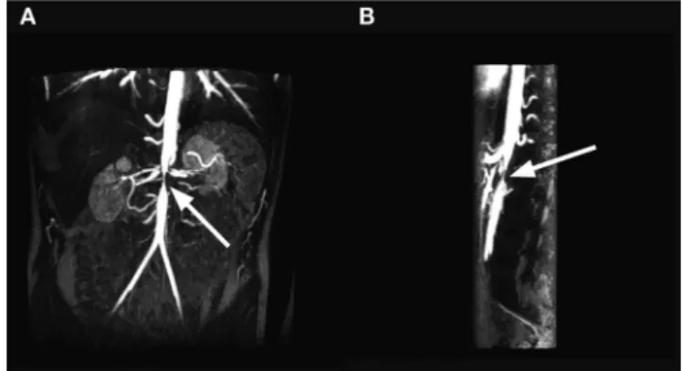
Figure 6. 15 year old with aortic and superior mesenteric artery stenosis
secondary to neurofibromatosis. Maximal intensity projections of the subtracted contrast-enhanced ViBe sequences show focal narrowing of the abdominal aorta (arrow) at the level of the renal arteries in coronal (A) and sagittal (B) projections. the sagittal image also shows focal
narrowing of the origin of the superior mesenteric artery, just caudal to the normal-sized celiac axis and cranial to the aortic narrowing. courtesy Professor R Bhargava.
Author Contributions
All authors contributed to the manuscript concept, review of data, writing, and critical review of the draft. All authors approved the final version of the text.
DISCLOSURES AND ETHICS
As a requirement of publication the authors have provided signed confirmation of their compliance with ethical and legal obligations including but not limited to compliance with icMJe authorship and competing interests guidelines, that the article is neither under consideration for publication nor published elsewhere, of their compliance with legal and ethical guidelines concerning human and animal research participants (if applicable), and that permission has been obtained for reproduction of any copy-righted material. this article was subject to blind, independent, expert peer review. the reviewers reported no competing interests.
REFERENCES
1. Brenner DJ. Estimating cancer risks from pediatric CT: going from the qualita-tive to the quantitaqualita-tive. Pediatr Radiol. 2002;32(4):228–231.
2. Brenner DJ, Elliston CD, Hall EJ, Berdon WE. Estimates of the cancer risks from pediatric CT radiation are not merely theoretical: comment on “point/ counterpoint: in x-ray computed tomography, technique factors should be selected appropriate to patient size. Against the proposition”. Med Phys. 2001; 28(11):2387–2388.
3. McCollough CH, Primak AN, Braun N, Kofler J, Yu L, Christner J. Strategies for reducing radiation dose in CT. Radiol Clin North Am. 2009;47(1):27–40.
4. ACR practice guideline for the performance and interpretation of pediatric mag-netic resonance imaging. American College of Radiology. http://www.acr.org/~/ media/6DA1E94ED99645CDB414AB325414F542.pdf. Accessed July 25, 2013. 5. Balassy C, Hormann M. Role of MRI in paediatric musculoskeletal conditions.
Eur J Radiol. 2008;68(2):245–258.
6. Hormann M. MR imaging of the gastro-intestinal tract in children. Eur J Radiol. 2008;68(2):271–277.
7. Vezina LG. Imaging of central nervous system tumors in children: advances and limitations. J Child Neurol. 2008;23(10):1128–1135.
8. Callen DJ, Shroff MM, Branson HM, et al. MRI in the diagnosis of pediatric multiple sclerosis. Neurology. 2009;72(11):961–967.
9. Battal B, Akgun V, Kocaoglu M. Diffusion-weighted MRI beyond the central nervous system in children. Diagn Interv Radiol. 2012;18(3):288–297. 10. Utsunomiya H. Diffusion MRI abnormalities in pediatric neurological
disor-ders. Brain Dev. 2011;33(3):235–242.
11. Aaen GS, Holshouser BA, Sheridan C, et al. Magnetic resonance spectros-copy predicts outcomes for children with nonaccidental trauma. Pediatrics. 2010;125(2):295–303.
12. Cecil KM. MR spectroscopy of metabolic disorders. Neuroimaging Clin N Am. 2006;16(1):87–116, viii.
13. Thomas B, Al DN, Widjaja E. MRI of childhood epilepsy due to inborn errors of metabolism. AJR Am J Roentgenol. 2010;194(5):W367–W374.
14. Chen J, Licht DJ, Smith SE, et al. Arterial spin labeling perfusion MRI in pediatric arterial ischemic stroke: initial experiences. J Magn Reson Imaging. 2009;29(2):282–290.
15. Triulzi F. Paediatric neuroimaging. Neurol Sci. 2008;29 Suppl 3:342–345. 16. Saunders DE, Thompson C, Gunny R, Jones R, Cox T, Chong WK. Magnetic
resonance imaging protocols for paediatric neuroradiology. Pediatr Radiol. 2007;37(8):789–797.
17. Sanchez CE, Richards JE, Almli CR. Age-specific MRI templates for pediatric neuroimaging. Dev Neuropsychol. 2012;37(5):379–399.
Figure 7. Child with Crohn’s disease with inflamed terminal ileum and inflamed duodenum demonstrated by MR contrast-enhanced enterography.
haste (A) axial images show thickening of the wall of the distal ileum (arrow) that is slightly brighter than muscle. the t1 transverse images pre- (B)
and post-contrast (C) show diffuse enhancement of the thickened wall along with prominence of the vascularity in the adjacent mesentery. this is
also seen in the coronal t1 contrast-enhanced images with fat saturation (arrow, D and E), along with similar abnormal enhancement of the thickened
18. Sanchez CE, Richards JE, Almli CR. Neurodevelopmental MRI brain templates for children from 2 weeks to 4 years of age. Dev Psychobiol. 2012;54(1): 77–91.
19. Bailliard F, Hughes ML, Taylor AM. Introduction to cardiac imaging in infants and children: techniques, potential, and role in the imaging work-up of vari-ous cardiac malformations and other pediatric heart conditions. Eur J Radiol. 2008;68(2):191–198.
20. Shenoy-Bhangle A, Nimkin K, Gee MS. Pediatric imaging: current and emerg-ing techniques. J Postgrad Med. 2010;56(2):98–102.
21. MacKenzie JD, Vasanawala SS. Advances in pediatric MR imaging. Magn Reson Imaging Clin N Am. 2008;16(3):385–402.
22. MacKenzie JD, Vasanawala SS. State-of-the-art in pediatric body and mus-culoskeletal magnetic resonance imaging [abstract]. Semin Ultrasound CT MR. 2010;31(2):86–99.
23. Vazquez E, Castellote A, Piqueras J, et al. Imaging of complications of acute mastoiditis in children. Radiographics. 2003;23(2):359–372.
24. McGee K. The role of a child life specialist in a pediatric radiology department. Pediatr Radiol. 2003;33(7):467–474.
25. Coté CJ, Wilson S. Guidelines for monitoring and management of pediatric patients during and after sedation for diagnostic and therapeutic procedures: an update. Pediatrics. 2006;118(6):2587–2602.
26. Machata AM, Willschke H, Kabon B, Kettner SC, Marhofer P. Propofol-based sedation regimen for infants and children undergoing ambulatory magnetic reso-nance imaging. Br J Anaesth. 2008;101(2):239–243.
27. Schulte-Uentrop L, Goepfert MS. Anaesthesia or sedation for MRI in children. Curr Opin Anaesthesiol. 2010;23(4):513–517.
28. Mahmoud M, Gunter J, Donnelly LF, Wang Y, Nick TG, Sadhasivam S. A comparison of dexmedetomidine with propofol for magnetic resonance imag-ing sleep studies in children. Anesth Analg. 2009;109(3):745–753.
29. Wachtel RE, Dexter F, Dow AJ. Growth rates in pediatric diagnostic imaging and sedation. Anesth Analg. 2009;108(5):1616–1621.
30. Heng Vong C, Bajard A, Thiesse P, Bouffet E, Seban H, Marec BP. Deep seda-tion in pediatric imaging: efficacy and safety of intravenous chlorpromazine. Pediatr Radiol. 2012;42(5):552–561.
31. Malviya S, Voepel-Lewis T, Eldevik OP, Rockwell DT, Wong JH, Tait AR. Sedation and general anaesthesia in children undergoing MRI and CT: adverse events and outcomes. Br J Anaesth. 2000;84(6):743–748.
32. Macmaster FP, Rosenberg DR. Preparing children for MRI. Pediatr Radiol. 2008;38(3):270.
33. Olsen OE. MRI: how to perform a pediatric scan. Acta Radiol. 2013; Feb 6. [Epub ahead of print].
34. Carter AJ, Greer ML, Gray SE, Ware RS. Mock MRI: reducing the need for anaesthesia in children. Pediatr Radiol. 2010;40(8):1368–1374.
35. de Amorim e Silva CJ, Mackenzie A, Hallowell LM, Stewart SE, Ditchfield MR. Practice MRI: reducing the need for sedation and general anaesthesia in children undergoing MRI. Australas Radiol. 2006;50(4):319–323.
36. de Bie HM, Boersma M, Wattjes MP, et al. Preparing children with a mock scanner training protocol results in high quality structural and functional MRI scans. Eur J Pediatr. 2010;169(9):1079–1085.
37. Fogel MA, Pawlowski TW, Harris MA, et al. Comparison and usefulness of cardiac magnetic resonance versus computed tomography in infants six months of age or younger with aortic arch anomalies without deep sedation or anesthesia. Am J Cardiol. 2011;108(1):120–125.
38. Golan A, Marco R, Raz H, Shany E. Imaging in the newborn: infant immobi-lizer obviates the need for anesthesia. Isr Med Assoc J. 2011;13(11):663–665. 39. Mathur AM, Neil JJ, McKinstry RC, Inder TE. Transport, monitoring,
and successful brain MR imaging in unsedated neonates. Pediatr Radiol. 2008;38(3):260–264.
40. Windram J, Grosse-Wortmann L, Shariat M, Greer ML, Crawford MW, Yoo SJ. Cardiovascular MRI without sedation or general anesthesia using a feed-and-sleep technique in neonates and infants. Pediatr Radiol. 2012;42(2):183–187. 41. Vasanawala SS, Alley MT, Hargreaves BA, Barth RA, Pauly JM, Lustig M.
Improved pediatric MR imaging with compressed sensing. Radiology. 2010;256(2):607–616.
42. Vasanawala SS, Lustig M. Advances in pediatric body MRI. Pediatr Radiol. 2011;41 Suppl 2:549–554.
43. Jaramillo D, Laor T. Pediatric musculoskeletal MRI: basic principles to optimize success. Pediatr Radiol. 2008;38(4):379–391.
44. Dagia C, Ditchfield M. 3T MRI in paediatrics: challenges and clinical applica-tions. Eur J Radiol. 2008;68(2):309–319.
45. Alfke K, Jensen U, Pool C, et al. Contrast-enhanced magnetic resonance angi-ography in stroke diagnostics: additional information compared with time-of-flight magnetic resonance angiography? Clin Neuroradiol. 2011;21(1):5–10. 46. Spuentrup E, Wiethoff AJ, Parsons EC, Spangenberg P, Stracke CP. High
spatial resolution magnetic resonance imaging of experimental cerebral venous thrombosis with a blood pool contrast agent. Eur J Radiol. 2010;74(3):445–452. 47. Hirsch W, Sorge I, Krohmer S, Weber D, Meier K, Till H. MRI of the lungs in
children. Eur J Radiol. 2008;68(2):278–288.
48. Olsen OE. Practical body MRI-A paediatric perspective. Eur J Radiol. 2008;68(2):299–308.
49. Mentzel HJ, Seidel J, Fitzek C, et al. Pediatric brain MRI in neurofibromatosis type I. Eur Radiol. 2005;15(4):814–822.
50. Runge VM, Muroff LR, Jinkins JR. Central nervous system: review of clinical use of contrast media. Top Magn Reson Imaging. 2001;12(4):231–263.
51. Hentsch A, Aschauer MA, Balzer JO, et al. Gadobutrol-enhanced moving-table magnetic resonance angiography in patients with peripheral vascular disease: a prospective, multi-centre blinded comparison with digital subtraction angiog-raphy. Eur Radiol. 2003;13(9):2103–2114.
52. Kastrup O, Wanke I, Maschke M. Neuroimaging of infections of the central nervous system. Semin Neurol. 2008;28(4):511–522.
53. Nickerson JP, Richner B, Santy K, et al. Neuroimaging of pediatric intracra-nial infection—part 1: techniques and bacterial infections. J Neuroimaging. 2012;22(2):e42–e51.
54. Mulkey SB, Glasier CM, El-Nabbout B, et al. Nerve root enhancement on spinal MRI in pediatric Guillain-Barré syndrome. Pediatr Neurol. 2010;43(4): 263–269.
55. Yikilmaz A, Doganay S, Gumus H, Per H, Kumandas S, Coskun A. Magnetic resonance imaging of childhood Guillain-Barre syndrome. Childs Nerv Syst. 2010;26(8):1103–1108.
56. Lobel U, Sedlacik J, Reddick WE, et al. Quantitative diffusion-weighted and dynamic susceptibility-weighted contrast-enhanced perfusion MR imaging analysis of T2 hypointense lesion components in pediatric diffuse intrinsic pon-tine glioma. AJNR Am J Neuroradiol. 2011;32(2):315–322.
57. Kan J, Young R, Yu C, Hernanz-Schulman M. Clinical impact of gadolinium in the MRI diagnosis of musculoskeletal infection in children. Pediatr Radiol. 2010;40(7):1197–1205.
58. Grattan-Smith JD, Little SB, Jones RA. MR urography in children: how we do it. Pediatr Radiol. 2008;38 Suppl 1:S3–17.
59. Vivier PH, Dolores M, Taylor M, Elbaz F, Liard A, Dacher JN. MR urog-raphy in children. Part 1: how we do the F0 technique. Pediatr Radiol. 2010;40(5):732–738.
60. Vivier PH, Dolores M, Taylor M, Dacher JN. MR urography in children. Part 2: how to use ImageJ MR urography processing software. Pediatr Radiol. 2010;40(5):739–746.
61. Green D, Parker D. CTA and MRA: visualization without catheterization. Semin Ultrasound CT MR. 2003;24(4):185–191.
62. ACR-NASCI-SPR practice guideline for the performance of body magnetic resonance angiography (MRA). American College of Radiology. http://www. acr.org/~/media/D1BC4FB23D4B4005872FDDAE018E0CE7.pdf. Accessed July 25, 2013.
63. Saleh RS, Singhal A, Lohan D, Duckwiler G, Finn P, Ruehm S. Assessment of cerebral arteriovenous malformations with high temporal and spatial resolution contrast-enhanced magnetic resonance angiography: a review from protocol to clinical application. Top Magn Reson Imaging. 2008;19(5):251–257.
64. Valsangiacomo ER, Levasseur S, McCrindle BW, MacDonald C, Smallhorn JF, Yoo SJ. Contrast-enhanced MR angiography of pulmonary venous abnormalities in children. Pediatr Radiol. 2003;33(2):92–98.
65. Zhong Y, Jaffe RB, Zhu M, Sun A, Li Y, Gao W. Contrast-enhanced magnetic resonance angiography of persistent fifth aortic arch in children. Pediatr Radiol. 2007;37(3):256–263.
66. Thoeny HC, De Keyzer F. Diffusion-weighted MR imaging of native and trans-planted kidneys. Radiology. 2011;259(1):25–38.
67. Tamrazi A, Vasanawala SS. Functional hepatobiliary MR imaging in children. Pediatr Radiol. 2011;41(10):1250–1258.
68. Bonnerot V, Sebag G, de Montalembert M, et al. Gadolinium-DOTA enhanced MRI of painful osseous crises in children with sickle cell anemia. Pediatr Radiol. 1994;24(2):92–95.
69. Burry MV, Cohen J, Mericle RA. Use of gadolinium as an intraarterial contrast agent for pediatric neuroendovascular procedures. J Neurosurg. 2004;100(2 Suppl Pediatrics):150–155.
70. Debatin JF, Nadel SN, Gray L, et al. Phase III clinical evaluation of gadoteridol injection: experience in pediatric neuro-oncologic MR imaging. Pediatr Radiol. 1992;22(2):93–98.
71. Kirchin MA, Pirovano G, Venetianer C, Spinazzi A. Safety assessment of gadobenate dimeglumine (MultiHance): extended clinical experience from phase I studies to post-marketing surveillance. J Magn Reson Imaging. 2001;14(3):281–294.
72. Baker JF, Kratz LC, Stevens GR, Wible JH, Jr. Pharmacokinetics and safety of the MRI contrast agent gadoversetamide injection (OptiMARK) in healthy pediatric subjects. Invest Radiol. 2004;39(6):334–339.
73. Breslau J, Jarvik JG, Haynor DR, Longstreth WT, Jr., Kent DL, Maravilla KR. MR contrast media in neuroimaging: a critical review of the literature. AJNR Am J Neuroradiol. 1999;20(4):670–675.
74. Wible JH, Jr., Tata PN, Napoli AM, Lowe LH, Kearns GL. Pharmacokinetics of gadoversetamide injection, a gadolinium-based contrast agent, in pediatric patients. Magn Reson Imaging. 2009;27(4):512–518.