80
Immune Network
항원제공능 증가
1가톨릭대학교 의과대학 미생물학교실, 2가톨릭조혈모세포은행
박정용
1ㆍ윤성희
1ㆍ김은경
1ㆍ윤선옥
1ㆍ손현정
2ㆍ김태규
1,2Enhancement of Proliferation and Antigen Presentation of Human B Cells in Vitro by K562 Cells Expressing CD40L
Jung-Yong Park
1, Sung Hee Yoon
1, Eun-Kyung Kim
1, Sun-Ok Yun
1, Hyun-Jung Sohn
2and Tai-Gyu Kim
1,21Department of Microbiology, 2Catholic Hematopoietic Stem Cell Bank, College of Medicine, The Catholic University of Korea, Seoul, Korea
ABSTRACT
Background: CD40-activated B (CD40-B) cells might be an attractive source of autologous antigen-presenting cells (APCs) for immunotherapy due to the convenience to obtain from peripheral blood and expand in vitro. Moreover, CD40-B cells were found to be comparable with DCs in their capacity to raise antigen-specific CD8+ T cells.
Here, we have established K562 cells expressing CD40L to expand CD40-activated B cells used for APCs. Methods: After activation of B cell by K562/CD40L, CD40-B cells were examined by counting B cell numbers. Surface expression of CD54, CD80, CD86 and HLA class II was measured by flow cytometry. The CD40-B cells were tested for its function as APC by mixed lymphocyte reactions (MLR) and by induction of T cell responses specific for pp65 peptide in vitro. Results: The expansion of B cells by K562/CD40L increased about 6-folds compared with anti-CD40 or K562. Furthermore, the expression of CD54, CD80, CD86 and HLA class II was up-regulated by K562/CD40L. B cells by K562/CD40L showed comparable antigen presentation activity with mature DCs as shown in MLR, INF-γ ELISPOT assay. Conclusion: These results suggest that K562/CD40L could be used to generate activated B cells as potent APCs which could be useful for cellular vaccination and adoptive immunotherapy. (Immune Network 2007;7(2):80-86)
Key Words: Antigen-presenting cell, B cell, CD40L, K562, immunotherapy
책임저자:김태규, 가톨릭대학교 의과대학 미생물학교실
137-701, 서울시 서초구 반포동 505번지 Tel: 02-590-1216, Fax: 02-3476-7355 E-mail: kimtg@catholic.ac.kr
본 연구는 보건복지부 암정복추진연구개발사업 지원으로 이루어 진 것임(0620320-1).
서 론
수지상세포(dendritic cell, DC)는 항원 포획, 처리, 제공 능력이 뛰어나 활성화된 B 세포 그리고 활성화된 대식 세포를 포함하는 항원제공세포들 사이에서 가장 강력한 항원제공세포로 알려져 왔다(1,2). 항원과 결합된 수지상
세포는 임상적으로 흑색종, 전립선암, 림프종, 신장세포 암과 같은 여러 암들의 치료를 위해 이용되었다(3-6). 하 지만 수지상세포가 T 세포 반응을 강력하게 유도함에도 불구하고 말초 혈액에서의 한정된 개체 수와 시험관내 증식의 어려움 등의 결점을 안고 있다. 수지상세포와는 대조적으로 활성화된 B 세포는 말초 혈액에서 쉽게 획 득할 수 있고 시험관 내에서 증식이 가능하여 면역요법 에 이점이 있다(7). 더구나 생체 밖에서 활성화된 B 세포 는 항원 펩타이드나 종양 용해물의 자극에서 수지상세 포와 유사하게 항원특이 T 세포 반응을 유도할 수 있는 능력을 가진다(2,8).
포에 발현하는 CD40 ligand (CD40L)에 의해 활성 신호를 받은 B 세포는 CD80, CD86, MHC class I과 class II가 수 지상세포와 비슷한 수준으로 발현한다(9). 이전 연구들 에서 수용성 CD40L, 항-CD40 항체 또는 CD40L 발현 세 포주같은 다양한 형태의 CD40-CD40L 결합을 통하여 시험관 내에서 B 세포 활성을 유도하였다(2,10,11).
CD40-CD40L 결합에 의해서 증식된 B 세포는 시험관 내 항원 특이 세포독성 T 세포의 생성을 위한 기능적인 APC로 사용할 수 있다. 수용성 형태의 CD40L를 이용한 B 세포 활성은 NIH3T3/CD40L나 293T/CD40L를 이용 한 세포성 CD40L보다 시험관 내에서 항원 특이 T 세포 를 생성시키는 능력이 낮았다. 그러나 이러한 세포성 CD40L를 이용한 자극법은 B 세포 자극 시 잠재적인 동 종반응이나 이종반응을 야기할 수도 있다(10,11).
본 연구는 이종반응을 회피하기 위해 HLA가 결핍된 K562 세포주를 사용하여 CD40L를 안정적으로 발현하 는 세포주(K562/CD40L)를 확립하였다. 이러한 K562/
CD40L 세포주를 B 세포 자극에 이용하여 B 세포의 활 성과 증식을 관찰하고 활성화된 B 세포의 APC 기능을 확인하였다.
재료 및 방법
공여자와 세포주. 백혈구 성분채집술(leukapheresis)과 비 등차 분획법(Ficoll-Paque density gradient)(Parmacia, Piscataway, NJ)을 이용하여 건강한 공여자의 말초혈액 단핵구세포(peripheral blood mononuclear cells, PBMC)를 획득하였다. MACS CD19 Microbeads isolation kit (Milte- nyi Biotec, Bergisch Gladbach, Germany)를 이용하여 CD19+ B 세포를 분리 하였다. B 세포의 순도는 유세포 분석(flow cytometry analysis)(Becton Dickinson, Immuno- cytometry system, CA, USA)으로 검사하였으며 모든 실 험들에서 90% 이상을 유지하였다(data not shown).
사람 CD40L 유전자 클로닝. 사람 CD40L의 PCR 증폭 주형을 위해 활성화된 CD4+ T 세포들에서 분리해낸 cDNA를 이용하였다. PCR은 95oC에서 5분 1회, 95oC에 서 30초, 57oC에서 30초, 72oC에서 40초를 30회 반복, 72oC에서 10분 1회 조건으로 실시하였다. PCR로 획득된 사람 CD40L DNA를 절단효소인 EcoR I과 Not I으로 절 단 후 pcDNA3/Neo (Invitrogen, Carlsbad, CA, USA)안으 로 클로닝을 하였다. 클로닝된 유전자는 염기서열분석 으로 확인하였다.
CD40L를 발현하는 K562 세포주 생성. pcDNA3/CD40L 는 K562 세포 안으로 NucleofectorTM kit V (Amaxa Biosys- tems, Kӧln, Germany)를 이용하여 형질 도입시켰다. 형질 도입된 세포들은 10% FBS (Sigma, St. Louis, Mo., USA), 2 mM glutamine (Gibco/BRL, Gaithersburg, MD), 100 U/ml
(Cambrex Bio Science, Walkersvile, MD USA)에서 배양되 었으며 1 mg/ml G418 (Duchefa, Hearlem, The Nethelands) 와 유세포 분류기(fluorescence-activated cell sorting, FACS) 에 의해 선별 되었다. 실험이 진행되는 동안 K562 세포 에서 CD40L의 발현은 안정적(±10%)이었다.
수지상세포의 생성. 수지상세포는 이전과 동일하게 생 성하였다(2). 간략히 기술하면, MACS CD14 Microbeads isolation kit (Miltenyi Biotec)을 이용하여 PBMC로부터 CD14+ 단핵구를 획득하였고, IL-4 (Endogen, Woburn, MA) 와 GM-CSF (Endogen, Rockford, IL, USA)와 함께 10% RPMI1640에서 7일 동안 배양하였다. 배양액은 4일 째에 추가적으로 공급하였으며 7일째에 50 ng/ml GM-CSF, 10 ng/ml tumor necrosis factor (TNF)-α (Endogen), 1μg/ml LPS (Sigma) 그리고 10 ng/ml IL-4를 이용해 24시간 동안 성숙을 유도하였다.
B 세포 증식. 분리된 B 세포 (5×10⁵ cells/ml)는 100 Gy로 조사된 K562/CD40L와 함께 12-well plates (Nunc, Roches- ter, NY, USA)에서 20 ng/ml IL-4를 첨가한 10% Iscove’s modified Dulbecco’s medium (IMDM)(Cambrex)를 이용하 여 배양하였다. 증식된 B 세포는 3∼4일에 채취하여 증 가된 수를 측정하였다. 그 후에 조사된 K562/CD40L와 활성화된 B 세포(5×10⁵ cells/well)를 새로운 배양액과 함 께 다시 배양하였다. 세포수와 생존율은 Trypan Blue 염 색법에 의해 측정되었고 비등차 분획법을 이용하여 사 멸된 B 세포와 K562/CD40L의 잔유물을 제거한 후 이용 할 때까지 액체질소에서 보관하였다.
표현형 분석. PE-conjugated anti-CD154/CD40L, anti-CD54, anti-CD80, anti-CD86, anti-HLA Class II (BD Pharmingen, San Diego, CA)를 이용하여 K562와 활성화된 B 세포의 보조자극분자들과 활성 표지자들의 발현을 측정하였다.
표현형은 유세포 분석기를 이용하여 이전과 같은 방법 으로 분석하였다(11). 간략히 기술하면, 100 ul PBS (Cambrex)안의 1백만개의 세포들은 2% FBS가 함유된 PBS로 두 번 세척한 후 0.5μg 단클론 항체를 20분 동안 4oC에서 염색시켰다. 염색된 세포를 세척 후 1%
Paraformaldehyde 함유된 PBS로 고정시켰다. 유세포 분 석은 위에서 언급한 유세포 분석기와 CellQuest software (BD Biosciences)를 이용하여 수행하였다.
혼합 림프구 반응(mixed lymphocyte reaction). MLR은 XTT assay (Roche Diagnostics GmbH, Mannheim, Ger- many)를 이용하여 이전과 같은 방법으로 생존한 T 세포 를 측정하였다(11). 간략히 기술하면, MACS CD4 Micro- beads isolation kit (Miltenyi Biotec)를 이용하여 CD4+ T 세포를 분리 하였다. 분리된 동종 T 세포(1×10⁵)는 동일 한 수의 활성화된 B 세포와 96-well flat-bottomed tissue culture plates (Falcon, Bedford, MA)에서 6일 동안 배양을
B
Human CD40 ligand
pcDNA3 5.4kb
80%
unstaining
CD40L
A
EcoR I
Not I
80%
B
Human CD40 ligand
pcDNA3 5.4kb
80%
unstaining
CD40L
A
EcoR I
Not I
80%
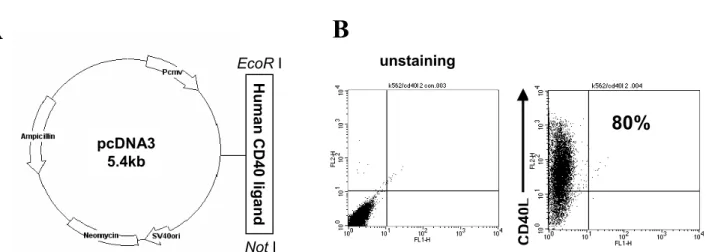
Figure 1. Stable K562 transfectant simultaneously expressing CD40L. (A) Constructive diagram of CD40L gene expression. The cDNA encoding human CD40L is derived from the activated CD4+ T cells by extracting RNA using with EcoR I and Not I restriction site hanging at the 5’ and 3’ terminal respectively. The amplified human CD40L is then inserted into the mammalian cell expression vector pcDNA3. (B) Expression of CD40L on K562 cell lines. CD40L expressing stable transfectant of K562 is sorted by combination of a drug selection and FACS sorting. The surface markers on K562/CD40L were tested with PE-conjugated human CD40L.
하였다. 흡광도는 490 nm에서 ThermoMax microplate reader (Molecular Devices, Palo Alto, CA, USA)로 측정하 였다.
ELISPOT에 의한 IFN-γ 발현 림프구 측정. ELISPOT assay는 이전과 같은 방법으로 수행하였다(8). 간략히 기 술하면, ELISPOT kit (BD biosciences)를 사용하였다. 7일 동안 K562/CD40L에 의해 활성화된 B 세포에 인간 거대 세포바이러스(human cytomegalovirus, HCMV)인 pp65 HLA-A*0201 결합 펩타이드 495∼503 (NLVPMVATV) (10μg/ml)를 결합시킨 후 자극세포로 이용하였다. MACS CD8 Microbeads isolation kit (Miltenyi Biotec)를 이용하여 CD8+ T 세포를 분리 하였다. 분리된 CD8+ T 세포 (5×10⁴ cells/well)는 활성화된 B 세포(5×10³ cells/well)와 함께 혼합하였다. 혼합된 세포들은 U-bottomed 96-well plate (Falcon)에서 100μl 배양액에 4시간 동안 배양되었 다. Spot들을 나타내기 위하여 streptavidin-HRP와 기질을 이용하였으며 ELISPOT reader (AID, Jessup, MD)를 이용 하여 측정하였다.
통계학적 처리. 모든 결과들은 mean±SE로 표현하였으 며, 각 실험군 간의 통계학적 유의성은 Student's t-test에 의하여 검정하였다. 통계적 유의성은 p-value <0.05인 경우에만 인정하였다. 통계분석은 SigmaPlot 2000 soft- ware (SPSS Inc. Chicago, IL, USA)를 이용하였다.
결 과
CD40L를 발현하는 K562 세포주 확립. B세포의 증식을 유도하기 위해 K562/CD40L 세포주를 확립하였다. 이러 한 K562 세포들은 프로모터와 neomycin 저항성 유전자 를 함유한 플라스미드에 제한효소를 처리하여 클로닝된 CD40L를 형질 도입시킴으로 만들어 졌다(Fig. 1A).
CD40L 형질도입 된 K562 세포는 G418 약제와 CD40L 항체를 이용한 유세포 분류기를 이용해 선별 하였다. 선 별된 K562/CD40L 세포주의 CD40L의 발현량은 6개월 이후에도 80% 이상을 유지하였다(Fig. 1B).
K562/CD40L에 의한 B 세포의 증식과 항원제공분자 발 현. K562/CD40L와 B세포의 비율에 따른 B 세포의 활성 화 정도를 측정하기 위하여 IL-4 (20 ng/ml)를 첨가한 상 태에서 고정된 수의 B 세포와 K562/CD40L의 비율을 1:0.25에서 1:4로 조절하여 자극시켰다. 모든 비율의 자극에서 K562/CD40L와 B 세포는 뭉치는 현상을 보였 다. 하지만 1:0.25나 1:0.5 비율에서 B 세포의 증식은 유도되지 못하였다. 흥미로운 점은 1:2와 1:4 비율에 서 B 세포에서 뭉치는 정도가 크고 증식도 강하게 유도 되었으나(Fig. 2A and B), 1:4 비율의 B 세포 자극에서 는 많은 K562/CD40L 잔유물이 생겨서 반복적인 자극에 는 부적합하여, 본 연구에서는 K562/CD40L를 1:2의 비 율로 B 세포를 자극 하였다. 14일 동안 K562/CD40L로 자극시킨 B 세포(B-K562/CD40L)는 6배정도 증가 되었 으나 말초혈액 B 세포와 K562만으로 자극 시킨 B (B-K562)세포는 모두 사멸되었다(Fig. 2C).
B 세포에서 APC 기능과 연관성이 있는 표현형의 변 화를 측정하기 위하여 자극시키지 않은 말초혈액 B 세 포와 7일 동안 활성 시킨 B 세포에서 CD54, CD80, CD86 그리고 HLA class II의 발현을 측정하였다. 말초혈액 B 세포와 비교하여 B-K562/CD40L 세포에서 CD54, CD80, CD86 그리고 HLA class II 발현량이 증가하였다(Fig. 3).
이들 발현은 자극 1일 후부터 증가되기 시작하였다(data not shown).
B-K562/CD40L 세포를 이용한 T 세포의 효과적인 자극.
B-K562/CD40L 세포의 항원 제공 능력을 확인하기 위하
Figure 2. B cells proliferation using K562/CD40L. (A) Optimal ratio of B cell to K562/CD40L. Various numbers of K562/CD40L (sIL-4; 20 ng/ml) were then added to each well. On day 3, cultures were re-stimulated by fresh K562 transfectants. B cells were expanded for 7 days. (B) Morphology of K562/CD40L-stimulated B cells (B-K562/CD40L) after 7 days of culture. Typical cluster for- mation of B-K562/CD40L that surround the K562/CD40L (original magnification: ×100). (C) Comparison of long-term expansion of B-K562/CD40L and B-anti-CD40. B cells were stimulated with control K562 cells, anti-CD40 mAb (10μg/ml) or K562/CD40L (sIL-4;
20 ng/ml) for 14 days. Cultures were harvested, counted and re-stimulated twice a week.
Figure 3. Phenotypical analysis of B and B-K562/CD40L. Peripheral Blood B cells (PBB) were activated for 7 days with K562/CD40L (sIL-4; 20 ng/ml). The ratio of B cells to K562/CD40L is fixed 1:2 and after coincubation for 7 days, the costimulatory molecule CD54, CD80, CD86 and HLA II molecule are analyzed on activated B cells, the dot line. Thin solid line indicates fluorescence of isotype matched antibody.
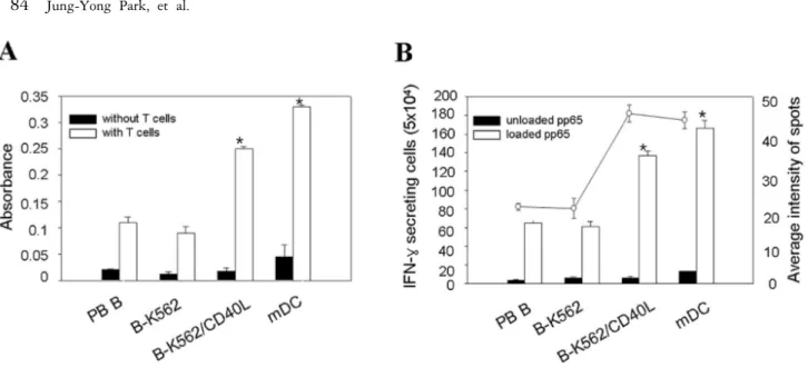
Figure 4. MLR or IFN-γ ELISPOT assay using HCMV pp65 peptide-pulsed activated B cells. (A) Proliferation of allogeneic T cells by activated B cells. Induction of T-cell proliferation was performed with PB B, B-K562, B-K562/CD40L or mature dendritic cells (mDCs) pre-cultured for 6 days. Purified allogeneic T cells were co-cultured with activated B cells in a final volume of 100μl. MLRs were cultured for 6 days and proliferating viable cells were analyzed by XTT assay. Data are shown as means±SE of 3 independent experiments, each performed is triplicate. *: p<0.05 for B-K562/CD40L vs. mDC. (B) Induction of IFN-γ secretion T cells by activated B cells. A*0201+ CD8 T cells from normal donors were stimulated with autologous activated B cells and subsequently analyzed for their immune response by ELISPOT. Stimulator cells were autologous activated B cells from donors pulsed with CMV antigen pp65 peptide. Open circle indicates average intensity of spots. Data are shown as means±SE of 3 independent experiments. *: p<0.05 for B-K562/CD40L vs. mDC.
여 혼합림프구 반응을 측정한 결과, Fig. 4A에서와 같이 T 세포의 증식은 말초혈액 B 세포나 B-K562 세포에서보 다 B-K562/CD40L 세포에 의해서 상당한 증가를 보였다.
하지만 이러한 B 세포를 이용한 T 세포 증식은 성숙 수 지상세포(mDC)보다는 낮은 수준이었다. 또한 B-K562/
CD40L 세포에 항원을 감작시킨 후 CD8+ T 세포를 자극 시켜 T 세포 면역반응의 점화(priming)를 유도하였다. 성 인 80∼90%가 면역화 되어있어 넓은 면역반응을 일으키 는 HCMV 항원인 pp65를 사용하였다. Fig. 4B의 ELISPOT 검사법에서 pp65가 감작된 B 세포는 CD8+ T (5×10⁴)에 서 성숙 수지상세포 만큼 IFN-γ 분비하는 T 세포의 높 은 증가를 유도하였다. 더구나 단일 T 세포에서 분비하 는 IFN-γ 양은 K562/CD40L-B 세포와 성숙 수지상세포 가 유사하였다.
고 찰
EBV 림프성 세포주를 이용해 영구화시킨 B 세포들은 항원 특이 T 세포들을 효과적으로 유도할 수 있었다 (13,14). 비록 EBV-LCL의 생성이 간단하고 항원제공세 포 기능을 유지하지만 살아있는 바이러스 이용에 관한 안정성의 문제점과 EBV-LCL를 확립하기 위해 4∼6주 간이 기간이 필요하다는 단점이 있었다. 이전 연구들에 서 B 세포 활성은 다양한 형태의 CD40L로 자극되었으 며, 활성화된 B 세포는 시험관 내에서 바이러스와 종양
항원 특이적인 세포독성 T 세포의 활성화와 면역반응을 유도하기 위한 항원제공세포로 이용되었다(2,8). 레트로 바이러스 형질도입, 펩타이드 감작과 RNA 형질전환을 이용하여 활성화된 B 세포를 자극하여 면역반응을 유도 하는 연구가 진행되었으며, 수지상세포와 비슷하게 항 원 특이 CD8+ T 세포를 유도하였다(7,8,12). 다양한 형태 의 B 세포 자극은 주로 수용성 CD40L와 세포성 CD40L 의 두가지 형태로 이용되어 왔다. 임상실험에서 시험관 내 항원 특이 T 세포 유도를 위해 수용성 CD40L를 B 세포 활성에 이용되었지만 세포성 CD40L보다 자극 수 준이 낮았다(2). 그러므로 세포성 CD40L를 발현하는 NIH3T3 세포주나 293T 세포주를 이용하여 B 세포의 증 식을 유도하였으나 NIH3T3 세포에서의 이종반응과 293T 세포에서 동종반응이 야기 되는 문제점이 있었다(10-12).
본 연구는 지금까지 연구들의 문제점을 보완하기 위 하여 K562 세포주를 이용하였다. K562 세포주는 사람 골수세포에서 유래되었고, HLA 분자가 발현되지 않으 며, 부유세포배양 되므로 배양이 용이한 이점을 가지고 있다(16). 최근 K562 세포주가 T 세포 리셉터와 CD28과 4-1BB 보조자극 분자들의 리간드를 발현하는 인공항원 제공세포(artificial antigen presenting cell, AAPC)로서 이 용되었다(16,17). K562/CD40L는 B 세포를 14일 동안 자 극하여 3배 증가시켰고 CD40 항체로 자극한 증식보다 높았다. 비록 기존 연구에서 사용된 NIH3T3/CD40L와
만 K562/CD40L로 자극한 B 세포와 비슷한 성장 곡선을 보였다(Fig. 2B). 또한 이전 CD40L를 발현하는 S2 세포 주를 이용한 자극에서 B 세포를 활성화시키긴 하였지만 높은 증식을 유도하지 못한 것과 달리 K562/CD40L는 높 은 증식을 유도하였다(11). 이러한 결과는 정확히 밝히 지는 못하였지만 K562 세포에는 CD54 (ICAM-1), CD58 (LFA-3)과 αVβ3 (vitronectin 수용체) 등과 같은 세포결합 에 관여하는 세포부착분자들이 발현하기 때문에 세포간 에 강한 결합력이 작용하여 효과적인 자극이 이루어진 것으로 생각된다(18). 항원제공세포로서 B-K562/CD40L 세포를 이용한 MLR 분석법을 통해서 K562/CD40L에 의 해 활성화된 B 세포가 성숙 수지상세포보다는 낮았지만 높은 T 세포 증식을 유한다는 것을 확인하였다(Fig. 4A).
활성화된 B 세포의 표면분자를 분석한 결과를 통해 K562/CD40L로 자극했을 때 수용성 CD40L나 다른 세포 성 CD40L로 자극했을 때와 비교하여 CD54와 CD80은 동일한 수준으로 발현하였으며 CD86과 HLA class II는 높게 발현하는 걸 확인하였다(2,10). 항체분비능을 측정 하기 위하여 활성화된 B 세포의 IgM과 IgG의 분비를 확 인한 결과 아무 반응을 보이지 않았다(data not shown).
K562/CD40L에 의해 자극된 B 세포는 항체를 생산하는 단계인 플라즈마로 분화되기 전 상태라는걸 확인하였 다. 또한 HCMV항원인 pp65 펩타이드를 이용한 ELISPOT 검사법을 통해서 K562/CD40L-B 세포가 성숙 수지상세 포보다 IFN-γ의 분비하는 세포수는 적었지만 단일세포 에서 분비하는 IFN-γ 양에선 큰 차이를 보이지 않았다 (Fig. 4B). 일반적으로 활성화된 B 세포는 성숙 수지상세 포보다 낮은 항원제공능을 가진다고 알려졌지만 K562/
CD40L에 의해 자극된 B 세포는 높은 항원제공능을 보 였다(19). 지금까지 K562/CD40L에 의해 활성화된 B 세 포가 가지는 증식능과 항원제공능을 알아 보았다. 비록 활성화된 B 세포가 높은 증식능과 항원제공능을 가지지 만 특정항원만을 제시하는 특성과 항체를 생산하는 세 포로 분화기에 생기는 항원제공능의 한계를 가진다. 전 문항원제공세포로서 완벽하게 수지상세포를 대체하기 위해서는 K562/CD40L뿐만 아니라 좀더 다양한 전략적 연구가 필요하다.
본 연구에서는 바이러스 항원인 pp65 특이 면역반응 만을 확인하였지만 다양한 종양 항원에 대한 추후 연구 가 수반 되어야 할 것이며, 수지상세포와 비교하여 활성 화된 B 세포의 생체 내에서의 이동과 유도된 면역반응 의 특성을 규명하는 것이 필요하다.
본 연구에서 K562/CD40L를 이용해 활성화된 B 세포 를 생성하고 활성화된 B 세포의 항원제공기능을 확인하 였으며, 향후 B 세포 매개 세포성 백신의 개발과 입양면 역치료에 기여할 수 있을 것으로 사료된다.
참 고 문 헌
1. Steinman RM, Witmer-Pack M, Inaba K: Dendritic cells: an- tigen presentation, accessory function and clinical relevance.
Adv Exp Med Biol 329;1-9, 1998
2. von Bergwelt-Baildon MS, Vonderheide RH, Maecker B, Hirano N, Anderson KS, Butler MO, Xia Z, Zeng WY, Wucherpfennig KW, Nadler LM, Schultze JL: Human pri- mary and memory cytotoxic T lymphocyte responses are effi- ciently induced by means of CD40-activated B cells as anti- gen-presenting cells: potential for clinical application. Blood 99;3319-3325, 2002
3. Thurner B, Haendle I, Roder C, Dieckmann D, Keikavoussi P, Jonuleit H, Bender A, Maczek C, Schreiner D, von den Driesch P, Brocker EB, Steinman RM, Enk A, Kampgen E, Schuler G: Vaccination with mage-3A1 peptide-pulsed ma- ture, monocyte-derived dendritic cells expands specific cyto- toxic T cells and induces regression of some metastases in advanced stage IV melanoma. J Exp Med 190;1669-1678, 1999
4. Heiser A, Coleman D, Dannull J, Yancey D, Maurice MA, Lallas CD, Dahm P, Niedzwiecki D, Gilboa E, Vieweg J:
Autologous dendritic cells transfected with prostate-specific antigen RNA stimulate CTL responses against metastatic prostate tumors. J Clin Invest 109;409-417, 2002 5. Maier T, Tun-Kyi A, Tassis A, Jungius KP, Burg G,
Dummer R, Nestle FO: Vaccination of patients with cuta- neous T-cell lymphoma using intranodal injection of autolo- gous tumor-lysate-pulsed dendritic cells. Blood 102;2338- 2344, 2003
6. Kugler A, Stuhler G, Walden P, Zoller G, Zobywalski A, Brossart P, Trefzer U, Ullrich S, Muller CA, Becker V, Gross AJ, Hemmerlein B, Kanz L, Muller GA, Ringert RH:
Regression of human metastatic renal cell carcinoma after vaccination with tumor cell-dendritic cell hybrids. Nat Med 6;332-336, 2000
7. Schultze JL, Michalak S, Seamon MJ, Dranoff G, Jung K, Daley J, Delgado JC, Gribben JG, Nadler LM: CD40- acti- vated human B cells: an alternative source of highly efficient antigen-presenting cells to generate autologous anti- gen-specific T cells for adoptive immunotherapy. J Clin Invest 100;2757-2765, 1997
8. Kondo E, Topp MS, Kiem HP, Obata Y, Morishima Y, Kuzushima K, Tanimoto K, Harada M, Takahashi T, Akat- suka Y: Efficient generation of antigen-specific cytotoxic T cells using retrovirally transduced CD40-activated B cells. J Immunol 169;2164-2171, 2002
9. Grewal IS, Flavell RA: CD40 and CD154 in cell-mediated immunity. Annu Rev Immunol 16;111-135, 1998 10. Ivanov R, Aarts T, Hagenbeek A, Hol S, Ebeling S: B-cell
expansion in the presence of the novel 293-CD40L-sCD40L cell line allows the generation of large numbers of efficient xenoantigen-free APC. Cytotherapy 7;62-73, 2005 11. Yoon SH, Cho HI, Kim TG: Activation of B cells using
Schneider 2 cells expressing CD40 ligand for the enhance- ment of antigen presentation in vitro. Exp Mol Med 37;567-574, 2005
12. Coughlin CM, Vance BA, Grupp SA, Vonderheide RH:
RNA-transfected CD40-activated B cells induce functional T-cell responses against viral and tumor antigen targets: im- plications for pediatric immunotherapy. Blood 103;2046- 2054, 2004
13. Okamura T, Uluhan S, Rooney CM: Antigen-presenting ca-
pabilities of retrovirus-transduced T cell blasts. Blood 102;
285a, 2003
14. Liu YN, Fuad S, Gehrz RC: Epstein-Barr virus-transformed lymphoblastoid cell lines as antigen-presenting cells and
"augmenting" cells for human CMV-specific Th clones. Cell Immunol 108;64-75, 1987
15. Ascher NL: Issues in xenotransplantation. Liver Transpl Surg 1;320-321, 1995
16. Maus MV, Thomas AK, Leonard DG, Allman D, Addya K, Schlienger K, Riley JL, June CH: Ex vivo expansion of poly- clonal and antigen-specific cytotoxic T lymphocytes by artifi- cial APCs expressing ligands for the T-cell receptor, CD28
and 4-1BB. Nat Biotechnol 20;143-148, 2002
17. Dessureault S, Alsarraj M, McCarthy S, Hunter T, Noyes D, Lee D, Harkins J, Seigne J, Jennings R, Antonia SJ: A GM-CSF/CD40L producing cell augments anti-tumor T cell responses. J Surg Res 125;173-181, 2005
18. Rainaldi G, Calcabrini A, Santini MT: Positively charged pol- ymer polylysine-induced cell adhesion molecule redistribution in K562 cells. J Mater Sci Mater Med 9;755-760, 1998 19. Schultze JL, Grabbe S, von Bergwelt-Baildon MS: DCs and
CD40-activated B cells: current and future avenues to cellular cancer immunotherapy. Trends Immunol 25;659-664, 2004