□
증례보고
□994
Third-generation Cryosurgery for Prostate Cancer Patient with a Penile Prosthesis
Myung Ki Kim1, Jun Cheon2, Hyung Jin Kim
From the Department of Urology, Chonbuk National University Medical School, Jeonju, 1Chonnam National University Medical School, Gwangju, 2Korea Universi- ty College of Medicine, Seoul, Korea
Treatment options for clinically localized prostate cancer include surgical extirpation, radiation therapy (external beam radiation therapy and/or brachytherapy), or watchful waiting. Cryosurgery has recently re-emerged as a developing technology and as a minimally invasive treatment option.
There may be technical difficulties in placing cryoprobes in patient with a penile prosthesis. To the best of our knowledge, this is the first report of cryosurgical ablation of the prostate in a prostate cancer patient with a penile prosthesis. (Korean J Urol 2007;48:994-996)
Key Words: Cryotherapy, Prostate cancer, Penile prosthesis
대한비뇨기과학회지 제 48 권 제 9 호 2007
전북대학교, 1전남대학교 및
2고려대학교 의과대학
비뇨기과학교실
김명기1ㆍ천 준2ㆍ김형진 접수일자:2007년 4월 19일 채택일자:2007년 6월 26일 교신저자: Hyung Jin Kim
Department of Urology, Chonbuk National
University Medical School, 634-18, Geumam-dong, Deokjin-gu, Jeonju 561-712, Korea TEL: 063-250-1568 FAX: 063-250-1564 E-mail: hjkim@
chonbuk.ac.kr
With the recent introduction of novel, minimally invasive procedures for the treatment of prostate cancer, cryotherapy has become a feasible option as a viable alternative to traditional radical surgery and radiation therapy. As a result of better understanding of tumor cryodestruction at a molecular level, refinements in cryotechniques and improved patient selection, the results of cryotherapy are becoming more promising.
Furthermore, the dramatic decrease in the number of complic- ations after modern cryotherapy leads to a better quality of life, which may be a preferable option, especially for elderly patients with comorbidities.1,2 Because the cyroprobes is inserted through the perineum, cryotherapy has been limited in patient with penile prosthesis. We report a case of cryosurgical ablation of the prostate in prostate cancer patient with penile prosthesis.
CASE REPORT
A 72-year-old man presented with one year history of obstructive voiding symptoms. He had suffered from cardiac arrhythmia for several years. 10-year ago, he underwent penile prosthetic surgery due to erectile dysfunction. Digital rectal
examination was not notable for a palpable abnormality of the prostate. Prostate-specific antigen (PSA) level was 16.22 ng/ml.
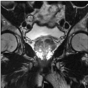
Basic laboratory values, urinalysis and cytology were within normal limit. Ultrasound guided prostate needle biopsy demon- strated prostatic adenocarcinoma, 3 cores-positive of 12 cores and Gleason score 5 (3+2) at left peripheral zone of the pros- tate. Estimated prostate volume was 18 cc. Contrast enhanced endorectal magnetic resonance imaging of the pelvis revealed a localized prostate cancer with no evidence of periprostatic infiltration or pelvic lymphadenopathy and prosthesis materials in other view. (Fig. 1, 2) Other imaging modalities showed no evidence of metastatic lesions.
With the patient under regional anesthesia, he was placed in lithotomy position. A transrectal ultrasound probe was posi- tioned using a holding device, and a brachytherapy template was positioned close to the perineum. The malleable penile prosthesis was palpated in the perineum, but there were no difficulties in placing the any cryoprobes. The ten cryoprobes were inserted through the perineum and positioned in the prostate under ultrasound guidance in an overlapping pattern to ensure complete coverage of the prostate. Three thermosensors
Myung Ki Kim, et al:Third-generation Cryosurgery for Prostate Cancer Patient with Penile Prosthesis
995
Fig. 1. Contrast enhanced endorectal magnetic resonance imaging of the pelvis shows about a 1.5 cm sized nodule in the left peri- pheral zone of the prostate.
Fig. 2. Contrast enhanced magnetic resonance imaging of the pelvis shows the prosthesis materials in the corpus cavernosum.
were also positioned to monitor the temperature near the urethra, at the external sphinter and along Denonvilliers’ fascia.
Flexible cystoscopy was performed, and it was ensured that no probes was penetrated the urethra or bladder. Next, a urethral warming catheter was placed and continuously irrigated with warm saline (42oC). The probes were activated from anterior to posterior so that the advancing edge of the iceball can be monitored with ultrasound. Two freeze-thaw cycles were used.
After cryosurgical ablation of prostate, urethral catheter was inserted and removed at 2 weeks later. Postoperative course was uneventful and the patient discharged at 2 days later.
Followup examination at 2 months later, PSA level was 0.24 ng/ml and voiding was well without irritative voiding symptoms.
DISCUSSION
Cryosurgery began in England in the 1840s when James Arnott used iced saline, was applied through hollow irrigating tubes, to treat gynecological tumors.3 It was not until the 1960s that cryosurgery was applied to urological conditions. Gonder et al.4 in New York began to use liquid nitrogen to treat bladder neck obstruction from both prostate cancer and benign prostatic hyperplasia (BPH). Improved techniques for percutaneous access to the prostate, coupled with the development of transrectal ultrasonography (TRUS) in the 1980s, led to renewed interest
in cryosurgery of the prostate.5 In 2000, the first use of gas-driven cryoprobes was reported.6 The changes to gas-driven systems caused two major changes to cryosurgical technique.
First adding a thaw capability with rapid transition from freezing to defrosting improved control of ice-ball formation and allowed faster procedure. However, more importantly the change permitted a dramatic reduction in the diameter of the cryoprobes.6 Ultra-thin probes with sharp tips allows direct transperineal penetration and placement without an insertion kit.7
There may be technical difficulties in placing the probes in patient with penile prosthesis. We use third-generation cryosu- rgery device (Seednet Gold System & Accessories, Galil Medi- cal, Yokneam, Israel), it enables us to do cryosurgery of the prostate in patient with penile prosthesis. There are no difficulties in placing cryoprobes in that patient.
We think that cryosurgical ablation of the prostate in patient with penile prosthesis is possible procedure and no more contraindication.
REFERENCES
1. Han KR, Belldegrun AS. Third-generation cryosurgery for primary and recurrent prostate cancer. BJU Int 2004;93:14-8 2. Mouraviev V, Polascik TJ. Update on cryotherapy for prostate
cancer in 2006. Curr Opin Urol 2006;16:152-6
3. Arnott J. Practical illustrations of the remedial efficacy of a
996 대한비뇨기과학회지:제 48 권 제 9 호 2007
very low or anaesthetic temperature. I. in cancer. Lancet 1850;2:257-9
4. Gonder MJ, Soanes WA, Shulman S. Cryosurgical treatment of the prostate. Invest Urol 1966;3:372-8
5. Onik G, Porterfield B, Rubinsky B, Cohen J. Percutaneous transperineal prostate cryosurgery using transrectal ultrasound
guidance: animal model. Urology 1991;37:277-81
6. Zisman A, Leibovici D, Seigel YI, Lindner A. Prostate cryoablation without an insertion kit using direct transperineal placement of ultrathin freezing probes. Tech Urol 2000;6:34-6 7. Rees J, Patel B, MacDonagh R, Persad R. Cryosurgery for
prostate cancer. BJU Int 2004;93:710-4