Blood Res2021;56:44-58. bloodresearch.or.kr
50 Letters to the Editor
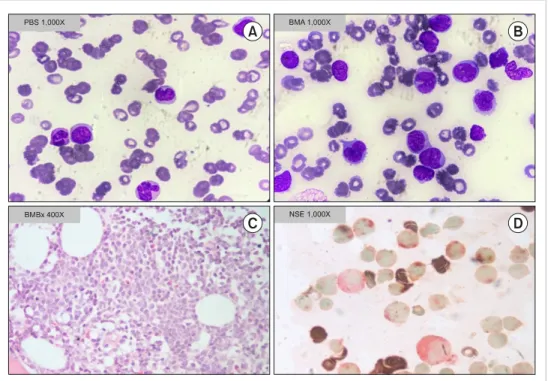
Fig. 1. Peripheral blood and bone marrow morphology. Peripheral blood smear showing blasts and monocytes (A). Bone marrow aspirate showing blasts and monocytic population (B). Bone marrow biopsy showing marrow spaces replaced by blasts (C). NSE positivity in blasts (D).
3. Chen JS, Lin DT, Chuu WM, Lin KH, Su IJ, Lin KS. Acute myelo- fibrosis terminating in acute lymphoblastic leukemia: report of one case. Zhonghua Min Guo Xiao Er Ke Yi Xue Hui Za Zhi 1992;33:136-43.
4. Dunphy CH, Kitchen S, Saravia O, Velasquez WS. Acute myelo- fibrosis terminating in acute lymphoblastic leukemia: case report and review of the literature. Am J Hematol 1996;51:85-9.
5. Abla O, Ye CC. Acute lymphoblastic leukemia with massive myelofibrosis. J Pediatr Hematol Oncol 2006;28:633-4.
6. Avcı Z, Malbora B, Gülşan M, Şahin FI, Celasun B, Özbek N.
Acute massive myelofibrosis with acute lymphoblastic leukemia.
Turk J Haematol 2009;26:204-6.
7. Gonzalez MM, Kidd L, Quesada J, Nguyen N, Chen L. Acute mye- lofibrosis and acute lymphoblastic leukemia in an elderly patient with previously treated multiple myeloma. Ann Clin Lab Sci 2013;43:176-80.
8. Friesenbichler W, Schumich A, Simonitsch-Klupp I, et al.
Concurrent acute myelofibrosis and acute lymphoblastic leuke- mia in childhood: case report and review of the literature. J Pediatr Hematol Oncol 2018;40:235-7.
9. Bain BJ, Catovsky D, O’Brien M, et al. Megakaryoblastic leuke- mia presenting as acute myelofibrosis -- a study of four cases with the platelet-peroxidase reaction. Blood 1981;58:206-13.
10. Verhoef GE, De Wolf-Peeters C, Ferrant A, et al. Myelodysplastic syndromes with bone marrow fibrosis: a myelodysplastic dis- order with proliferative features. Ann Hematol 1991;63:235-41.
11. Norén-Nyström U, Roos G, Bergh A, et al. Bone marrow fibrosis in childhood acute lymphoblastic leukemia correlates to bio- logical factors, treatment response and outcome. Leukemia 2008;22:504-10.
12. Yokoyama K, Amamiya T, Kawai Y, et al. Acute lymphocytic leu- kemia associated with severe myelofibrosis. Keio J Med 1997;
46:196-204.
13. Ray R, Garewal G, Marwaha RK, Marwaha N. Childhood myelo- fibrosis: transformation to acute lymphoblastic leukemia.
Pediatr Hematol Oncol 1993;10:175-8.
14. Nordan UZ, Humbert JR. Myelofibrosis and acute lymphoblastic leukemia in a child with Down syndrome. J Pediatr 1979;94:253-5.
15. Marino R, Altshuler G, Humphrey GB. Idiopathic myelofibrosis followed by acute lymphoblastic leukemia. Am J Dis Child 1979;133:1194-5.
16. Eliacik E, Isik A, Aydin C, et al. Bone marrow fibrosis may be an effective independent predictor of the ‘TKI drug response level’
in chronic myeloid leukemia. Hematology 2015;20:392-6.
A paradigm shift: lineage switch from T-ALL to B/myeloid MPAL
TO THE EDITOR: Lineage switch in acute leukemia is de- fined as a complete immunophenotypic change either at relapse or during therapy. Lineage switch from acute lym- phoblastic leukemia (ALL) to acute myeloid leukemia (AML) is observed in most cases; the reverse is uncommon [1, 2].
The exact mechanism underlying lineage switch is unclear [3]. Lineage switch from T-ALL to mixed-phenotype acute leukemia of B/myeloid lineage (B/myeloid MPAL) is excep- tionally rare. We report a case of T-ALL at initial pre- sentation, which showed lineage switch to B/myeloid MPAL at relapse. We believe that this is the first such case to be reported.
A 15-year-old boy was initially presented to our hospital in 2007 with fever and breathing difficulty. Clinical exami-
bloodresearch.or.kr Blood Res 2021;56:44-58.
Letters to the Editor 51
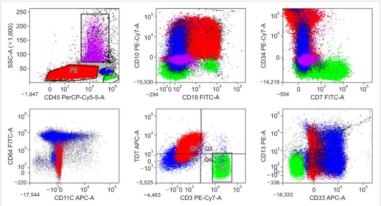
Fig. 2. Flow cytometric immunophenotyping plots. Flow cytometric immunophenotyping identified two abnormal populations, i.e., B-lymphoblasts (red) and monoblasts/promonocytes (blue), indicating B/myeloid MPAL.
nation showed bilateral cervical lymphadenopathy and hepatosplenomegaly. Contrast-enhanced computed tomog- raphy (CECT) showed mediastinal lymphadenopathy with bilateral pleural effusion. The hemogram showed a white blood cell (WBC) count of 19.5×10⁹/L, a hemoglobin level of 11.0 g/dL, and a platelet count of 171×109/L. No blasts were observed in the peripheral blood film. The bone mar- row aspirate and biopsy showed >80% blasts. These blasts were negative for myeloperoxidase (MPO), Sudan Black B (SBB), non-specific esterase (NSE), and periodic acid-Schiff (PAS) stain. Flow cytometric immunophenotyping of the bone marrow was performed at an external diagnostic cen- ter, which showed an immunophenotype consistent with precursor T-ALL. The blasts were positive for CD34, cyto- plasmic CD3, CD5, CD7, and CD45 and negative for CD19, CD20, CD10, CD117, CD22, CD33, and MPO. Karyotype analysis was unsuccessful as no metaphase chromosomes were detected in the culture. Genetic studies were negative for 11q23 (MLL) gene rearrangement (11q23 probe; Cancer Genetics Inc., Rutherford, NJ, USA). The central nervous system was not involved. The patient was treated with an appropriate chemotherapy protocol (hyper-CVAD regi- men). He achieved remission after 8 cycles of chemotherapy with complete blood count recovery. Subsequently, he was lost to follow-up at our hospital and reportedly continued treatment elsewhere. He was doing well post-treatment un- til mid-2020.
The patient was again admitted to our hospital in mid-2020 (13 years after the initial presentation in 2007)
with complaints of fever and inguinal lymphadenopathy.
No hepatosplenomegaly was observed on clinical examination.
He had a WBC count of 16.0×10⁹/L with 18% blasts and monocytosis (Fig. 1A). Hemoglobin and platelet counts were 5.3 g/dL and 60×109/L, respectively. The bone marrow aspi- rate showed two distinct abnormal populations on morpho- logical examination; the first population consisted of me- dium-sized, round agranular blasts (65% of all nucleated cells), and the second population consisted of monoblasts, promonocytes, and abnormal monocytes (30% of all nucleat- ed cells) (Fig. 1B, C). Flow cytometric immunophenotyping performed on the bone marrow sample showed two distinct abnormal cell populations (Fig. 2). The first population (45%
of all viable singlets) showed negative to moderate CD45 expression and low side scatter (progenitor population). The cells expressed CD19, CD10, cCD79a, CD34, TdT, HLA-DR, CD13, and CD64 and were negative for c-MPO, CD117, CD33, cytoCD3, CD7, CD11c, CD2, and CD5. The second population was detected in the monocyte region and in- cluded 20% of all viable singlets. The monocytic population expressed CD34, CD64, and CD11c (dim). On cytochemistry, these blasts were positive for NSE (Fig. 1D) and negative for MPO and PAS stain. A diagnosis of B/myeloid MPAL was made. Cytogenetic analysis by conventional karyotyp- ing showed no structural chromosomal anomalies. Targeted next-generation sequencing (NGS) showed mutations in ASXL1 (c.1926_1927insG), ETV6 (c.313_314insTGGGCCCT), and RUNX1 (c.592G>A, IKZF1c.476A>C) with mutant allele percentages of 32.8%, 41.5%, and 45.2%, respectively.
Blood Res2021;56:44-58. bloodresearch.or.kr
52 Letters to the Editor
The patient was treated using an augmented Berlin- Frankfurt-Münster (BFM) chemotherapy protocol. On day 29 of treatment (post-induction therapy), the bone marrow showed complete remission with incomplete platelet recov- ery (platelet count, 25×109/L).
MPAL accounts for 2–4% of all acute leukemia cases, and prognosis is poor compared with that for ALL or AML [4]. MPAL is genetically extremely heterogeneous. According to the World Health Organization (WHO), a diagnosis of MPAL requires the expression of lineage-specific markers in at least two lineages [4]. Lineage switch occurs in 6–9%
of acute leukemia cases during relapse [3]. Switching from ALL to AML is associated with MLL gene rearrangement in most cases, especially in cases of pediatric leukemia [5, 6].
These cases are associated with poor survival. Lineage switch may occur due to a highly plastic original clone or the emergence of a new clone; however, the exact mechanism remains unclear [3]. One of the several hypotheses suggests that the original committed neoplastic clone is highly plastic, and phenotypic changes may be observed with or without changes in the genotype [3]. The possible role of the micro- environment in the switch is also suggested [3]. In clonal selection theory, the emergence of resistant subclones with different lineages at relapse has been hypothesized [7]. In B-ALL to myeloid conversion or vice versa, the role of a common B-myeloid progenitor is suggested. Myeloid switch from B-ALL can occur either by trans-differentiation, de-differentiation, or re-differentiation [8]. Lineage switch in acute leukemia is uncommon, partly because repeat im- munophenotyping is not always performed at relapse. Most of the information found in the published literature de- scribes a switch from B-ALL to AML or the reverse. Some studies reporting a switch from T-ALL to AML have been published [9-11]. However, lineage switch from T-ALL to B/myeloid MPAL appears to be extremely rare. To the best of our knowledge, this has not been published previously.
Relapse in childhood acute leukemia usually occurs with- in 2 years and is thought to arise from the same clone as that at the initial presentation. Late relapse may indicate a completely new clone, which is characteristic of secondary leukemia and associated with a prior history of cytotoxic therapy [12]. The presence of therapy-related myeloid neo- plasms (t-MNs) is a late effect of chemotherapy (alkylating agents/topoisomerase II inhibitors) and/or radiation after the treatment of a primary disease (post-transplant lympho- proliferative disorders following solid organ transplantation) [13]. t-MNs are chemoresistant and have an aggressive course with a survival time of several months [13]. t-MNs associated with treatment using alkylating agents almost always have a dysplastic component in the marrow with cytogenetic anomalies such as del5q, del7q, or monosomy 7 on karyotyping [14]. Secondary ALL has been suggested to be constitutional and not related to any prior therapy [15]. In this case, targeted NGS analysis did not show any findings suggestive of germline mutations or constitutional abnormalities. The patient did not have any persistent symp-
toms attributable to cytopenias of myelodysplastic syndrome (MDS) prior to the second presentation. Karyotyping analy- sis at relapse did not reveal any MDS-defining cytogenetic abnormality, which is common in therapy-related myeloid neoplasms following the use of alkylating agents. Therefore, constitutional genetic abnormalities, MDS, and t-MNs were less likely in this case. The emergence of a resistant subclone at relapse or a highly plastic original clone may be respon- sible for the lineage switch in this case.
Cases of leukemia with lineage switch may have distinct biological characteristics and clinical features and may be associated with a poor prognosis and response to therapy [1]. Changes in the blast morphology and immunophenotype suggested lineage switch in this case. The recognition of this event may help to guide appropriate investigation and planning for therapy. Overall, the findings demonstrated the importance of repeat immunophenotyping even with acute leukemia relapse to identify relevant cases for proper management.
Asish Rath, Tribikram Panda, Rishi Dhawan, Jasmita Dass, Manoranjan Mahapatra, Ganesh Kumar Viswanathan Department of Hematology, All India Institute of Medical
Sciences, New Delhi, India
Correspondence to: Ganesh Kumar Viswanathan Department of Hematology, All India Institute of Medical
Sciences, Room no. 206, New Private Ward, AIIMS, New Delhi 110029, India E-mail: [email protected]
Received on Oct. 27, 2020; Revised on Dec. 26, 2020; Accepted on Jan. 19, 2021 https://doi.org/10.5045/br.2021.2020268
AuthorsÊ Disclosures of Potential Conflicts of Interest No potential conflicts of interest relevant to this article were reported.
REFERENCES
1. Rossi JG, Bernasconi AR, Alonso CN, et al. Lineage switch in childhood acute leukemia: an unusual event with poor outcome.
Am J Hematol 2012;87:890-7.
2. Wu B, Jug R, Luedke C, et al. Lineage switch between B-lympho- blastic leukemia and acute myeloid leukemia intermediated by
“occult” myelodysplastic neoplasm: two cases of adult patients with evidence of genomic instability and clonal selection by chemotherapy. Am J Clin Pathol 2017;148:136-47.
3. Dorantes-Acosta E, Pelayo R. Lineage switching in acute leuke- mias: a consequence of stem cell plasticity? Bone Marrow Res 2012;2012:406796.
4. Charles NJ, Boyer DF. Mixed-phenotype acute leukemia: diag- nostic criteria and pitfalls. Arch Pathol Lab Med 2017;141:
1462-8.
5. Sakaki H, Kanegane H, Nomura K, et al. Early lineage switch in an infant acute lymphoblastic leukemia. Int J Hematol 2009;
bloodresearch.or.kr Blood Res 2021;56:44-58.
Letters to the Editor 53
Table 1. Platelet cross-match, platelet antibody screening and HPA genotyping results.
Tests Results
Platelet antibody screening with baby’s
serum Positive
Platelet antibody screening with
mother’s serum Positive
Mother’s serum cross-matched with
baby’s platelets Incompatible
Mother’s serum cross-matched with
husband’s (father’s) platelets Incompatible Baby’s serum cross-matched with
father’s platelets Incompatible
Incompatibility % during platelet
cross-matching: from day 1 to day 20 50% (8/16) Incompatibility % during platelet
cross-matching: from day 21 to day 40 30% (6/20) Incompatibility % during platelet
cross-matching: from day 41 to day 61 18.2% (4/22) HPA genotyping of baby and parents to
detect HPA incompatibility in the baby HPA-15b incompatibility detected in the baby Abbreviation: HPA, human platelet antigen.
90:653-5.
6. Heidenreich O, Tirtakusuma R, Bomken S, et al. The genomic landscape of lineage switch acute leukemia. Blood (ASH Annual Meeting Abstracts) 2013;122(Suppl):2552.
7. Jiang JG, Roman E, Nandula SV, Murty VV, Bhagat G, Alobeid B. Congenital MLL-positive B-cell acute lymphoblastic leuke- mia (B-ALL) switched lineage at relapse to acute myelocytic leu- kemia (AML) with persistent t(4;11) and t(1;6) translocations and JH gene rearrangement. Leuk Lymphoma 2005;46:1223-7.
8. Ruiz-Delgado GJ, Nuñez-Cortez AK, Olivares-Gazca JC, Fortiz YC, Ruiz-Argüelles A, Ruiz-Argüelles GJ. Lineage switch from acute lymphoblastic leukemia to myeloid leukemia. Med Univ 2017;19:27-31.
9. Aujla A, Hanmantgad M, Islam H, Shakil F, Liu D, Seiter K.
Lineage switch from T-cell lymphoblastic leukemia/lymphoma to acute myeloid leukemia and back to T-cell lymphoblastic leu- kemia/lymphoma in a patient diagnosed during pregnancy. Stem Cell Investig 2019;6:12.
10. Ittel A, Jeandidier E, Helias C, et al. First description of the t(10;11)(q22;q23)/MLL-TET1 translocation in a T-cell lympho- blastic lymphoma, with subsequent lineage switch to acute mye- lomonocytic myeloid leukemia. Haematologica 2013;98:e166-8.
11. Higuchi Y, Tokunaga K, Watanabe Y, et al. Lineage switch with t(6;11)(q27;q23) from T-cell lymphoblastic lymphoma to acute monoblastic leukemia at relapse. Cancer Genet 2016;209:267-71.
12. Babić A, Kurić L, Dubravčić K, et al. A case of an unusual lineage switch in late relapse ALL—is it actually a secondary leukemia?
J Hematop 2020;13:51-5.
13. Ganser A, Heuser M. Therapy-related myeloid neoplasms. Curr Opin Hematol 2017;24:152-8.
14. McNerney ME, Godley LA, Le Beau MM. Therapy-related mye- loid neoplasms: when genetics and environment collide. Nat Rev Cancer 2017;17:513-27.
15. Ganzel C, Devlin S, Douer D, Rowe JM, Stein EM, Tallman MS.
Secondary acute lymphoblastic leukaemia is constitutional and probably not related to prior therapy. Br J Haematol 2015;170:
50-5.
Utility of platelet cross-matching in a case of neonatal alloimmune
thrombocytopenia associated with a de novo MECOM variant
TO THE EDITOR: Neonatal alloimmune thrombocytopenia (NAIT) is the most important cause of thrombocytopenia in term neonates. It was estimated to occur at a frequency of 1 in 1,000–2,000 live births [1]. Platelet destruction is caused by a maternal antibody directed against a fetal human platelet antigen (HPA) inherited from the father and lacking in the platelets of the mother. In the Caucasian population, 80% alloimmunization occurs predominantly due to anti- bodies to the HPA-1a antigen [1]. Although less common,
the HPA-15 antigen system is also implicated in serologically confirmed NAIT cases [2]. The HPA-15a15b system is lo- cated on the glycosyl-phosphatidylinositol-anchored pro- tein, CD109, and is defined by the single amino acid sub- stitution of Ser703Tyr (Ser682Tyr of mature CD109 protein) [3]. The diagnosis of NAIT can be established by document- ing the presence of a platelet-specific antigen incompatibility between mother and infant (or mother and father) and the presence of maternal antiplatelet allo-antibodies di- rected against the incompatible HPA antigen [4]. The mono- clonal antibody-specific immobilization of platelet antigen (MAIPA) assay is considered as the ‘gold-standard’ test to detect maternal anti-HPA antibodies [4]. However, it is not available for routine use in India. Platelet cross-match, which is commonly used to provide platelet transfusion support in platelet refractoriness, may be considered as an alternative strategy to manage emergency cases of NAIT in resource-limited facilities where a platelet-donor registry is still unavailable [5].
In 2015, heterozygous mutations in the MECOM (MDS1 and EVI1 complex locus) were first identified as a cause of amegakaryocytic thrombocytopenia in neonates [6].
There are many novel mutations in the MECOM gene that have been identified recently, and a new name, ‘MECOM- associated syndrome,’ has been proposed for this disease [7]. Herein, we report a rare case of NAIT associated with HPA-15b incompatibility in a baby who was managed ini- tially with the transfusion of cross-matched platelets, but due to persistent transfusion-dependent thrombocytopenia and inadequate response to appropriate therapy such as IVIG, whole exome sequencing was performed, which re-